


Murilo Augusto Ferreira; Luiz Gustavo Ferreira Fressatti; Luís Henrique Barbizan de Moura; Samira Yarak
Submitted on: 11/04/2023
Approved on: 02/05/2023
Financial support: None.
Conflict of interest: None
How to cite this article: Ferreira MA, Fressatti LGF, Moura LHB, Yarak S. Case series: variation of the Mercedes flap in the scalp. Surg Cosmet Dermatol. 2023;15:e20230247.
Reconstruction of the scalp after suspected skin lesions excision is a challenge. Tamir et al. described the three-point-advancement (Mercedes flap) to close circular wounds. These tissue advancements create triangles in the edges, which must be removed. However, extensive lesions do not allow the three points to join, preventing central closure. Valesky et al. described the central closure using total skin grafts made from the triangles. The literature lacks these case reports, making it difficult to incorporate them into surgical practice. We applied this modified technique to five patients with good aesthetic and functional results.
Keywords: Scalp; Surgical flaps; Plastic surgery procedures; Skin transplantation; Skin neoplasms
Scalp reconstruction after the excision of skin lesions is a surgical challenge due to the low elasticity of the galea aponeurotica, convexity of the skull, and reduced local vascularization.1,2 To close circular lesions in challenging locations such as the scalp, Tamir et al. described the three-points-advancement that meets in the center, known as the Mercedes flap.3,4 The advancement of the tissue invariably creates everted marginal cones, which must be excised.
However, in surgical practice, extensive wounds do not allow the three points to meet, preventing central closure. In this context, Valesky et al. described a variation of the Mercedes flap with central closure from a total skin graft made with Burrow’s triangles from excised cones.5 Despite the promising results, the literature lacks cases describing this technique, making its dissemination and incorporation into surgical practice difficult.6
This study aims to report the applicability of the Valesky technique in a series of five patients and to encourage its use in relevant cases.
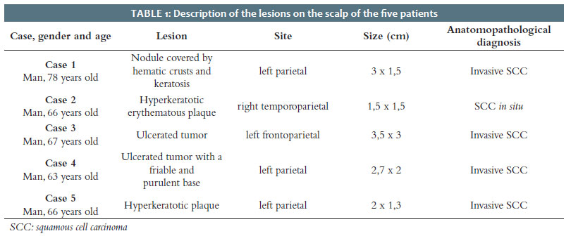
We describe five cases that underwent removal of squamous cell carcinomas from the scalp, in which the modified Mercedes flap technique was used.
The project was submitted to the Research Ethics Committee and approved under registration CAAE 65768322.6.0000.5505.
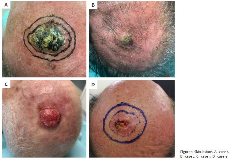
Case 1: A 78-year-old man presented a nodular lesion measuring 3 x 1.5 cm on the scalp, covered by hematic crusts and keratosis (Figure 1A).
Case 2: A 66-year-old man, kidney transplant recipient, presented a 1.5 x 1.5 cm hyperkeratotic tumor in the right temporoparietal region (Figure 1B).
Case 3: A 67-year-old man, a kidney transplant recipient, presented an ulcerated tumor measuring 3 x 3.5 cm in the left frontoparietal region and a history of multiple non-melanoma skin neoplasms. The patient had metastasis to cervical lymph nodes, lungs, and pleura, resulting in death (Figure 1C).
Case 4: A 63-year-old man, kidney transplant recipient, presenting an ulcerated tumor with a friable bottom and purulent secretion, measuring 2.7 x 2 cm in the left parietal region (Figure 1D).
Case 5: A 66-year-old man, a kidney transplant recipient, presented a hyperkeratotic plaque in the left parietal region measuring 2 x 1.3 cm (Table 1).
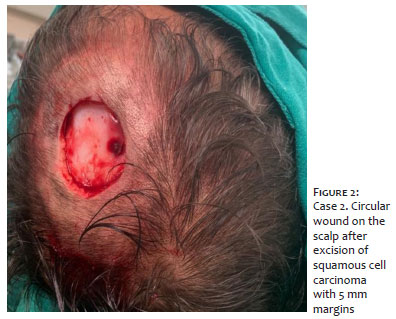
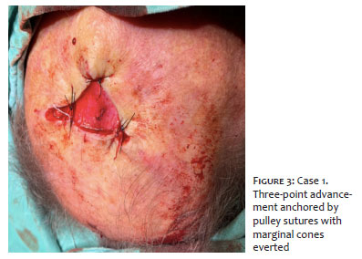
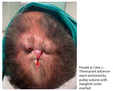
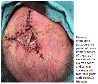
After scalp lesions resection, we dissected the plane between the galea aponeurotica and the periosteum to add mobility to the adjacent tissue (Figure 2). We performed a three-point-advancement using three pulley sutures parallel to the tangent of three points equidistant from the circumference, leaving the center without skin coverage (Figures 3 and 4). Then, we excised the everted cones at the three ends into Burrow’s triangles and sutured the resulting edges with simple stitches. Central closure was performed with total skin grafts meshed with Burrow’s triangles (Figures 5 and 6). We made Brown’s dressing for better adherence and nutrition of the graft and removed it a week later.
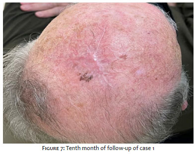
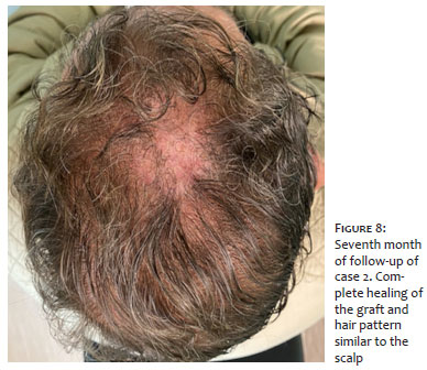
All patients had excellent functional and aesthetic results (Figures 7 and 8). Follow-up of one of the patients was not possible due to his death.
Several techniques can be used for scalp synthesis. Primary closure represents the simplest, but it promotes everted cones at the margins and generates significant unidirectional tension, causing deformities. The total skin graft requires additional incisions in other sites, in addition to being susceptible to losses due to poor nutrition of the periosteum and not being aesthetic on the scalp due to differences in the hair pattern between the donor and recipient areas. Distance pedicled flaps and free flaps are restricted to cases of major tissue loss or significant damage to the periosteum, given their high complexity and great metabolic and anesthetic demand. These factors should be considered especially for elderly patients.2
The local flap offers excellent aesthetic results due to hair similarity. Also, it is less invasive, reducing the morbidity of the procedure. In extensive wounds, however, its use may be unfeasible, especially in wounds larger than 100 cm.2,7
In this context, the modified Mercedes flap represents a good alternative for closing extensive circular wounds on the scalp. Its advantages are less complexity and less metabolic demand, absence of additional incisions to capture the graft, good aesthetic results due to capillary similarity, and division of wound tension in three directions.8 The central coverage with Burrow’s triangle prevents closure by secondary intention. Also, it works as a biological dressing and allows the formation of granulation tissue with, theoretically, reduced risk of infection. The application of the technique is not restricted to the scalp, and different authors have described its usefulness in other challenging regions, such as the heel, malleolus, eyebrow, and temporal region.4,5
The study limitations are limited evidence power of the case series, subjectivity in the analysis, and lack of a control group.
The Mercedes flap combined with a total skin graft with Burrow’s triangles proves to be a good option for scalp synthesis, especially in treating transplant patients or elderly people who have wounds without the possibility of central closure with simple three-point advancement.
Murilo Augusto Ferreira
ORCID: 00-0002-9588-5678
Approval of the final version of the manuscript; study design and planning; preparation and writing of the manuscript; collecting, analyzing, and interpreting data; critical literature review; critical review of the manuscript.
Luiz Gustavo Ferreira Fressatti
ORCID: 00-0002-9449-1351
Approval of the final version of the manuscript; study design and planning; preparation and writing of the manuscript; collecting, analyzing, and interpreting data; critical literature review; critical review of the manuscript.
Luís Henrique Barbizan de Moura
ORCID: 00-0002-5714-8386
Study design and planning; collecting, analyzing, and interpreting data; intellectual participation in propaedeutic and/or therapeutic conduct of studied cases.
Samira Yarak
ORCID: 00-0002-5657-6645
Statistical analysis; approval of the final version of the manuscript; study design and planning; collecting, analyzing, and interpreting data; effective participation in research orientation; intellectual participation in propaedeutic and/or therapeutic conduct of studied cases; critical literature review; critical review of the manuscript.
1. Russo F. Mercedes flap with releasing incisions for scalp defects. Ann Plast Surg. 2017;79(2):149-55.
2. Ehrl D, Brueggemann A, Broer PN, Koban K, Giunta R, Thon N. Scalp reconstruction after malignant tumor resection: an analysis and algorithm. J Neurol Surg B Skull Base. 2020;81(2):149-57.
3. Tamir G, Birkby CS, Berg D. Three point-advancement closure for skin defects. J Cutan Med Surg. 1999;3(6):288-92.
4. Xue S, Mutesi R, Rong M, Liu J. The Mercedes flap: a modified closure for circular skin defects around the eyebrow. Clin Exp Dermatol. 2013;38(7):816-7.
5. Valesky EM, Kaufmann R, Meissner M. The Mercedes flap and its new variants: a 'workhorse' flap for the dermatological surgeon? J Eur Acad Dermatol Venereol. 2016;30(8):1332-5.
6. Chow M, Swift R, Higgins S, Wysong A. Triple advancement flap for the lateral upper forehead and temple. J Cutan Med Surg. 2018;22(5):533-4.
7. Denewer A, Khater A, Farouk O, Hegazy M, Mosbah M, Hafez M, et al. Can we put a simplified algorithm for reconstruction of large scalp defects following tumor resection? World J Surg Oncol. 2011;9:129.
8. Kaufman AJ. Adjacent-tissue skin grafts for reconstruction. Dermatol Surg. 2004;30(10):1349-53.
 All content the journal, except where identified, under the Creative Commons Attribution 4.0 International licence - ISSN-e 1984-8773


 Read in Portuguese
 Portuguese PDF
 Print
Print
 Send this article by email
 How to cite this article
 Submit a comment
 Mendeley
 Pocket