


Srinjoy Saha
Financial support: none
Conflict of interest: none
How to cite this article: Saha S. Composite fat grafts for correcting facial dystrophy in a case of localized scleroderma. Surg Cosmet Dermatol. 2023;15:e20230181.
Localized scleroderma (linear morphea) is a rare disease that causes facial dystrophy and asymmetry in young women. We report the case of a 20-year-old female patient who was treated with novel composite fat grafts. After extracting lipoaspirate from deep subcutaneous fat, the microfat and the unfiltered nanofat were processed separately, and amalgamated, obtaining composite fat grafts. These were meticulously transplanted layer by layer to rebuild the subcutaneous tissue of the patient's face. After one year, a good volume of fat retention was observed with successful correction of asymmetry and restoration of facial balance.
Keywords: Ambulatory surgical procedures; Abdominal subcutaneous fat; Facial asymmetry; Subcutaneous injections; Localized scleroderma
Localized scleroderma is a rare autoimmune disorder that primarily affects young women. It is also known as linear morphea and is characterized by progressive skin fibrosis. There is currently no cure. Medical treatments can slow the disease progression and keep patients in remission, but they cannot prevent complications such as skin fibrosis and facial asymmetry.1 In some young women, the psychological effects of facial disfigurement can be devastating, as it can severely impair self-image and cause depression. Currently available surgical options involve adding volume to the affected facial areas. Microsurgical free tissue transfers, derma-fat grafts, bone grafts, implants, artificial dermis, dermal fillers, and autologous fat grafts are all plastic surgeries that correct volume deficits. Fat grafting is a simple but effective option for large-volume filling of facial deformities with minimal complications.2
A woman in her twenties with Fitzpatrick skin type V presented to our plastic surgery clinic with facial dystrophy involving the left side, leading to facial asymmetry. She had previously been diagnosed with localized scleroderma and had completed medical treatment to induce remission in scleroderma progression.
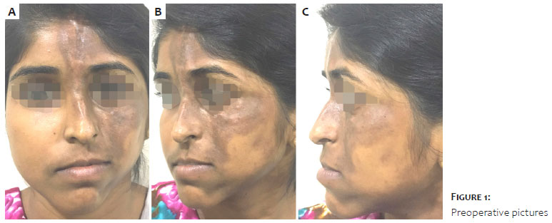
Another surgeon performed iliac crest bone grafting on her two years ago to correct the “coupe de sabre” defect over her forehead and elevate the left malar eminence. She was, however, dissatisfied with the results of her previous surgery and desired improved facial soft-tissue contouring and symmetry. On examination, she had hyperpigmented fibrotic skin patches with atrophic subcutaneous tissues on her left forehead, cheek, nose, and chin. Scarred, thin, hyperpigmented skin adhered to previously applied bone grafts in patchy areas of her forehead and cheek, with no subcutaneous tissues beneath (Figures 1A-C). Her mouth opening was adequate, and she did not have dry mouth. There was no evidence of significant dystrophy or deviation of the mandible on radiography. She declined other reconstructive surgeries, such as microvascular free tissue transfer and cosmetic dermal fillers, after extensive counseling about her treatment options, preferring fat grafts instead.
We infiltrated the deep subcutaneous layers of abdominal fat with a tumescent solution (1000 ml of normal saline solution mixed with 25 ml of 2% lidocaine, 1 ml of 1:1000 epinephrine, and 10 ml of 8.4% sodium bicarbonate) while the patient was sedated. Lipoaspirate was extracted using a low-suction technique with a 3 mm cannula with 1 mm side holes and a 10 ml Luer lock syringe. It was gravity sedimented for 15 minutes to allow fat in the middle layer to separate from the upper oil and lower blood layers, which were discarded.3
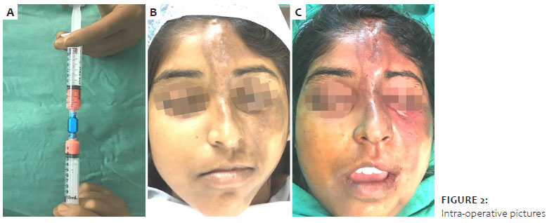
Then, some of the harvested lipoaspirates were converted to unfiltered nanofat grafts by repeated mechanical flocculation.4 Lipoaspirate was forced through fat-transfer adapters with progressively smaller diameters of 2.4 mm, 1.4 mm, and 1.2 mm until it became completely fluidic (Figure 2A). Microfat and unfiltered nanofat were thoroughly mixed in a 4:1 ratio and transferred into several 1 ml syringes attached to a 1 mm blunt-tip cannula for facial fat grafting to form composite fat grafts.
Drop by drop and layer by layer, 25 ml of combined fat grafts were carefully injected into the affected forehead, cheek, nose, and chin.3,4 Fat injections continued until her fibrotic facial skin allowed no more stretch. Bony contours over her left cheek, nose, and chin were augmented, while the sharp edges of a previously placed bone graft over her forehead were contoured. The immediate postoperative results were satisfactory, with the adequate restoration of facial symmetry and balance (Figures. 2B-C). However, due to the tension on the overlying skin, it was not possible to overfill the affected side as is commonly done. The patient’s postoperative recovery was uneventful, with no complications.
The patient was followed up on for a year after surgery, which was uneventful.
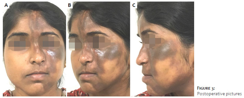
Before and one year after the surgery, we used a patient-reported outcomes questionnaire to document treatment satisfaction through a visual analog scale. The patient perceived a significant improvement in all of the individually treated areas of her face, but especially in the cheeks, chin, and overall facial appearance (Table 1). After one year, there was a good volume of fat graft retention and an improvement in skin quality (Figures 3A-C).
Immunologically overactive fibroblasts play a key role in scleroderma pathogenesis, secreting collagen, and extracellular matrix excessively within the affected dermis, resulting in fibrosis.5 Facial stiffness, asymmetry, and deformity are all symptoms of progressive cutaneous fibrosis. Loss of elasticity and thickening of the skin around the mouth can cause mouth narrowing and lead to difficulties in eating, speaking, brushing, maintaining oral hygiene, and dental treatments.6 All these factors, when combined, severely impair a patient’s daily life.
Autologous fat grafting was beneficial in two ways. First, as a large-volume filler, it corrected the facial asymmetry. Second, because of the presence of pre-adipocytes and adipose-derived stem cells, the regenerative potential of fat grafts improved the quality of overlying skin.7 Fat grafting has been shown to reverse fibrosis of “coupe de sabre” lesions over the forehead in patients with localized scleroderma, improving mouth opening and reducing fibrosis in patients with systemic sclerosis.8,9
Autologous fat grafting is commonly used worldwide for facial soft tissue augmentation, with generally positive and satisfying results.10 A variant produced after repeated flocculation, called unfiltered nanofat, was used to satisfactorily rejuvenate postburn scars on the face in an earlier study.11 It contained pre-adipocytes and adipose-derived regenerative cells, as well as better regeneration capabilities.12
A gradual decrease in the volume of transplanted fat remained an area of concern, necessitating follow-up procedures every six months. The initial fat graft’s take was determined by the size of the fat droplets and whether it was placed like a string of beads, allowing vascularity to develop between the deposited fat parcels.3 Higher amounts of a stromal vascular fraction rich in pre-adipocytes and adipose-derived stem cells increased revascularization. Previously, nanofat processing with repeated mechanical flocculation revealed a high concentration of stem cells in unfiltered nanofat grafts.4,12
In this case, it was hypothesized that composite fat grafts combining microfat and unfiltered nanofat would increase the retention volume after fat transplant.
High levels of patient satisfaction and a high volume of grafted fat retention one year after surgery demonstrated that composite fat grafts were beneficial. It reduced the need for fat grafts while improving the quality of the atrophic skin and subcutaneous tissues. A larger study with a longer patient follow-up is needed to investigate the benefits of such a combination of fat grafts.
In a patient with linear scleroderma, combining microfat with unfiltered nanofat to form composite fat grafts allowed the grafted fat to survive longer. It significantly improved the appearance and quality of the affected facial skin and had high satisfaction rates on patient-reported outcomes questionnaires.
Srinjoy Saha 0000-0001-8971-148X
Statistical analysis; approval of the final version of the manuscript; study design and planning; preparation and writing of the manuscript; data collection, analysis, and interpretation; active participation in research orientation; intellectual participation in propaedeutic and/or therapeutic conduct of studied cases; critical literature review; critical revision of the manuscript.
1. Zulian F, Culpo R, Sperotto F, Anton J, Avcin T, Baildam EM, et al. Consensus-based recommendations for the management of juvenile localised scleroderma. Ann Rheum Dis. 2019;78(8):1019-24.
2. Strong AL, Rubin JP, Kozlow JH, Cederna PS. Fat grafting for the treatment of scleroderma. Plast Reconstr Surg. 2019;144(6):1498-1507.
3. Shauly O, Gould DJ, Ghavami A. Fat Grafting: basic science, techniques, and patient management. Plast Reconstr Surg Glob Open. 2022;10(3):e3987.
4. Lombardo GAG, Tamburino S. The unfiltered nanofat: a great source of staminal cells. Ann Plast Surg. 2019;83(4):488.
5. Kumánovics G, Péntek M, Bae S, Opris D, Khanna D, Furst DE, et al. Assessment of skin involvement in systemic sclerosis. Rheumatology (Oxford). 2017;56(suppl 5):v53-v66.
6. Lauesen SR, Daugaard-Jensen J, Lauridsen EF, Kjær I. Localised scleroderma en coup de sabre affecting the skin, dentition and bone tissue within craniofacial neural crest fields. Clinical and radiographic study of six patients. Eur Arch Paediatr Dent. 2019;20(4):339-50.
7. Magalon G, Daumas A, Sautereau N, Magalon J, Sabatier F, Granel B. Regenerative approach to scleroderma with fat grafting. Clin Plast Surg. 2015;42(3):353-64
8. Barin EZ, Cinal H, Cakmak MA, Tan O. Treatment of linear scleroderma (en Coup de Sabre) with dermal fat grafting. J Cutan Med Surg. 2016;20(3):269-71.
9. Scuderi N, Ceccarelli S, Onesti MG, Fioramonti P, Guidi C, Romano F, et al. Human adipose-derived stromal cells for cell-based therapies in the treatment of systemic sclerosis. Cell Transplant. 2013;22(5):779-95.
10. Gornitsky J, Viezel-Mathieu A, Alnaif N, Azzi AJ, Gilardino MS. A systematic review of the effectiveness and complications of fat grafting in the facial region. JPRAS Open. 2018;19:87-97.
11. An SN, Bashir MM, Khan FA, Hidayat Z, Ansari HH, Sohail M, et al. Unfiltered Nanofat Injections Rejuvenate Postburn Scars of Face. Ann Plast Surg. 2019;82(1):28-33.
12. Lo Furno D, Tamburino S, Mannino G, Gili E, Lombardo G, Tarico MS, et al. Nanofat 2.0: experimental evidence for a fat grafting rich in mesenchymal stem cells. Physiol Res. 2017;66(4):663-71.
 All content the journal, except where identified, is under a Creative Commons Attribution-NonCommercial 4.0 International license - ISSN-e 1984-8773


 Read in Portuguese
 Portuguese PDF
 Print
Print
 Send this article by email
 How to cite this article
 Submit a comment
 Mendeley
 Pocket