


Grasiela Cassia Monteiro1; Carolina Matté-Dagostini2; Pedro Henrique Lodi2; Samantha Lia Ziotti-Bohn-Gonçalves-Soares2; Fernando De-Marco-Dos-Santos1
Submitted on: 21/10/2021
Approved on: 27/01/2022
Financial support: None
Conflict of interest: None
Acknowledgments: The authors thank all the professionals who assisted in the care of patients in the skin cancer awareness campaign in December 2016, 2017, and 2018, at the University of Caxias do Sul, especially the coordinator of CECLIN-UCS, Mr. Jose Henrique Teixeira
How to cite this article: Monteiro GC, Matté-Dagostini C, Lodi PH, Ziotti-Bohn-Gonçalves-Soares SL, De-Marco-Dos-Santos F. Skin cancer awareness campaign in Southern Brazil: A retrospective cohort study. Surg Cosmet Dermatol. 2022;14:e20220105
INTRODUCTION: Skin cancer is the most frequent malignant neoplasm in Brazil. Its prognosis is directly related to early diagnosis and institution of adequate treatment. The Skin Cancer National Awareness Campaign (SCNAC) is an essential tool to prevent and detect malignant skin lesions.
OBJECTIVES: To investigate the incidence of skin cancer and the accuracy of dermoscopy in patients assessed at the SCNAC.
METHODS: We conducted a retrospective cohort study using directly the data collected from the population assisted at the SCNAC day in 2016, 2017, and 2018 at Clinical Center of the Universidade de Caxias do Sul (CECLIN-UCS).
RESULTS: Of the 634 patients included, 105 were referred for biopsy with histopathological study of the lesion. Dermoscopy was suggestive of a malignant lesion in 55 cases. Biopsy diagnosed malignant lesions in 43 patients and benign lesions in 32 patients. Thus, the sensitivity and specificity of the test were 86% and 50%, respectively. The accuracy of dermoscopy in identifying malignant lesions over the three years of the campaign was around 70%.
CONCLUSIONS: Dermoscopy in SCNAC has a good level of sensitivity and specificity when correlated with final histopathological results.
Keywords: Dermatology; Dermoscopy; Observational study; Skin neoplasms; Health promotion
Melanoma and non-melanoma skin cancer (squamous cell carcinoma and basal cell carcinoma) are the most frequent neoplasm in Brazil, corresponding to 27% of all malignant tumors.1 It is estimated that the number of new cases will increase in the coming years, with a higher incidence in Santa Catarina (SC) and Rio Grande do Sul (RS) in the southern region of Brazil.1 Current evidence indicates that early diagnosis with dermoscopic evaluation and timely treatment reduces skin cancer mortality and morbidity.2,3
Dermoscopy is a non-invasive test to assess skin lesions.4 However, histopathological examination is necessary for diagnostic confirmation.2 Previous studies suggest that dermoscopy increases the diagnostic accuracy of skin neoplasms compared to the naked eye examination.5-7 Previous meta-analysis indicated that the sensitivity of dermoscopy associated with clinical examination for the diagnosis of melanoma was 90% (95% CI 80-95) with a specificity of 90% (95%CI 57-98).8
Primary and secondary prevention of skin cancer in the adult population through interventions in skin cancer awareness campaigns has been proven to improve the diagnostic rate and can reduce the mortality of these neoplasms.9-11 Thus, the Brazilian Society of Dermatology (Sociedade Brasileira de Dermatologia - SBD) has promoted, since 1999, the National Skin Cancer Awareness Campaign (NSCAC). It consists of a day in December reserved for a free examination of the population by dermatologists, through dermoscopy and biopsies, associated with guidelines on sun exposure habits and skin cancer prevention.
The data collected at the NSCAC can be analyzed to present an overview of the prevention and detection of skin cancer in this population. Therefore, this study aims to investigate the incidence of skin cancer and the accuracy of dermoscopy of patients treated in the National Skin Cancer Awareness Campaign held at the University of Caxias do Sul (RS) during the years 2016, 2017, and 2018.
Study design
A multidisciplinary team of dermatologists, clinicians, surgeons, pathologists, nurses, and academics from the Medical School develops the National Skin Cancer Prevention Campaign, promoted by the Brazilian Society of Dermatology. It takes place annually at the Clinical Center of the University of Caxias do Sul (CECLIN-UCS) in Caxias do Sul (RS), Brazil. The campaign encompasses dermatological examination, histopathological diagnosis, clinical and surgical treatment, guidance, and patients’ follow-up.
The evaluators are responsible for filling out a standard form for each patient seen on the day of the campaign and, later, forwarding it to the Brazilian Society of Dermatology.
We conducted a retrospective cohort study using the data collected from the population assisted on the NSCAC day in 2016, 2017, and 2018 at CECLIN-UCS. The information obtained from the standard forms completed on the days of the campaigns was transcribed into a Microsoft Excel spreadsheet. The anatomopathological examinations were transcribed by consulting the medical records of patients undergoing skin biopsies.
Standard form of the National Skin Cancer Prevention Campaign
The standard form presented a table for recording age, biological sex, and skin phototype according to the Fitzpatrick classification; participant’s degree of sun exposure; the presence of previous pathological history, family history, or risk factors for the development of skin cancer; how the individual learned about the campaign; clinical hypothesis according to dermoscopy; evolution time or location if lesion present; treatment conducted from the first service in the campaign; histopathological result after biopsy of a suspicious lesion.
Research Ethics Committee
The Research Ethics Committee of the University of Caxias do Sul (CEP-UCS) previously approved this study under the number 24951659.3.0000.5.341. The principles of the Declaration of Helsinki were followed.
Population studied
To be included, patients had to: (1) be participating in the NSCAC of the respective year; (2) be ≥18 years of age. Incomplete forms were excluded from the analysis. In total, 634 patients were included in the study.
Endpoints
The primary endpoint was the accuracy of dermoscopy for the diagnosis of skin lesions. Secondary endpoints were: (1) skin cancer incidence by type; (2) analysis of the epidemiological characteristics of the participants of the NSCAC.
Statistical analysis
The authors used IBM's SPSS version 23.0 for Microsoft Windows. Frequency and accuracy measurements were obtained.
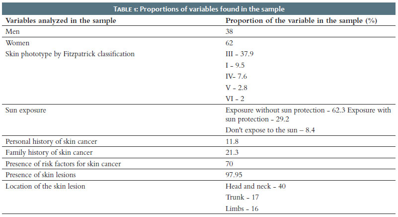
Table 1 shows the sample variables. The study included 634 patients, 62.1% women and 37.9% men. In the consultations, the responsible dermatologist classified all individuals according to their skin phototypes following the Fitzpatrick classification. Skin phototype II represented 48.1% of the sample, and it was the most identified, followed by: skin phototype III (37.9%), I (9.5%), IV (7.6%), V (2.8 %), and VI (2%). Only 8.4% of patients reported not exposing to the sun. About 62.3% of the subjects usually exposed to the sun without sun protection, and only 29.2% exposed to the sun using protection. Of the participants, 11.8% had a previous pathological history of skin cancer, and 21.3% had a positive family history of skin cancer. Thus, approximately 70% of the sample was classified as having recognized risk factors for malignant skin neoplasms.
We expected the participants to have skin lesions and seek clarification, guidance, and conduct about them when necessary. This demand was present in 98% of the sample. We asked the patients about the evolution time of their main lesion: 30.4% reported ≤1 year; 31.4%, between 1 and 2 years; and 30.4%, ≥3 years. Of the total sample, the examining physician did not describe the lesion site in 52 patients. Of those who did, 40% of lesions were located in the head, 17% in the trunk, and about 16% in the limbs.
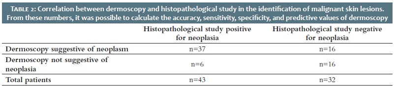
Of the 634 patients included in the research, 105 were referred for biopsy with the histopathological study of the lesion. Among these, we had access to the results of 75 patients, which represents 71% of the sample. Dermoscopy was suggestive of a malignant lesion in 55 cases and non-suggestive in 20 cases. Biopsy diagnosed malignant lesions in 43 patients and benign lesions in 32 patients. Thus, in the study, the sensitivity and specificity of the test were 86% and 50%, respectively. The positive and negative predictive values were 69% and 72%, respectively. We concluded that the accuracy of dermoscopy in identifying malignant lesions over the three years of the campaign was around 70%. In this context, table 2 demonstrates the correlation between dermoscopy and histopathological study in identifying malignant skin lesions.
NSCACs are essential for early diagnosis as they enable better monitoring of skin lesions and greater adherence to health promotion. Consequently, it is suggested that they act to reduce morbidity and mortality and trigger a more favorable prognosis.
The number of exams performed each year shows a growing trend, indicating higher adherence to NSCAC and its importance in controlling this diasease.12 On the other hand, unfortunately, the proportion of skin cancer in the population has increased over the years. In the present study, adherence to NSCAC was higher in individuals who already had skin lesions or risk factors for neoplasms and had a family history of cutaneous neoplasms.
The estimate of new cases of skin cancer is higher among women in Brazil.1 Studies suggest that women also have more significant participation in skin cancer prevention campaigns. Likewise, in the sample assessed, greater adherence of the female population to NSCAC is suggested. Also, this population is likely to be more attentive to protection strategies and more careful with sun exposure and the use of sunscreen.12 However, women are also more likely than men to sunbathe and use tanning methods.13
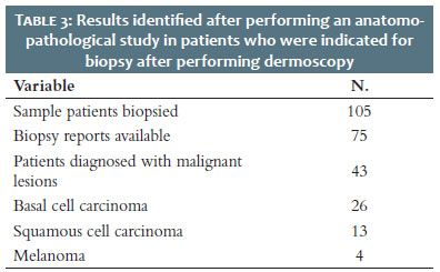
Regarding the subtypes of skin cancer, non-melanoma is the most frequent, and basal cell carcinoma is the most frequent in this group. Melanoma skin cancer represents 3% of malignant skin tumors in Brazil. However, it has the worst prognosis, with high invasive and metastatic power.1,14,15 In the evaluated sample, 60.5% of cases were diagnosed with basal cell carcinoma, 30.2% squamous with cell carcinoma, and 9.3% with melanoma, according to table 3. Simultaneously with the demographics of skin cancer,1 our research showed that the older the age, the greater the probability of having a diagnosis in NSCAC. That is, the incidence of cutaneous neoplasms increases with advancing age. According to an American analysis that followed 30 years of skin cancer screening in the United States, the diagnosis of melanoma doubled in individuals with an average age of 60 years and older. 16,17
The use of sun protection is still lower than necessary, according to a survey in the city of Rio Grande (RS).18 The study showed that the percentages of individuals in the NSCAC who did not use sunscreen in 2010 and 2011 were 53.01% and 45.58%, respectively. In this context, the present research showed that most patients (62.3%) of NSCAC, when exposed to the sun, did not use sun protection.
Regarding the relationship between skin phototype and suspected skin cancer, literature shows it is more frequent in skin phototypes I and II.19 Skin phototype II patients were the most adept at NSCAC performed in the southern region of Brazil, with a percentage of 48.1%, followed by patients with skin phototype III, with 31.9%. Therefore, the probability of NSCAC diagnosing a neoplasm in these patients would be increased due to their greater participation.
Based on the data collected in three years of the campaign, the present study compared the ability of dermoscopy to identify malignant lesions to the histopathological study, considered the gold standard. The physician who performed the dermoscopy made his main clinical hypothesis and requested a biopsy for diagnostic confirmation. The comparison was established by relating these two variables to the malignancy or not of the lesion and presented an accuracy of 70%. Similar European studies propose a detection rate for melanoma skin cancer ranging from 0.1% to 2%. Dermoscopy was used in around 78-80% of examinations with clinical suspicion of melanoma, and full-body skin examination was performed in 72-86% of patients.19, 20 By our results, research suggests that dermoscopy has high discriminatory power, reiterating that this technique also facilitates the detection of skin lesions at an early stage.9,19,21 Also, an Australian meta-analysis showed that the odds of finding a melanoma were nine times greater with dermoscopy than with naked eye examination alone.8
In our study, the physician who performed the dermoscopy developed his chief clinical hypothesis and requested a biopsy for diagnostic confirmation. The comparison was established by relating these two variables to the malignancy or not of the lesion. It is known that several dermatologists participated in the three-year campaign, that dermoscopy is a component of the physical examination of the dermatologist and that, as it is examiner-dependent, its accuracy tends to improve with clinical practice. Therefore, it is noteworthy that we did not use criteria to determine how much the physician's experience improves the accuracy of the exam, although the authors recognize that this would be relevant in future studies.
Strengths and limitations
To the authors’ knowledge, this is a differentiated study since it assesses the accuracy of dermoscopy for the first time in the southern region of Brazil. Furthermore, the number of participants was high (n=634), which provided statistical significance to the results. On the other hand, our study is also prone to bias. As this was a retrospective analysis, data on neoplasm subtypes and biopsy results could not be accessed. By including campaigns conducted in three consecutive years, we assumed that the evaluators comprised a heterogeneous group, which impacts the results of the accuracy of dermoscopy since the examiner experience has a direct relationship with the dermoscopy performance.
This study enabled us to conclude that dermoscopy in national skin cancer awareness campaigns has a good level of sensitivity and specificity when correlated with the final histopathological results. It is one of the pioneering studies on the accuracy of dermoscopy in skin campaigns in Brazil. Thus, it confirms that a skin examination including dermoscopy increases diagnostic accuracy. Also, it was conducted in one of the regions with the highest prevalence of skin cancer in the country. Therefore, its results can guide future research and health promotion measures.
Grasiela Cassia Monteiro 0000-0001-5110-8626
Statistical analysis; approval of the final version of the manuscript; study design and planning; preparation and writing of the manuscript; data collection, analysis, and interpretation; active participation in research orientation; intellectual participation in propaedeutic and/or therapeutic conduct of studied cases; critical literature review; critical revision of the manuscript.
Carolina Matté-Dagostini 0000-0003-4189-7339
Statistical analysis; approval of the final version of the manuscript; study design and planning; preparation and writing of the manuscript; data collection, analysis, and interpretation; active participation in research orientation; intellectual participation in propaedeutic and/or therapeutic conduct of studied cases; critical literature review; critical revision of the manuscript.
Pedro Henrique Lodi 0000-0002-9981-0964
Statistical analysis; approval of the final version of the manuscript; study design and planning; preparation and writing of the manuscript; data collection, analysis, and interpretation; active participation in research orientation; intellectual participation in propaedeutic and/or therapeutic conduct of studied cases; critical literature review; critical revision of the manuscript.
Samantha Lia Ziotti-Bohn-Gonçalves-Soares 0000-0003-1061-2580
Statistical analysis; approval of the final version of the manuscript; study design and planning; preparation and writing of the manuscript; data collection, analysis, and interpretation; active participation in research orientation; intellectual participation in propaedeutic and/or therapeutic conduct of studied cases; critical literature review; critical revision of the manuscript
Fernando De-Marco-Dos-Santos 0000-0002-0366-7068
Statistical analysis; approval of the final version of the manuscript; study design and planning; preparation and writing of the manuscript; data collection, analysis, and interpretation; active participation in research orientation; intellectual participation in propaedeutic and/or therapeutic conduct of studied cases; critical literature review; critical revision of the manuscript.
1. INCA. Câncer de pele: saiba como prevenir, diagnosticar e tratar. 2020. Available at: https://www.inca.gov.br/noticias/cancer-de-pele-saiba-como-prevenir-diagnosticar-e-tratar.
2. Kim JYS, Kozlow JH, Mittal B, Moyer J, Olenecki T, Rodgers P. Guidelines of care for the management of cutaneous squamous cell carcinoma. J Am Acad Dermatol. 2018;78(3):560-78.
3. Waldman A, Schmults C. Cutaneous squamous cell carcinoma. Hematology/oncology clinics of North America. 2019;33(1):1-12.
4. Argenziano G, Soyer HP. Dermoscopy of pigmented skin lesions--a valuable tool for early diagnosis of melanoma. Lancet Oncol. 2001;2(7):443-9.
5. Kittler H, Pehamberger H, Wolff K, Binder M. Diagnostic accuracy of dermoscopy. Lancet Oncol. 2002;3(3):159-65.
6. Bafounta ML, Beauchet A, Aegerter P, Saiag P. Is dermoscopy (epiluminescence microscopy) useful for the diagnosis of melanoma? Results of a meta-analysis using techniques adapted to the evaluation of diagnostic tests. Arch Dermatol. 2001;137(10):1343-50.
7. Dinnes J, Deeks JJ, Chuchu N, Ferrante di Ruffano L, Matin RN, Thomson DR, et al. Dermoscopy, with and without visual inspection, for diagnosing melanoma in adults. Cochrane Database Syst Rev. 2018;12(12):CD011902.
8. Vestergaard ME, Macaskill P, Holt PE, Menzies SW. Dermoscopy compared with naked eye examination for the diagnosis of primary melanoma: a meta-analysis of studies performed in a clinical setting. Br J Dermatol. 2008;159(3):669-76.
9. Brunssen A, Waldmann A, Eisemann N, Katalinic A. impact of skin cancer screening and secondary prevention campaigns on skin cancer incidence and mortality: a systematic review. J Am Acad Dermatol. 2017;76(1):129-39.e10.
10. Cohen SA, Cohen LE, Tijerina JD. The impact of monthly campaigns and other high-profile media coverage on public interest in 13 malignancies: a Google Trends analysis. Ecancermedicalscience. 2020;14:1154.
11. Criado PR, Ocampo-Garza J, Brasil ALD, Belda Junior W, Di Chiacchio N, de Moraes AM, et al. Skin cancer prevention campaign in childhood: survey based on 3676 children in Brazil. J Eur Acad Dermatol Venereol. 2018;32(8):1272-7.
12. Análise de dados das campanhas de prevenção ao câncer da pele promovidas pela Sociedade Brasileira de Dermatologia de 1999 a 2005. An Bras Dermatol. 2006;81:533-9.
13. Stanton WR, Janda M, Baade PD, Anderson P. Primary prevention of skin cancer: a review of sun protection in Australia and internationally. Health Promot Int. 2004;19(3):369-78.
14. Moraes CO, Beltrão ES, Fernandes AA, Castelo LN, Rocha DAP. Skin cancer prevention – self examination as strategy acessible to everybody. Rev Extendere. 2016;9:63-75.
15. Silva LCeP, André Cesar; Saito, Daniela Terumi; Mota, Isabella Cardoso da; Steiner, Denise. Diagnostic index of cutaneous neoplasia in a campaign to fight skin cancer at a dermatologic service located in the Brazilian State of São Paulo's midlands. Surg cosmet dermatol. 2017;9(4):311-312.
16. Okhovat JP, Beaulieu D, Tsao H, Halpern AC, Michaud DS, Shaykevich S, et al. The first 30 years of the American Academy of Dermatology skin cancer screening program: 1985-2014. J Am Acad Dermatol. 2018;79(5):884-91.e3.
17. Andrulonis R, Secrest AM, McGuire ST, Geskin LJ, Ferris LK. The influence of age and sex on reasons for seeking and expected benefits of skin cancer screening. Arch Dermatol. 2010;146(10):1097-102.
18. Clavico LST, Gilma Santos; Rodrigues, Obirajara; Trindade, Régis Augusto Reis. Campanha de Prevenção ao Câncer da Pele (Rio Grande - RS): Perfil Epidemiológico dos Atendidos. Saúde e Pesquisa. 2015;8.
19. Suppa M, Altomare G, Cannavò SP, Capizzi R, Catricalà C, Colombo E, et al. The Italian Euromelanoma Day: evaluation of results and implications for future prevention campaigns. Int J Dermatol. 2014;53(6):699-706.
20. van der Leest RJ, de Vries E, Bulliard JL, Paoli J, Peris K, Stratigos AJ, et al. The Euromelanoma skin cancer prevention campaign in Europe: characteristics and results of 2009 and 2010. J Eur Acad Dermatol Venereol. 2011;25(12):1455-65.
21. Soyer HP, Argenziano G, Talamini R, Chimenti S. Is dermoscopy useful for the diagnosis of melanoma? Arch Dermatol. 2001;137(10):1361-3.
 All content the journal, except where identified, under the Creative Commons Attribution 4.0 International licence - ISSN-e 1984-8773


 Read in Portuguese
 Portuguese PDF
 Print
Print
 Send this article by email
 How to cite this article
 Submit a comment
 Mendeley
 Pocket