


Norami de Moura Barros; Karin Milleni Araujo; Juan Piñeiro-Maceira; Carlos Baptista Barcaui
Submitted on: 01/12/2021
Approved on: 09/12/2021
Financial support: None
Conflict of interest: None
How to cite this article: Barros NM, Araujo KM, Piñeiro-Maceira J, Barcaui CB. The accuracy of mobile teledermatoscopy in the assessment of pigmented skin lesions. Surg Cosmet Dermatol. 2022;14:e20220076
In this cross-sectional study, we compared the diagnosis made through teledermatoscopy with histopathological diagnosis. Conventional and dermoscopic photos of 31 pigmented lesions were taken and sent to an experienced dermatologist using the WhatsApp® Messenger application. A dermatopathologist excised and examined all lesions. The global accuracy of mobile teledermatoscopy was 90.32%. Regarding the ability of teledermatoscopy to define malignancy of the pigmented lesion, the specificity was 81.8% and the sensitivity was 100%. Our results provide additional evidence of the reliability of mobile teledermatoscopy with high sensitivity and accuracy.
Keywords: Dermoscopy; Teledermatology; Telemedicine
Due to the peculiar characteristic of visual diagnosis, Dermatology is suitable for the development of new diagnostic tools such as teledermatology and, more recently, teledermatoscopy.1
In this context, mobile teledermatoscopy stands out, where clinical and dermoscopic images are captured and transmitted using mobile devices.2 Despite the importance of the topic, there are technical, legal, ethical, regulatory, and cultural aspects that limit the routine use of teledermatology.3
Considering the continental dimensions of countries such as Brazil, India, and China, among others, and the difficulty of accessing specialized centers, mobile teledermatoscopy can positively impact the definition of conduct, reducing costs and diagnosis time and avoiding referrals and unnecessary displacements.4,5
However, studies in the literature comparing mobile teledermatoscopy to histopathological diagnosis (gold standard) are scarce.1,4,5
This study aims to assess the accuracy of the diagnosis of pigmented lesions using mobile teledermatoscopy. It is a cross-sectional study that consecutively selected patients at the Dermatology Service of Hospital Universitário Pedro Ernesto from September 2018 to June 2019.
We included patients with pigmented lesions, melanocytic or not, with an indication for excision, after signing a consent form for the use of their data and images. Two images of each skin lesion were obtained with a cell phone camera (16 megapixels, resolution 4608 x 3456 pixels; Samsung Galaxy, model A8).
The clinical image was standardized at approximately 15 centimeters from the patient’s skin. The image used the autofocus tool without flash or zoom.
The interface gel and a DermLite DL4 (3Gen, CA, USA) dermoscope attached to the cell phone (universal adapter; 3Gen) were used to capture the dermoscopic image; position 0 with polarized light. Thus, we defined the standard dermoscopic image.
The clinical and dermoscopic images were sent to a single dermatologist with experience in dermoscopy using the WhatsApp® Messenger application. Teledermatoscopic diagnoses were stored for further analysis. A dermatopathologist excised and examined all lesions, who had access to the clinical characteristics of the patients but no information related to the teledermatoscopic diagnosis.
We calculated the accuracy and sensitivity of mobile teledermatoscopy. The kappa coefficient was used to analyze the levels of agreement between teledermatoscopic and histopathological diagnoses. The Landis and Koc criterion was adopted to interpret the results: k=0.61–0.80 and k≥0.81 were considered as substantial agreement and perfect agreement, respectively. The standard error was calculated to measure the accuracy of the kappa estimate: the smaller the standard error, the more accurate the estimate.
The p-value for the kappa coefficient was also calculated to measure evidence against the null hypothesis (agreement between teledermatoscopic diagnosis and standard is due to chance). A value of p≤0.05 rejected the null hypothesis. SPSS 26.0 software (IBM, USA) was used.
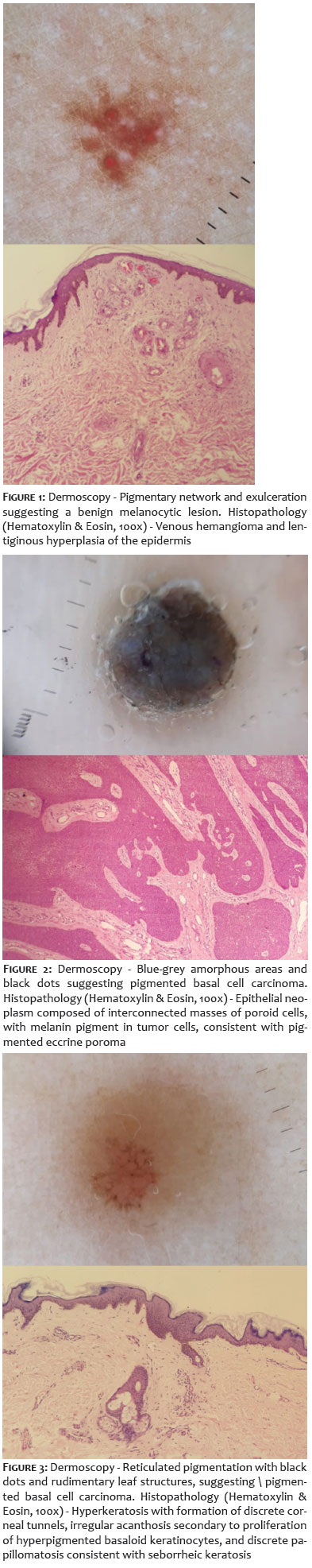
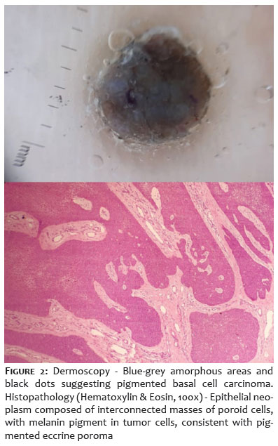
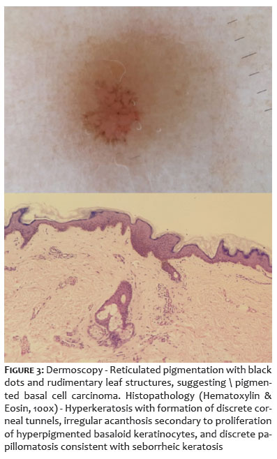
We assessed 26 (57.7% women, aged 66.1±16.1 years) with a total of 31 lesions. In the evaluation of all pigmented lesions, the global accuracy of mobile teledermatoscopy was 90.32% (28 lesions). The agreement between mobile teledermatoscopy for pigmented skin lesions and histopathology was perfect (kappa = 0.850, standard error = 0.080, p<0.0001). There was diagnostic disagreement in three lesions (Figures 1-3).
Regarding the teledermatoscopy ability to define malignancy of the pigmented lesion, the specificity was 81.8%, and the sensitivity was 100%. Two lesions were misdiagnosed as malignant lesions during teledermatoscopy analysis. Subsequently, the histopathological analysis showed that they were two benign lesions (Figures 2 and 3), resulting in an accuracy of 93.5%. The agreement between teledermatoscopy and histopathology was also perfect (kappa = 0.853, standard error = 0.099, p<0.0001).
The sensitivity for melanoma diagnosis (four lesions on histopathology), basal cell carcinoma (16), and benign melanocytic lesions (five) was 100%. For the hemangioma diagnosis (three lesions on histopathology), seborrheic keratosis (two), and pigmented eccrine poroma (one), it was 66.7%, 50%, and 0%, respectively.
Previous investigations have demonstrated that teledermatoscopy is an effective, accurate, and reliable tool for assessing pigmented lesions.2,4,5
Also, earlier studies revealed that mobile teledermatoscopy had an 81-90% agreement with the face-to-face dermatological examination.4,5
Our results provide additional evidence of the reliability of mobile teledermatoscopy with high sensitivity and accuracy. The risk of a pigmented malignant lesion going unnoticed by mobile teledermatoscopic evaluation is low due to the high sensitivity of this detection (100%).
As dermoscopic and cellular devices are widely used by dermatologists,2 we believe that mobile teledermatoscopy can be adopted as an additional tool to the diagnostic arsenal of dermatological practice.
Due to inconsistencies in the interobserver analyses,3 future investigations should assess the influence of the examiner’s experience on the accuracy of mobile teledermatoscopy.
Norami de Moura Barros 0000-0001-9765-602X
Statistical analysis; approval of the final version of the manuscript preparation and writing of the manuscript; data collection, analysis, and interpretation; active participation in research orientation; critical literature review; critical revision of the manuscript.
Karin Milleni Araujo 0000-0003-2421-3978
Statistical analysis; approval of the final version of the manuscript preparation and writing of the manuscript; data collection, analysis, and interpretation; active participation in research orientation; critical literature review; critical revision of the manuscript.
Juan Piñeiro-Maceira 0000-0002-8021-2374
Approval of the final version of the manuscript preparation and writing of the manuscript; data collection, analysis, and interpretation; intellectual participation in propaedeutic and/or therapeutic conduct of studied cases.
Carlos Baptista Barcaui 0000-0002-3303-3656
Approval of the final version of the manuscript; study design and planning; preparation and writing of the manuscript; data collection, analysis, and interpretation; active participation in research orientation; intellectual participation in propaedeutic and/or therapeutic conduct of studied cases.
1. Piccoli MF, Amorim BD, Wagner HM, Nunes DH. Teledermatology protocol for screening of skin cancer. An Bras Dermatol. 2015;90(2):202-10.
2. Yildiz H, Bilgili ME, Simsek HA. The diagnostic accuracy of the mobile phone teledermatoscopy. J Surg Dermatol. 2018;3:178.
3. Finnane A, Dallest K, Janda M, Soyer HP. Teledermatology for the diagnosis and management of skin cancer: a systematic review. JAMA Dermatol. 2017;153(3):319-27.
4. Barcaui CB, Lima PMO. Application of teledermoscopy in the diagnosis of pigmented lesions. Int J Telemed Appl. 2018:1624073.
5. Silveira CEG, Carcano C, Mauad EC, Faleiros H, Longatto-Filho A. Cell phone usefulness to improve the skin cancer screening: preliminary results and critical analysis of mobile app development. Rural Remote Health. 2019;19(1):4895.
 All content the journal, except where identified, under the Creative Commons Attribution 4.0 International licence - ISSN-e 1984-8773


 Read in Portuguese
 Portuguese PDF
 Print
Print
 Send this article by email
 How to cite this article
 Submit a comment
 Mendeley
 Pocket