


Deborah Heloisa Cezar Dourado1; Nathália Bacni Garcia1; Marilda Aparecida Milanez Morgado de Abreu1; Vinícius de Souza2; Ana Claudia Cavalcante Esposito1,2
Submitted on: 19/08/2021
Approved on: 16/09/2021
Financial support: None
Conflict of interest: None
How to cite this article: Dourado DHC, Garcia NB, Abreu MAMM, Souza V, Esposito ACC. Agminated blue nevus over acne scars: coincidence or pathophysiological relationship? Surg Cosmet Dermatol. 2022;14:e20220088
Blue nevus is a benign lesion arising from dermal melanocytes containing large amounts of melanin. The agminated variant presents a cluster of blue nevus lesions with linear or blaschkoid distribution. We report two cases of patients with agminated blue nevus that developed on previous acne scars, and dermoscopy helped a lot in the differential diagnosis. This is the first occurrence found in the literature of agminated blue nevus that appeared on a previous scar, which may have occurred by chance or as a result of the collagen remodeling process inherent to the healing process.
Keywords: Scar; Collagen; Dermoscopy; Nevus blue
The blue nevus is a benign lesion, usually asymptomatic, originated from dermal melanocytes that present large amounts of melanic pigment.1 It can be congenital or acquired, and the literature describes several histological variants.2-4 The most common clinical form is the solitary (single lesion), but it can also manifest as a cluster of lesions with a linear or blaschkoid distribution usually less than 10 cm in length, referred to as agminated blue nevus.5
We report two rare cases of patients with agminated blue nevus that arose over previous acne scars on the face.
A 39-year-old woman, Fitzpatrick’s skin phototype IV, sought dermatological care reporting acne on the face between 12 and 19 years old with the formation of pustules and nodules. At the time, she did not undergo treatment and evolved with the emergence of multiple scars. Ten years ago, she reported the appearance of a blackened and asymptomatic spot in the right buccinator muscle region, which concerned her. She denied recent growth of the lesion and comorbidities or the use of any medications.
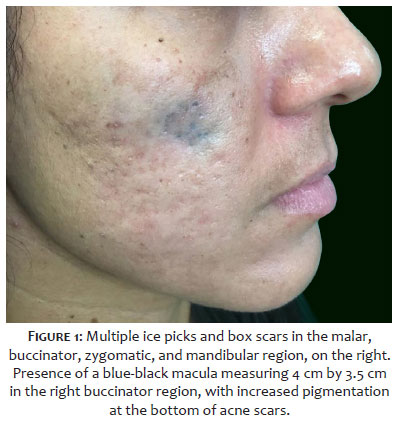
On dermatological examination, the patient presented multiple icepicks and box acne scars in the malar, buccinator, zygomatic, and mandibular regions, bilaterally. In the right buccinator muscle region, there was a blue-black macula measuring 4 cm by 3.5 cm, with increased pigmentation at the bottom of the acne scars (Figure 1). There were no palpable lymph nodes in the head and neck chains.
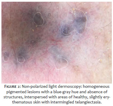
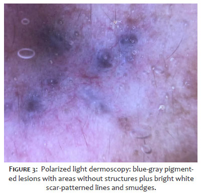
Non-polarized light dermoscopy showed homogeneous pigmented lesions with a blue-gray color and absence of structures, interspersed with areas of healthy, slightly erythematous skin with intermingled telangiectasia (Figure 2). Polarized light dermoscopy showed blue-gray pigmented lesions with areas without structures in addition to bright white lines and smudges with a scar pattern, probably resulting from the scar reorganization typical of the scarring process (Figure 3).
The clinical hypotheses were agminated blue nevus, with a differential for extrinsic pigmentation (tattoo-like), exogenous ochronosis, superficial extensive melanoma, and Reed nevus. Incisional biopsy was performed under local anesthesia (4 mm punch) in two areas. The anatomopathological examination showed normal epidermis, with elongated melanocytes in the dermis, with a variable quantity of cytoplasmic melanin, without nuclear atypia or mitosis. The histological findings were defined to diagnose blue nevus, agminated variant, which developed over previous acne scars.
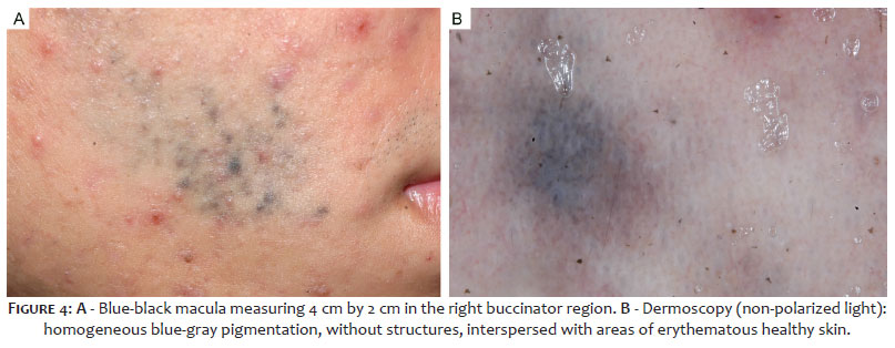
An 18-year-old man reported acne since he was 12 years old. Two years ago, he noticed a blue-black spot in the right buccinator muscle region. On dermatological examination, the patient presented closed comedones, erythematous papules, rare pustules, and icepicks and box acne scars (in smaller amounts) on the face, especially in the zygomatic, buccinators, and mandibular areas. In the right buccinator region, he had a blue-black macula measuring 4 cm by 2 cm (Figure 4A). At dermoscopy (non-polarized light), the lesion was composed of homogeneous blue-grey pigmentation, with no structures, interspersed with areas of erythematous healthy skin (Figure 4B). The clinical and dermoscopic hypothesis was agminated blue nevus that developed over acne-affected skin (including scars). The patient is under clinical follow-up without any surgical approach to the pigmented lesion.
Blue nevi are bluish macules, papules, plaques, or nodules. The agminated form was described for the first time only in 1947. Since then, the literature has gathered just over 30 cases of this variant, which justifies the rarity of the present case.5
Agminate lesions have a linear or blaschkoid distribution, and their pathogenesis is unknown. There are different theories to justify the segmental distribution, such as the development of lesions from peripheral nerves; dermal melanocytes resulting from an interruption during neural crest migration towards the epidermis; and, finally, clonal growth from a single cell.2,5,7
While the common blue nevus has a predilection for the limbs and face, the agminated form has a similar distribution between the trunk, extremities, head, and neck, in addition to affecting men and women equally.1,2,5,8 One hypothesis is that the agminated lesion tends to appear in an area of cutaneous trauma, with implantation of melanocytic cells in the deep dermis, or excessive sun exposure.3 In the reported patients, the lesion developed in a photoexposed area previously subjected to an intense inflammatory process resulting from acne.
The literature describes blue nevus agminated associated with nevus spilus, melanoma, dermatomyositis, Darier disease, and Carney complex. However, these are the first reports of the lesion appearing over the scar area.3-5,8,12 The lesion on the previous site of an acne scar may have occurred by chance, or the inflammatory process and collagen remodeling, typical of scar formation, may have facilitated the migration of melanocytes to the upper dermis.
Histologically, the agminated blue nevus more frequently corresponds to the common variant, where melanocytes – bipolar dendritic melanocytes or stellar with long dendrites – are located in the dermis, and may also aggregate around the skin appendages and neurovascular bundles.6,9
Activation mutations in GNAQ or GNA11, whose protein products signal through the MAPK pathway, are present in more than 83% and 7%, respectively, of blue nevus cases.10,11 In the case of agminated blue nevus, only one article assessed mutations identified in GNAQ.5 The two present reports did not perform mutation research.
The diagnosis of agminated blue nevus, in general, is a major clinical challenge.6 Given the complexity of the lesions, dermoscopy becomes a crucial tool, especially to differentiate melanoma from ochronosis.4,13 Dermoscopic findings in blue nevus include a homogeneous pattern melanocytic lesion, with few structures, bluish-gray amorphous areas, and there may be globules and spots.14 The blue color results from the Tyndall effect caused by the incident light in the deep melanin deposits melanin (dermis), which refracts and spreads.15 Despite the significant contribution of dermoscopy, the definitive diagnosis of blue nevus is histological.
The rarity of agminated blue nevus and its similarity to other dermatological lesions make its diagnosis challenging. These are the first reported cases of agminated blue nevus that have developed over a previous scar area. The inflammatory process and collagen remodeling, typical of the healing process, may have facilitated the migration of melanocytes to the upper dermis.
We thank Eliete Soares, photographer of the Dermatology discipline at FMB-Unesp, for documenting one of the cases. To Dr. Hamilton Ometto Stolf, for the shared medical attention in one of the cases cited in this article and for his commitment to the study of Clinical and Surgical Dermatology.
Deborah Heloisa Cezar Dourado 0000-0003-3611-5048
Approval of the final version of the manuscript; study design and planning; preparation and writing of the manuscript; data collection, analysis, and interpretation; critical literature review; critical revision of the manuscript.
Nathália Bacni Garcia 0000-0002-2539-5163
Approval of the final version of the manuscript; study design and planning; preparation and writing of the manuscript; data collection, analysis, and interpretation; critical literature review; critical revision of the manuscript.
Marilda Aparecida Milanez Morgado de Abreu 0000-0001-9099-6013
Approval of the final version of the manuscript; study design and planning; preparation and writing of the manuscript; data collection, analysis, and interpretation; critical literature review; critical revision of the manuscript.
Vinícius de Souza 0000-0001-8819-6906
Approval of the final version of the manuscript; preparation and writing of the manuscript; analysis, and interpretation; active participation in research orientation; intellectual participation in propaedeutic and/or therapeutic conduct of studied cases; critical literature review; critical revision of the manuscript.
Ana Claudia Cavalcante Esposito: 0000-0001-9283-2354
Approval of the final version of the manuscript; study design and planning; preparation and writing of the manuscript; data collection, analysis, and interpretation; active participation in research orientation; intellectual participation in propaedeutic and/or therapeutic conduct of studied cases; critical literature review; critical revision of the manuscript.
1. Fachal C, Pérez-Pérez LC, Allegue F, Calviño S. Subungual blue nevus. Actas Dermosifiliogr (Engl Ed). 2018;109(1):88-90.
2. Murali R, McCarthy SW, Scolyer RA. Blue nevi and related lesions: a review highlighting atypical and newly described variants, distinguishing features and diagnostic pitfalls. Adv Anat Pathol. 2009;16(6):365-82.
3. Lisboa AP, Silvestre KJ, Pedreira RL, Alves NR, Obadia DL, Azulay-Abulafia L. Agminated blue nevus - Case report. An Bras Dermatol. 2016;91(5):658-60.
4. Chen T, Kurwa HA, Trotter MJ, Haber RM. Agminated blue nevi in a patient with dermatomyositis. J Am Acad Dermatol. 2013;68(2):e52-3.
5. Eichenfield DZ, Cotter D, Thorson J, Hinds B, Sun BK. Agminated blue nevus with a GNAQ mutation: a case report and review of the literature. J Cutan Pathol. 2019;46(2):130-3.
6. Hunjan MK, Mohandas D, Bridges AG, Tollefson M. Agminated segmental plaque-type blue nevus associated with hypertrichosis and soft tissue hypertrophy: report of a case and review of the literature. Pediatr Dermatol. 2018;35(1):e22-e8.
7. Spring P, Perrier P, Erba P, Hagmann P, Mihm MC, Hohl D. Large agminated cellular 'plaque-type' blue nevus surrounding the ear: a case and review. Dermatology. 2013;227(1):21-5
8. Milkova L, Treudler R, Simon JC, Kunz M. Agminated blue naevi in a patient with EMO syndrome. Acta Derm Venereol. 2013;93(1):104-5.
9. Koba S, Mori M, Misago N, Narisawa Y. Agminated blue naevus on the sole. J Eur Acad Dermatol Venereol. 2016;30(2):334-5.
10. Van Raamsdonk CD, Bezrookove V, Green G, Bauer J, Gaugler L, O'Brien JM, et al. Frequent somatic mutations of GNAQ in uveal melanoma and blue naevi. Nature. 2009;457(7229):599-602.
11. Van Raamsdonk CD, Griewank KG, Crosby MB, Garrido MC, Vemula S, Wiesner T, et al. Mutations in GNA11 in uveal melanoma. N Engl J Med. 2010;363(23):2191-9.
12. Yoneyama K, Kamada N, Mizoguchi M, Utani A, Kobayashi T, Shinkai H. Malignant melanoma and acquired dermal melanocytosis on congenital nevus spilus. J Dermatol. 2005;32(6):454-8.
13. Ferrara G, Soyer HP, Malvehy J, Piccolo D, Puig S, Sopena J, et al. The many faces of blue nevus: a clinicopathologic study. J Cutan Pathol. 2007;34(7):543-51.
14. Oliveira AHK, Shiraishi AFMC, Kadunc BV, Sotero PC, Stelini RF, Mendes C. Blue nevus with satellitosis: case report and literature review. An Bras Dermatol. 2017;92(5 Suppl 1):30-3.
15. Prum RO, Torres R. Structural colouration of avian skin: convergent evolution of coherently scattering dermal collagen arrays. J Exp Biol. 2003;206(Pt 14):2409-29.
 All content the journal, except where identified, under the Creative Commons Attribution 4.0 International licence - ISSN-e 1984-8773


 Read in Portuguese
 Portuguese PDF
 Print
Print
 Send this article by email
 How to cite this article
 Submit a comment
 Mendeley
 Pocket