


Marina Emiko Yagima Odo; Lilian Mayumi Odo; Estele Yumi Odo Toledo de Barros
Received on: 03/03/2021
Approved on: 14/03/2021
Financial support: None
Conflict of interest: None
Acknowledgement: We thank the employees of the Odo Clinics
Study conducted at the Odo Clinics, São Paulo (SP), Brazil
A woman with type I diabetes and Hashimoto’s disease presented necrobiosis lipoidica at 15 years of age. Cyclosporine 100 mg/day was prescribed; however, an abscess in the inguinal region led to treatment interruption. Then, she tried dimethyl fumarate at increasing doses up to 120 mg/day. The lack of results led to the discontinuation of the therapy. The third attempt was isotretinoin 40 mg/day for eight months, with wound healing. Recently, after a knee scar repair surgery, necrobiosis lipoidica lesions appeared at the suture site. The patient received isotretinoin 40 mg/day again, and the lesion regressed in three months.
Keywords: Necrobiosis Lipoidica; Diabetes Mellitus, Type 1; Bacterial Infections
A 36-year-old woman reported the onset of an exulcerated skin lesion with a brown crust adhered to its surface in the bilateral pre-tibial region at 15 years of age after blunt local trauma. Three years ago, she already had type I diabetes mellitus comorbidity under control using insulin. The patient sought dermatological assistance, and an incisional biopsy was performed. The pathology was compatible with necrobiosis lipoidica. On this occasion, the use of cyclosporine 100 mg/day was indicated, with no significant improvement over a month. The appearance of an abscess in the inguinal region led to treatment interruption.
The second therapeutic option was dimethyl fumarate, in the recommended dose used for psoriasis of 30 mg/day during the first week, 60 mg/day in the second week, 90 mg/day in the third week, and then 120 mg/day. The patient used the drug for three months. However, as there was no improvement and importing the drug was complicated, she gave up treatment.
Improvement in necrobiosis lipoidica was only observed with the third therapeutic option, isotretinoin. The dosage was 40 mg/day for eight months, culminating in the lesion healing. The patient presented severe dysfunctional myalgia during treatment.
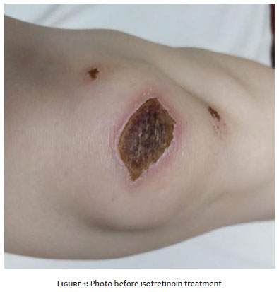
Three months ago, she sought our service. An unsightly scar had been removed from the knee, with the development of a lesion with characteristics of necrobiosis lipoidica in the suture area confirmed histologically (Figure 1). The patient uses levothyroxine sodium to control Hashimoto’s disease.
Because previously she had presented a good response to oral isotretinoin, pre-treatment control tests were requested, resulting in:
Blood glucose 361 mg/dl and glycated Hb 6.7%; Total cholesterol 198 mg/dl with HDL 88 mg/dl;
Thyroid peroxidase antibody 214.80 IU/ml (Normal up to 34 IU/ml);
Anti-thyroglobulin antibody 487.4 UI/ml (Normal up to 115 UI/ml);
TSH 1.370 uUI ml (Normal 0.400 to 4.500 uUI/ml);
Serum creatinine 0.82 (Normal:0.50 to 0.90 mg/dl).
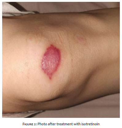
After 12 weeks of treatment with isotretinoin 40 mg/day, there were no more necrobiosis lipoidica lesions (Figure 2).
According to Burgdorf et al.,1 necrobiosis lipoidica is similar to necrobiotic xanthogranuloma with paraproteinemia, presenting as hard yellowish plaques, with central atrophy and telangiectatic vessels or blue veins, large and visible due to atrophy. Histopathologic findings reveal sally, granulomatous masses, intersecting bands of the dermis and subcutaneous tissue, and extensive necrobiosis. Granulomas contain histiocytes and foam cells. Frequently, it has a mixture of inflammatory cells and numerous foreign-body and Touton giant cells, with a peripheral ring of foamy cytoplasm. Cholesterol aggregates in wounds are common.
In most patients, Protein electrophoresis shows monoclonal IgG, a gammopathy that usually consists of light chain kappa. Bone marrow examination has revealed multiple myeloma in several patients.
Lipoid necrobiosis2 is a rare idiopathic, granulomatous pathology that affects diabetic individuals in 0.3% to 1.2% of cases. It can precede diabetes in more than 14% of cases, appear simultaneously in 24%, or appear after diagnosis in 62%. Other diseases may be associated, such as thyroid diseases (Graves, Hashimoto), Crohn's disease, ulcerative colitis, rheumatoid arthritis, and sarcoidosis; as well as other skin diseases, such as granuloma annulare, erythema nodosum, necrobiotic xanthogranuloma, and chronic venous ulcers. Multiple myeloma can accompany this condition or appear in the future. The discussion of etiopathogenesis is open. Deposition of immunocomplexes in the vessels or microangiopathic degeneration leading to collagen alteration is discussed. The abnormality of collagen is observed in the fibrils. The absence of transverse stretch marks3 is the most important finding.
In some cases, the complete collagen and elastin loss or the increase in collagen crosslinking caused by higher lysyl oxidase levels, typical of diabetic patients, have been suggested to contribute to the basement membrane thickening observed in necrobiosis lipoidica. Deposits of fibrin and immunoglobulin M (IgM) and C3 at the junction of blood vessels have also been found. In 30% of cases, vasculitis was demonstrated in the dermis by antibodies, leading to vascular occlusion. The most common finding in necrobiosis is the thickening of the vascular walls due to the endothelial enlargement, responsible for occlusion in the deeper layers of the dermis. The decreased flow is a potential factor that can also result from the deposition of glycoproteins in the vascular wall and an increase in Glut-1 (human erythrocyte glucose transporter).
The exact mechanism for increasing Glut-1 receptors is still under discussion. Researchers continue to consider the inflammatory factor because there are similar blood flows inside and outside the lesion. A case report described the simultaneous occurrence of ulcerated necrobiosis lipoidica and granuloma annulare.4
Several therapies have already been described: topical and intralesional corticosteroids if the lesion is closed, dapsone, injections of gold and bismuth, isoniazid, colchicine, clofazimine, topical nitrogen mustard, imiquimod, bovine collagen, PUVA, UVA-1, photodynamic therapy, glycemic control, dressings with animal gels, GM-CSF (Recombinant Human Granulocyte Macrophage Colony - Stimulant Factor), chloroquine, hydroxychloroquine, cyclosporine, fumaric acid ester, adalimumab, systemic and intralesional etanercept, systemic and intralesional infliximab, tacrolimus, pentoxifylline, aspirin, ticlopidine, CO2 laser, pancreatic transplant, thalidomide, platelet-rich plasma, topical tretinoin, intravenous immunoglobulin, pioglitazone, pulsed dye laser, surgical treatments such as grafts, cryotherapy, anti-TNF drugs, hyperbaric oxygen with steroid.5,6,7
Necrobiosis lipoidica is a disease with still unclear etiology and pathogenesis. The multiple therapeutic options for this entity demonstrate its complex treatment. We report a case of necrobiosis lipoidica where oral isotretinoin had effective control, but it did not eliminate the possibility of recurrence.
Marina Emiko Yagima Odo | 0000-0003-1982-8388
Active participation in research orientation.
Lilian Mayumi Odo | 0000-0001-7341-8924
Intellectual participation in propaedeutic and/or therapeutic conduct of studied cases.
Estele Yumi Odo Toledo de Barros | 0000-0002-9339-2407
Preparation and writing of the manuscript.
1. Burgdorf, WHC, The histiocytoses, in lever's histopathology of the skin, chapter 27, p. 591-605.
2. Sibbald C, Reid S, Alavi A. Necrobiosis lipoidica. Dermatologic Clinics. 2015;33(3):343-60.
3. Cunliffe WJ. Necrobiotic Disorders, Necrobiosis lipoidica. London: Oxford Blackwell Scientific publications. v. 3, p. 2033-7.
4. Homem de Mello e Souza F, Ribeiro CF, Pereira MAC, Mesquita L, Fabrício L. Ocorrência simultânea de necrobiose lipoídica ulcerada e granuloma anular em um paciente. An Bras Dermatol. 2011;86(5).
5. Peckruhn M, Tittelbach J, Elsner P. Update: treatment of necrobiosis lipoidica. J Deutsch Dermatol Ges. 2017;15(2):151-7.
6. Nguyen K, Washenik K, Shupack J. Necrobiosis lipoidica diabeticorum treated with chloroquine . J Am Acad Dermatol. 2002;46(2):34-6.
7. Medeiros KB, Torre DS, Jordão JM, Nogueira AC, Thumé T. Tratamento de necrobiose lipoídica no antebraço esquerdo com associação entre luz intensa pulsada e laser Erbium-YAG 2940nm. Surg Cosm Dermatol. 2020;12(4).
 All content the journal, except where identified, under the Creative Commons Attribution 4.0 International licence - ISSN-e 1984-8773


 Read in Portuguese
 Portuguese PDF
 Print
Print
 Send this article by email
 How to cite this article
 Submit a comment
 Mendeley
 Pocket