


Beatriz Poliseli Cernescu1; Mayara Teixeira Cruz1; Cássio Rafael Moreira1; Lígia Márcia Mário Martin1; Nikolai Cernescu Neto2
Received on: 13/09/2020
Approved on: 04/03/2021
Financial support: None
Conflict of interest: None
Acknowledgments: We thank Dr. Amanda Pelegrine Herculiani for kindly providing us with photos of the immunohistochemistry slides
Study conducted at the Municipal Health Department of Apucarana, Apucarana (PR), Brazil
Sarcomas são neoplasias mesenquimais malignas, raras, que acometem, principalmente, crianças e adolescentes. O rabdomiossarcoma, subtipo oriundo da musculatura esquelética, é condição incomum em adultos, acometendo sítios de localização não habitual, crescimento rápido e de difícil tratamento. Apresenta-se caso de adulto jovem com nodulação em lóbulo auricular esquerdo, cuja análise histopatológica e imuno-histoquímica confirmou tratar-se de rabdomiossarcoma alveolar, o qual foi conduzido em conjunto com a Oncologia.
Keywords: Rabdomiossarcoma Alveolar; Miogenina; Desmina; Excisão de Linfonodo
Rhabdomyosarcoma is an uncommon malignant neoplasm occurring mainly in children. It affects most commonly the head and neck (35%), genitourinary system, and extremities (40%). Less frequently, the condition affects the trunk, orbit, intrathoracic region, and retroperitoneum. The alveolar subtype is the most aggressive, with the worst prognosis.
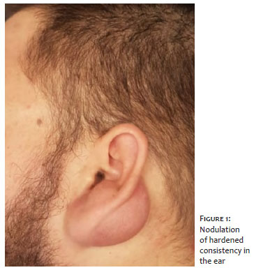
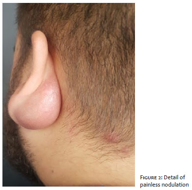
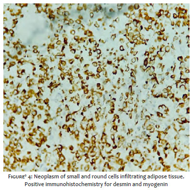
A twenty-one-year-old man reported a lesion on the left ear lobe for about a month, with a progressive increase in size. He denied other associated symptoms such as pain, fever, or weight loss. The general physical examination was normal. The dermatological examination revealed presence of hardened nodulation, painless on palpation, and without phlogistic signs at the anterior region of the left lobe, extending to the posterior region (Figures 1 and 2). An attempt to perform fine-needle aspiration (FNA) biopsy was unsuccessful. After an incisional biopsy, the anatomopathological examination showed neoplasia of small and round cells, infiltrating adipose tissue.
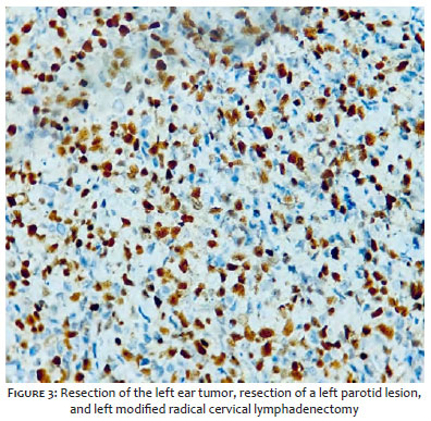
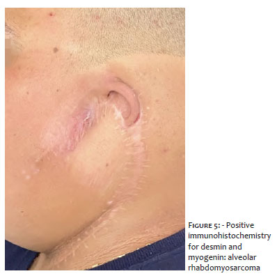
The immunohistochemistry was positive for desmin and myogenin (Figures 3 and 4), confirming the diagnosis of alveolar rhabdomyosarcoma. The patient was then referred to the Oncology Service. Computed tomography of the neck to detect metastasis evidenced an intraparotid lymph node enlargement on the left, with suspicion for secondary neoplastic involvement. Therefore, we conducted the resection of the left ear tumor, left parotid lesion, and left modified radical cervical lymphadenectomy (Figures 5). Histopathological analysis of tumor resection showed alveolar rhabdomyosarcoma, stage pT3 pN1. The resection product from parotid lesion showed infiltration by such sarcoma. On the left cervical lymphadenectomy, rhabdomyosarcoma metastasis was confirmed in four of the thirty resected lymph nodes. The patient underwent four chemotherapy sessions and 28 radiotherapy courses and is currently being followed up.
Sarcomas are rare malignant mesenchymal neoplasms that mainly affect children and adolescents.
Rhabdomyosarcoma, a subtype originating from skeletal muscle, is the most common type of soft tissue sarcoma in children, corresponding to approximately 50% of these tumors. The head and neck region are the most affected site, followed by the genitourinary tract, extremities, chest, and retroperitoneum.1 There are few reports in the literature of primary cutaneous rhabdomyosarcoma since this is an extremely rare condition. In adults, the occurrence is even more unusual – about 90% of all rhabdomyosarcomas occur in individuals under 25 years old – affecting sites of unusual location, presenting fast growth, and being difficult to treat.2
A biopsy is necessary to conclude the diagnosis. The biopsy can be surgical or with thick or fine needle aspiration (FNA).
Differential diagnoses of cutaneous lesions of the head and the neck include: hemangioma, lymphoma, lymphangioma, leukemia cutis, angiofibroma, neuroblastoma, cutaneous myofibroma, glioma, cellulitis, abscess, mastoiditis, and other sarcomas.2
A dermal infiltrate of small and oval-shaped cells with an acidophilic cytoplasm characterizes the histopathology of cutaneous alveolar rhabdomyosarcoma. Such changes can occur in various pathological conditions, and immunohistochemistry is essential to define the diagnosis. It is possible to find rhabdomyoblasts and characteristic neoplastic multinucleated giant cells in well-differentiated types, although immunohistochemistry is always an integral part of the diagnosis.2
There are four histopathological types, with their variants: embryonal, alveolar, pleomorphic, and sclerosing. The alveolar subtype is the most aggressive, with rapid progression, which causes early metastasis and increasing mortality rates. In this histological type, there is no association with environmental factors. Chromosomal translocations – t (2;13) and t (1;13) – are fundamental pieces in its development. There are also reports of involvement of individuals with other changes in the central nervous system, urogenital and gastrointestinal tract, and melanocytic nevi; thus it is believed these groups have to have a genetic predisposition.1
The first choice treatment is surgical, always combined with adjuvant chemotherapy to avoid metastasis. Radiotherapy is necessary when total lesion resection is not possible.
The prognosis depends on the site of origin, size of the lesion, clinical stage, age of the patient, and histological type. Good prognostic factors include early age at diagnosis, primary site in the genitourinary tract and orbit, and embryonal and botryoid histological types.2,3
The totally resected lesions are associated with a 90% survival in five years, which evidences the importance of diagnosis and treatment institution as early as possible.2,4
Beatriz Poliseli Cernescu | 0000-0001-5417-1815
Approval of the final version of the manuscript; study design and planning; preparation and writing of the manuscript; data collection, analysis, and interpretation; intellectual participation in propaedeutic and/or therapeutic conduct of studied cases; critical literature review; critical revision of the manuscript.
Mayara Teixeira Cruz | 0000-0002-5069-0519
Preparation and writing of the manuscript.
Nikolai Cernescu Neto | 0000-0001-5018-2672
Preparation and writing of the manuscript; critical literature review.
Cássio Rafael Moreira | 0000-0001-8710-6585
Approval of the final version of the manuscript; active participation in research orientation; intellectual participation in propaedeutic and/or therapeutic conduct of studied cases; critical revision of the manuscript.
Lígia Márcia Mário Martin | 0000-0002-0891-0813
Approval of the final version of the manuscript; critical revision of the manuscript.
1. Dziuba I, Kurzawa P, Dopierała M, Larque A, Januszkiewicz-Lewandowska D. Rhabdomyosarcoma in children - current pathologic and molecular classification. Polish J Pathol. 2018;69(1):20-32.
2. Lima LL, Rodrigues CAC, Pereira PMR, Schettini APM, Tupinambá WL. Rabdomiossarcoma alveolar cutâneo primário em paciente pediátrico. An Bras Dermatol. 2011;86(2):363-5.
3. Farias TP, Rangel LG, Dias FL, Castro ALC, Peryassú BC, Costa RM, et al. Impacto prognóstico do subtipo histológico na sobrevida de pacientes com sarcomas de cabeça e pescoço. Rev. Bras. Cir. Cabeça Pescoço. 2008;37(4):224-7.
4. Daya H, Chan HS, Sirkin W, Forte V. Pediatric rhabdomyosarcoma of the head and neck. Is there a place for surgical management? Arch Otolaryngol Head Neck Surg. 2000;126(4):468-72.
 All content the journal, except where identified, under the Creative Commons Attribution 4.0 International licence - ISSN-e 1984-8773


 Read in Portuguese
 Portuguese PDF
 Print
Print
 Send this article by email
 How to cite this article
 Submit a comment
 Mendeley
 Pocket