


Paula Colling Klein; Juliana Mazzoleni Stramari
Received on: 30/09/2020
Approved on: 04/03/2021
Financial support: None
Conflict of interest: None
Acknowledgments: I thank Dr. Juliana Stramari for allowing me to participate in this case and assist in developing this report
Study conducted at the Universidade Federal da Fronteira Sul, Passo Fundo (RS), Brazil
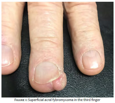
Superficial acral fibromyxoma is a rare and benign mesenchymal tumor. It mainly affects middle-aged men; however, it can occur in any gender and age group. It has a slow growth, with a preference for nail and periungual areas.
Keywords: Ambulatory surgical procedures; Fibroma; Antigens, CD34; Fingers
Superficial acral fibromyxoma is a rare and benign mesenchymal tumor. It mainly affects middle-aged men; however, it can occur in any gender and age group. It has slow growth, with a preference for nail and periungual areas.
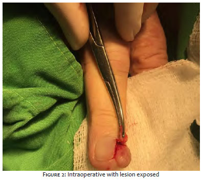
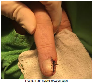
A 66-year-old man presented a nodular lesion on the lateral face of the third right finger. The lesion was asymptomatic and non-mobile, with fibroelastic consistency and progressive growth for five years. The patient was ex-alcoholic, ex-smoker, diabetic, hypertensive, and had ischemic heart disease. The tumor was resected with free margins after nail abrasion to access the lesion topography (Figures 1,2,3, and 4).
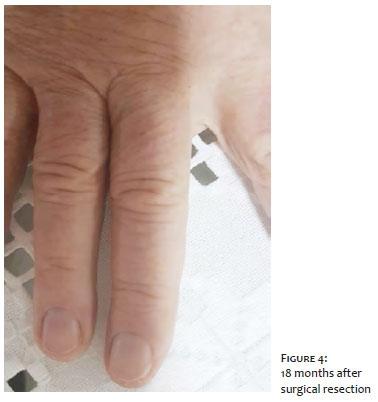
The histological examination showed dermal fusocellular proliferation amid myxoid stroma. The immunohistochemical examination was positive for the CD34 marker. Both results suggest the diagnosis of superficial acral fibromyxoma (Figure 4). At 28 months of follow-up after surgical resection, there were no signs of recurrence of the lesion. There is no description in the literature of malignant or aggressive behavior; however, focal atypia in some reported cases makes this tumor’s malignant potential uncertain.2
Fetsch et al.1 first described superficial acral fibromyxoma in 2001. Since then, there are just over 340 cases reported in the literature.2 The disease affects men and women in a 2:1 ratio and has a preference for the involvement of the feet.1 Although it is predominantly an ungual and periungual tumor, it can also affect the heel, ankle, and palms.
Typically, the condition is painless or little symptomatic and has slow growth. Still, it can cause nail deformities due to nail bed involvement and is not usually related to previous trauma.
Histopathological examination shows a well-delimited, circumscribed tumor, without a capsule, composed of a proliferation of fibroblasts in the myxoid stroma, sometimes accompanied by mast cells. On the other hand, immunohistochemical examination reveals characteristic positivity for the CD34 marker; however, CD99, CD10, and epithelial membrane antigen (EMA) may be present.1 Also, negativity is expected for cytokeratin, melanocytic markers, smooth muscle actin (SMA), and desmins.
The differential diagnosis must be made with ungual and periungual fibroma, neurofibromas, fibrokeratomas, sarcomas, and angiokeratomas, in addition to other benign conditions, such as onychocriptosis and cutaneous myxoma. Complete surgical resection is always indicated, given the lesion’s high recurrence rate.2-5
Given the relatively recent description of this tumor, the diagnosis of superficial acral fibromyxoma is still a challenge. Although it is not mandatory to perform imaging tests such as simple radiography and ultrasound, these tests are helpful tools for diagnostic clarification. The currently available rule out generic names such as myxoma, fibroma, and dermatofibroma as a histopathological diagnosis since the immunohistochemical examination differentiates the lesion.
The correct diagnosis indicates the best treatment. Currently, the literature recommends the surgical resection of the block lesion with free margins, contrasting with the past, which included even the amputation of the affected limb given the disease’s uncertainty of diagnosis and prognosis.
Although the dermatologist is trained to resolve such a condition, patients commonly seek orthopedists and hand surgeons. We report the present case due to the low incidence of this type of lesion and the need for suspicion in the dermatological consultation. The pathology seems to be not so rare, but it is still underdiagnosed and little reported.
Paula Colling Klein | 0000-0001-8969-4210
Approval of the final version of the manuscript; active participation in research orientation; critical literature review.
Juliana Mazzoleni Stramari | 0000-0002-7162-9856
Approval of the final version of the manuscript; active participation in research orientation; critical literature review.
1. García AM. Superficial Acral fibromyxoma involving the nail's apparatus. Case report and literature review. An Bras Dermatol. 2014;89(1):147-9.
2. Crepaldi BE. Superficial acral fibromyxoma: literature review. Rev Bras Ortop. 2019;54(5).
3. Pache S. Superficial acral fibromyxoma: a case report of an uncommon tumor of the foot. J Orthop Res Physio. 2018;5.
4. Porto ACS. Fibromixoma acral superficial em paciente do sexo feminino: um relato de caso. Surg Cosmet Dermatol. 2014;6(3):297-9.
5. Souza BGS. Fibromixoma acral superficial do polegar: relato de caso. Rev Bras Ortop. 2013;48(2):200-3.
 All content the journal, except where identified, under the Creative Commons Attribution 4.0 International licence - ISSN-e 1984-8773


 Read in Portuguese
 Portuguese PDF
 Print
Print
 Send this article by email
 How to cite this article
 Submit a comment
 Mendeley
 Pocket