


Raquel de Melo Carvalho; Thaiana Botarelli; Nilton Carlos dos Santos Rodrigues; Juliana Marques da Costa
Approved on: 01/06/2020
Received on: 04/03/2021
Financial support: None
Conflict of interest: None
Study conducted at the Hospital Naval Marcílio Dias, Rio de Janeiro (RJ), Brazil
Aneurysmal dermatofibroma (DF) is a rare benign tumor originating in the dermis, considered a variant of dermatofibroma. It is more prevalent in women over 30 years of age, in the lower limbs. Its etiology is unknown and histopathological examination confirms the diagnosis. This study reports two cases of aneurysmal DF in young patients, both men, presenting similar lesions: single hyperchromic nodule, with approximately 1.5 cm and progressive growth. This report aims to demonstrate an uncommon variant of dermatofibroma and to highlight the possible differential diagnosis with other tumors through dermoscopic examination.
Keywords: Dermoscopy; Histiocytoma, Benign Fibrous; Dermatology
Aneurysmal dermatofibroma (DF) is a benign tumor that originates in the dermis and represents less than 2% of dermatofibromas.1-5 Its etiology is unknown, and it is more prevalent in women over 30 years old. Histopathology provides a definitive diagnosis.
DF is generally more extensive than the classic DF and has an erythematous-brown or violet color. It can be painful if the lesion grows rapidly. Dermoscopy can identify any of the patterns already known to classical DF. However, the features that will suggest an aneurysmal (DF) are the linear white patterns, vascular structures, and delicate pigmented network on the periphery.
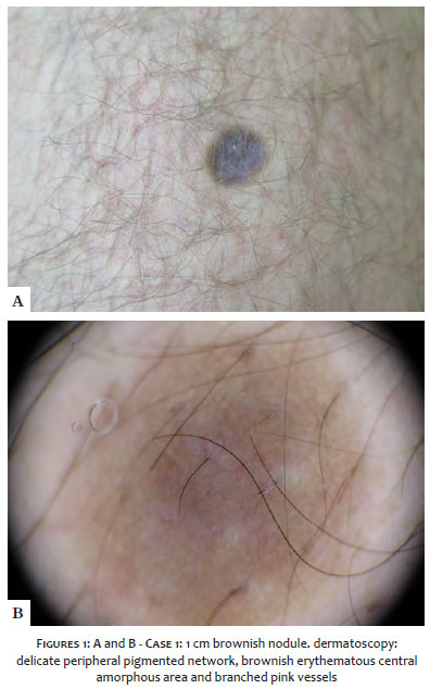
Case 1: A healthy, 25-year-old man presented with a hyperchromic, violaceous nodule, measuring an inch and a half, painful and with progressive growth. The nodule appeared for three years and had a positive dimple sign. Dermoscopy identified a delicate peripheral pigmented network, central wine-red, and bright-white areas (Figures 1 A and B).
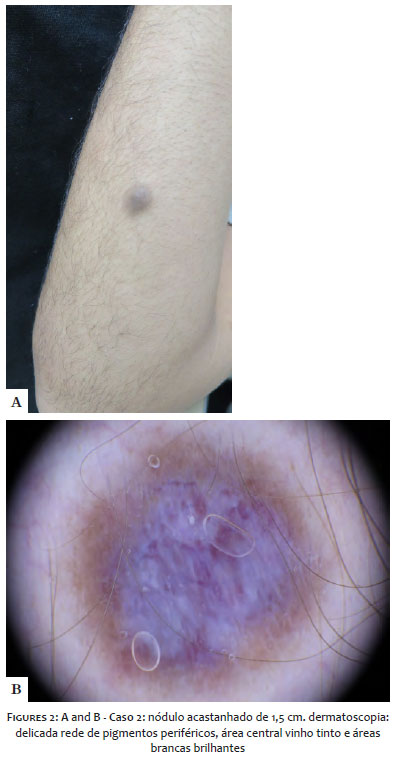
Case 2: A healthy man of similar age, complaining of an arm injury, with progressive increase and starting two years ago. On examination, he presented a pigmented nodular lesion, measuring 1cm in the right forearm. Dermoscopy showed a delicate peripheral pigment network, central erythematous brownish amorphous area, and pinkish branching vessels (Figures 2 A and B).
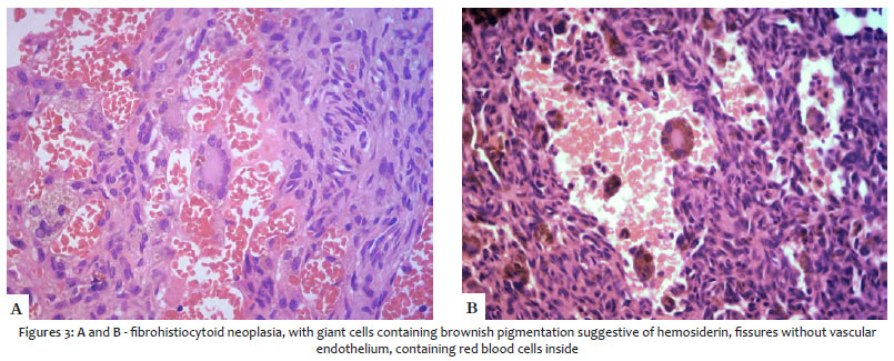
Histopathology of both cases showed acanthotic epidermis and hypercellularity in the center of the lesion, occupying the entire dermis up to the subcutaneous. It was forming a fibrohistiocytic neoplasm with the presence of giant cells containing brownish pigmentation suggestive of hemosiderin. The exam also showed gaps without vascular endothelium containing red blood cells in its interior. We observed incarceration of preexisting collagen fibers by newly formed collagen on the periphery of the lesion (Figures 3 A and B).
The variants of the DFs are cellular, epithelioid, hemangiopericytoma, atrophic, fibrocollagenous, pseudosarcomatous, and aneurysmal.4,5 Aneurysmal DF is a benign tumor that originates in the dermis and represents less than 2% of DFs.1-5 Its etiology is unknown, although some authors suggest that local trauma triggers the onset. It is more prevalent in women over 30 and has a recurrence rate of 19% when excised.
Histopathology is essential for the definitive diagnosis and may show neoformation composed of spindle-starred cells that preclude new fibrillar collagen, acanthosis, elongation of epidermal cones, multinucleated cells, and gaps containing red blood cells.6 Immunohistochemistry can help differentiate the most doubtful cases: aneurysmal DF is negative for S100, HMD45, and CD34.
Clinically, aneurysmal DF is generally larger than classic dermatofibroma, has an erythematous-brown or violet color, and can be painful when the lesion grows rapidly.8 As a differential clinical diagnosis, Kaposi’s sarcoma, vascular tumors, and melanoma can be highlighted.7 Dermoscopy can identify white linear patterns, vascular structures, and a delicate pigmented network on the periphery. Thus, this subtype can have any of the features already known to classical DFs, such as pigmented network, white area, vascular structures, homogeneous region, white network, globule-like structures, and irregular crypts. However, what suggests aneurysmal DF is the central erythematous-wine color.8-10 Therefore, we can conclude that dermoscopy is a helping tool for the dermatologist to differentiate aneurysmal dermatofibroma from its possible differential diagnoses, especially with malignant tumors.
Raquel de Melo Carvalho | 0000-0002-3991-4569
Study design and planning; data collection, analysis, and interpretation; ; intellectual participation in propaedeutic and/or therapeutic conduct of studied cases; critical literature review.
Thaiana Botarelli | 0000-0001-7619-7696
Study design and planning; data collection, analysis, and interpretation; intellectual participation in propaedeutic and/or therapeutic conduct of studied cases; critical literature review.
Nilton Carlos dos Santos Rodrigues | 0000-0002-5290-3063
Intellectual participation in propaedeutic and/or therapeutic conduct of studied cases.
Juliana Marques da Costa | 0000-0003-0401-1068
Approval of the final version of the manuscript; study design and planning; active participation in research orientation; intellectual participation in propaedeutic and/or therapeutic conduct of studied cases; critical literature review; critical revision of the manuscript
1. Zaballos P, Llambrich A, Ara M, Olazarán Z, Malvehy J, Puig S. Dermoscopic findings of haemosiderotic and aneurysmal dermatofibroma: report of six patients. Br J Dermatol. 2006;154:244-50.
2. Ramos-e-Silva M, Carmo GC, Costa JM. Fundamentos da Dermatoscopia: atlas dermatológico. 2 ed,; 2016. 423p.
3. Espasandín-Arias M, Moscarella E, Mota-Buçard A, et al. The dermoscopic variability of dermatofibromas. J Am Acad Dermatol. 2015;72(1 suppl):S22-4.
4. Santa Cruz DJ, Kyriakos M. Aneurysmal ("Angiomatoid") fibrous histiocitoma of the skin. Cancer. 1981;47(8):2053-61.
5. Pegas JR, Santos BA, Tebcherani AJ, Cade KV. Dermatofibroma aneurismático. Surg Cosmet Dermatol. 2010;2:225-7.
 All content the journal, except where identified, under the Creative Commons Attribution 4.0 International licence - ISSN-e 1984-8773


 Read in Portuguese
 Portuguese PDF
 Print
Print
 Send this article by email
 How to cite this article
 Submit a comment
 Mendeley
 Pocket