


Paulo Henrique Teixeira Martins; Natália Andressa Buss Venier; Laura Luzzatto; Fernando Eibs Cafrune
Received on: 20/11/2019
Approved on: 12/08/2020
Financial support: None Conflict of interest: None
Acknowledgement: We thank for the opportunity granted to write this clinical case, being possible only after much study and dedication. Values, which add a lot to professional training
Study conducted at the Santa Casa de Misericórdia de Porto Alegre Hospital, Porto Alegre (RS), Brazil
Although several techniques have been described for upper lip reconstruction, functional reconstruction of total upper lip defects remains a challenge. We report a case of a significant size squamous cell carcinoma excised in the upper lip region using the double-advancement technique, with positive functional and aesthetic results.
Keywords: Carcinoma, Squamous Cell; Lip Neoplasms; Surgical Flaps
Over the years, several techniques for lip reconstruction with different levels of complexity have been developed, given the organ’s peculiarity and functions. Each case is different from the other, with each patient’s characteristics, anatomy, sex, comorbidities, and smoking. It is essential to consider the size of the lesion and its location in the different labial subunits. The main objectives are to maintain speech ability, adequate nutrition, and symmetry and aesthetics, since it is located on the face and directly related to its personal image.
This report aims to show the approach on a large and deforming lesion with a surgical technique that preserved the patient’s lip and function.
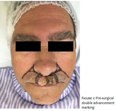
A 59-year-old man, without pathological history, in his first dermatological consultation, with a history of a lesion on the upper lip with five months of evolution, presented difficulty in speech and suction movement. The clinical examination observed a tumor with raised erythematous borders, an ulcerated keratotic center with whitish areas, and a hyperchromic center in the upper lip’s medial region measuring more than a third of the upper lip (Figure 1). Dermoscopy was limited by the keratotic component of the lesion, with few structures being observed. The rest of the facial skin did not have photodamage or other lesions suspected of malignancy.
Incisional biopsy and anatomopathological examination of the fragment were performed, describing follicular comedones and chronic inflammation. Due to the exuberant and rapidly progressing lesion, we opted for complete excision of the lesion and a new anatomopathological exam.
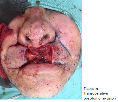
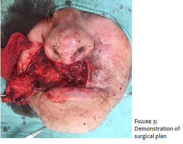
We performed total excision of the lesion with a 6 mm surgical limit and correction of the defect with bilateral advancement flap in the lateral subunits of the upper lip (Figures 2 and 3), with satisfactory results both aesthetically and functionally, preserving blood supply through the upper labial arteries, mucosa, and the orbicularis oris muscle of the mouth.
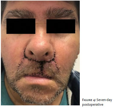
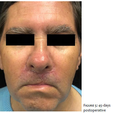
The result of the anatomopathological examination of the specimen with surgical limits was squamous cell carcinoma with peripheral and deep surgical limits free of neoplasia. The patient returned with an operative wound with good healing and satisfied with the preservation of the functionality of the lips (Figures 4 and 5).
Techniques for upper lip reconstruction are poorly described in the literature since tumors in this region are uncommon, with only 5% of lip tumors, and the most common histological type is squamous cell carcinoma, as in the case.1,2
The lip is divided into four subunits with two laterals and one medial at the top, the filter, and a single subunit at the bottom. The patient’s lesion was in the medial subunit, progressing to the left lateral subunit. There is no mandatory technique for reconstructing the upper lip. Professionals must analyze the size and location of each defect and know its anatomical structure. Thus, its division into units facilitates the reconstruction plan.3
With the flap with medial advancement of the cheeks, we managed to maintain tissue perfusion through the upper labial artery’s blood supply and the perforating arteries subcutaneously in the pedicle, preventing necrosis.4 As neighborhood skin is used, it is possible to maintain texture, hair, and skin color.
As we performed the excision of part of the vermilion lip, we also promoted the vermilion mucosa’s advancement to maintain the aesthetics and anatomical functionality.4 Small cutaneous branches of the infraorbital nerves are cut during the surgery, leaving the skin with reduced sensitivity, which is usually recovered spontaneously in the postoperative period.5
An option for surgery would be the renowned Abbe flap, described in the literature and performed more than 100 years ago. Nevertheless, we chose to perform a bilateral advancement flap to perform the surgery in just one surgical time, avoiding the need for patient collaboration to care for the surgical wound and the risks of being submitted to another surgical time.5
For upper lip defect correction, advancement flaps are a good option with satisfactory functional and aesthetic results.
Paulo Henrique Teixeira Martins | 0000-0003-2138-2741
Statistical analysis; approval of the final version of the manuscript; study design and planning; preparation and writing of the manuscript; data collection, analysis, and interpretation; active participation in research orientation; intellectual participation in propaedeutic and/or therapeutic conduct of studied cases; critical literature review; critical revision of the manuscript.
Natália Andressa Buss Venier | 0000-0001-8743-3631
Study design and planning; preparation and writing of the manuscript; active participation in research orientation; intellectual participation in propaedeutic and/or therapeutic conduct of studied cases; critical literature review.
Laura Luzzatto | 0000-0002-4193-6943
Data collection, analysis, and interpretation.
Fernando Eibs Cafrune | 0000-0002-6645-0122
Statistical analysis; approval of the final version of the manuscript; study design and planning; preparation and writing of the manuscript; data collection, analysis, and interpretation; active participation in research orientation; intellectual participation in propaedeutic and/or therapeutic conduct of studied cases; critical literature review; critical revision of the manuscript.
1. Alves PJ, Alves SST. Reconstrução labial superior com retalho de pedículo subcutâneo. Rev. Bras. Cir. Plást. 2011;26(2):254-8.
2. Denadai R, Sarmento GS, Buzzo CL, Raposo-Amaral CE, Raposo-do-Amaral CA. Retalho de Bernard-Webster para reconstrução do lábio inferior após exérese de carcinoma espinocelular: uma análise dos resultados funcionais. Rev. Bras. Cir. Plást. 2015;30(1):8-17.
3. Petrarolha SMP, Simões ASF, Oliveira JPC, Castro MAF, Devitis RA. Reconstrução de lábio superior com retalho nasogeniano em ilha. Rev. Bras. Cir. Cabeça Pescoço. 2016;45(1):25-7.
4. Faveret PLS. Reconstrução labial após ressecção de tumores. Rev. Bras. Cir. Plást. 2015;30(2):206-18.
5. Sanniec KJ, Carboy JA, Thornton JF. Simplifying lip reconstruction: an algorithmic approach. Semin Plast Surg. 2018;32(2):69-74.
 All content the journal, except where identified, under the Creative Commons Attribution 4.0 International licence - ISSN-e 1984-8773


 Read in Portuguese
 Portuguese PDF
 Print
Print
 Send this article by email
 How to cite this article
 Submit a comment
 Mendeley
 Pocket