


Bruna Ramos da Silva1; Flávia Thomé França1; Nathalia Fahl Cicotti2; Maria Paula Barbieri D’elia2; Jorge Logan Furtado Costa3
Receipt date: 21/07/2020
Approval date: 07/09/2020
Financial Support: None
Conflict of Interest: None
Acknowledgments: We thank Dr. Vinicius de Souza for his dermoscopic description, photographer Eliete Soares from the Department of Dermatology at Universidade Estadual Paulista, and Dr. Hamilton Stolf for their encouragement.
Study conducted ant the Clinical Hospital of the Universidade Estadual de Campinas, Campinas (SP), Brazil.
The nevus spilus (NS), also known as speckled lentiginous nevus or nevus on nevus, is represented by a brownish macule on which small macules of darker shades appear. It is more common on the trunk and lower limbs. The general population's prevalence is 0.2% to 2.3%, and they have a benign character. The exact risk for malignant transformation is still unknown; thus, it demands a routine clinical-dermoscopic follow-up. We present a case of malignant melanoma on acquired nevus spilus, in which early an excision was performed, with no recurrence, highlighting the importance of follow-up of these patients.
Keywords: Melanoma; Nevi and melanomas; Nevus
Nevus spilus (NS), also known as nevus on nevus or speckled lentiginous nevus, presents small macules and/or hyperchromic papules on a larger and slightly brownish macula. It is usually located on the trunk and lower limbs. A single or multiple lesion clinically characterizes NS, and this lesion may acquire a zosteriform aspect on the dermatome. It can be congenital or acquired, being more common in childhood, but there are reports of its appearance at any age, and there is no predisposition for a skin type.1 Its prevalence in the general population is 0.2% to 2.3%, and it is benign. Although malignant transformation is rare, NS must be monitored. This article aims to report an 84-year-old patient with nevus spilus with malignant transformation to melanoma.
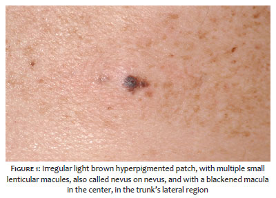
An 84-year-old man sought help at the Dermatology Clinic of the School of Medicine of Botucatu to treat four pancellular carcinomas on the face. In 2005, we observed an irregular stain measuring 15 x 10 cm in length, hyperpigmented, with a café-au-latte color in the trunk’s lateral region during the consultation. Upon the stain, there were multiple dark brown, lenticular macules.
In the stain’s center, we noticed a dark brown, asymmetrical macula, with irregular edges, measuring 15 mm in the largest diameter (Figures 1 and 2). Dermoscopy revealed an irregular pigmentation area, containing an atypical pigment network with thickening and abrupt termination (Figure 3). Physical examination presented the absence of palpable lymph nodes.
Because this was a lesion suspected of malignancy, surgical excision was performed. The anatomopathological examination revealed extensive superficial malignant melanoma with evident lymphocytic infiltrate, Clark level III, and Breslow index 0.4 mm, associated with junctional nevus (Figures 4 and 5).
The patient underwent clinical follow-up with a physical examination, dermoscopy, and tests for tumor staging, which resulted in no changes. There was no change in the remaining nevus and/or evidence of local recurrence or distant metastasis during seven years of follow-up. The patient was lost to follow-up in the Dermatology Clinic from 2012. In 2016, he died at 95 years of age from pneumonia, decompensated heart failure, and acute chronic kidney failure.
The nevus spilus (NS) is a hyperpigmented stain, resulting from lentiginous melanocytic hyperplasia. Smaller macules from 1 mm to 3 mm, with darker shades, compose the lesion, resulting in a mottled appearance. There is no preference for gender or race. It can be congenital or acquired. However, its etiology remains unknown.5 The first case of melanoma in NS was reported in 1957.2 Since then, less than 40 cases have been published.4 In Brazil, two case reports of malignant melanoma were found on nevus spilus.3,4 Some studies believe that the risk of malignancy can vary from 0.13% to 0.2%.13,14 Under microscopy, the darkest spots reflect junctional nevus cells nests, compound and intradermal, and more rarely Spitz nevus and blue nevus.6
It is crucial to detect clinical elements that suggest a higher risk of developing of melanoma in these patients’ follow-up. Rhodes and Mihm assumed that clinically irregular lesions could be associated with atypical histopathological features, designating them as “dysplastic” nevus spilus, and thus differentiating them from the “typical” nevus spilus.10 Our patient presented a nevus spilus of the acquired type. The lesion was suspected of malignancy, represented by the asymmetric macula, blackened, with irregular edges over the nevus. Dermoscopy showed an irregular pigmented network, with thickening and abrupt termination points on the lesion’s periphery. Histopathological examination confirmed the hypothesis of malignancy, showing extensive superficial melanoma in the area corresponding to the macula, with several shades of black, observed clinically.
There is a probability that the nevus spilus have a higher risk of developing into a melanoma.7,8,9 An increased risk of malignancy would be theoretically possible since nevus spilus is a subtype of congenital melanocytic nevus (CMN), that is, a hamartomatous proliferation of melanocytes. There is still no evidence that the presence of hair predisposes to melanoma.6
However, the nevus spilus appears to have a noticeably lower risk of malignant transformation than other classic CMNs of the same size. One explanation would be that the CMN's nevus cells are found in deeper layers of the dermis. Also, it is known that the greater the number of melanocytes, the greater the potential for malignant degeneration.6 There is still no protocol in the literature for the management or follow-up of nevus spilus.4 In the case presented, there was no recurrence or metastasis during the follow-up. This report and the other publications teach the importance of clinical follow-up associated with dermoscopy for the early detection and treatment of malignant lesions. Self-examination is advisable for patients with nevus spilus, paying attention to changes in color or irregular elements. Excisional biopsy in lesions suspected of malignancy on the nevus is essential for the early diagnosis of melanoma.9,12,15
Bruna Ramos da Silva | 0000-0002-2912-0474
Preparation and writing of the manuscript; critical literature review; critical revision of the manuscript.
Nathalia Fahl Cicotti | 0000-0001-9893-8184
Preparation and writing of the manuscript; data collection, analysis, and interpretation.
Jorge Logan Furtado Costa | 0000-0003-4312-7506
Preparation and writing of the manuscript.
Flávia Thomé França | 0000-0003-0830-7317
Critical literature review; critical revision of the manuscript.
Maria Paula Barbieri D’elia | 0000-0003-1524-721X
Approval of the final version of the manuscript; study design and planning; preparation and writing of the manuscript; active participation in research orientation; critical literature review; critical revision of the manuscript.
1. Fernandez-Flores A. Eponyms, morphology, and pathogenesis of some less mentioned types of melanocytic nevi. Am J Dermatopathol; 2012;34(6):607-18.
2. Perkinson NG. Melanoma arising in a café au lait spot of neurofibromatosis. Am J Surg. 1957;93(6):1018-20.
3. Tavoloni Braga JC, Gomes E, MacEdo MP, Pinto C, Duprat J, Begnami MD, et al. Early detection of melanoma arising within nevus spilus. J Am Acad Dermatol. 2014;70(2):e31-2.
4. De Brito MHTS, Fernandes CMBM, Rosa MJM de PM da C, Dionísio CSN de M, Ferreira JCM, Garcia MMAP da S. Synchronous melanomas arising within nevus spilus. An Bras Dermatol. 2017;92(1):107-9.
5. Vaidya DC, Schwartz RA, Janniger CK. Nevus spilus. Cutis. 2007;80(6):465-8.
6. Gathings RM, Reddy R, Bhatia AC, Brodell RT. Nevus spilus: is the presence of hair associated with an increased risk for melanoma?. Cutis. 2016;98(3):171-4.
7. Schaffer JV, Orlow SJ, Lazova R. Nevo lentiginoso salpicado: dentro do espectro de nevos melanocíticos congênitos. Arch Dermatol. 2001;137:172-8.
8. Singh S., Jain N., Khanna N. Nevo peludo spilus: uma série de casos. Pediatr Dermatol . 2013;30:100-4.
9. Haenssle HA, Kaune KM, Buhl T. Melanoma surgindo no nevo spilus segmentar: detecção por dermatoscopia digital seqüencial. J Am Acad Dermatol . 2009;61:337-41.
10. Yoneyama K, Kamada N, Mizoguchi M, Utani A, Kobayashi T, Shinkai H. Malignant melanoma and acquired dermal melanocytosis on congenital nevus spilus. J Dermatol. 2005;32(6):454-8.
11. Karam SL, Jackson SM. Malignant melanoma arising within nevus spilus. Skinmed. 2012;10(2):100-2.
12. Meguerditchian AN, Cheney RT, Kane JM. Nevus Spilus with synchronous melanomas: case report and literature review. J Cutan Med Surg. 2009;13(2):96-101.
13. Corradin MT, Giulioni E, Fiorentino R, Santeufemia DA, Re GL, Vettorello A. In situ malignant melanoma on nevus spilus in an elderly patient. Acta Dermatovenerol Alp Pannonica Adriat. 2014;23(1):17-9.
14. Corradin MT, Zattra E, Fiorentino R, Alaibac M, Belloni-Fortina A. Nevus spilus and melanoma: case report and review of the literature. J Cutan Med Surg. 2010;14(2):85-9.
15. Piana S, Gelli MC, Grenzi L, Ricci C, Gardini S, Piana S. Multifocal melanoma arising on nevus spilus. Int J Dermatol. 2006;45(11):1380-1.
 All content the journal, except where identified, under the Creative Commons Attribution 4.0 International licence - ISSN-e 1984-8773


 Read in Portuguese
 Portuguese PDF
 Print
Print
 Send this article by email
 How to cite this article
 Submit a comment
 Mendeley
 Pocket