


Vanessa Gheno1; Rogério Nabor Kondo2; Clarissa Patias Lena3
Received on: 21/11/2019
Approved on: 26/05/2020
Financial support: None
Conflict of interest: None
Research performed at the Dermatology, Hospital Universitário Regional do Norte do Paraná, Universidade Estadual de Londrina, Londrina (PR), Brazil
INTRODUCTION: Squamous cell carcinoma (SCC) is the second most frequent malignant tumor of the epidermis. When located in the labial region, it can present a great challenge for the reconstruction, since it’s located in the center of the inferior third of the face, causing scars and distortions that negatively affect the quality of life.
OBJECTIVE AND METHODS: We report two cases of SCC in the lower lip reconstructed with Gilles fan flap associated with zetaplasty.
RESULTS AND CONCLUSIONS: In both cases, the result was satisfactory, with tumor resolution, absence of microstomia, preservation of the functionality, and good aesthetic acceptance.
Keywords: Carcinoma Squamous Cell; Case Reports; Lip; Surgical Flaps
Squamous cell carcinoma (SCC) is the second most frequent malignant tumor of the epidermis. The tumor originates from atypical proliferation of cells in the squamous layer of the epidermis.1 It is more frequent in individuals 50 years or older, photo types I and II, and in skin areas with photoaging. Oral SCC in particular is related to smoking and alcohol consumption. Early diagnosis decreases the odds of cervical lymph node metastases, which can occur in 5-20% of cases.1,2 The lip defects pose a major challenge for reconstruction, since they are located at the center of the lower third of the face, and that the labial scars and deformations negatively affect the patient’s’ quality of life. Surgeons have studied numerous reconstruction techniques, aimed at good functional and aesthetic results.3,4
Two patients with diagnosis of SCC of the lower lip were treated:
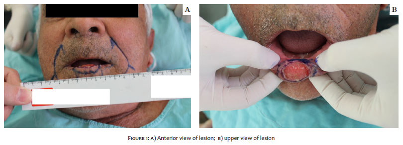
PATIENT 1: Male patient, 75 years, photo type III, from Londrina, Paraná State, Brazil, a farmer and construction worker, reported a painful lesion on the lower lip that appeared approximately seven months before. There was no report of local trauma or insect bite. The patient was previously healthy, with no history of smoking or alcohol use. Physical examination showed an ulcerated lesion, well-demarcated, with slightly elevated edges, on the lower lip. Palpation revealed infiltration of the tissues adjacent to the lesion (Figures 1a and 1b). Incisional biopsy was performed, revealing moderately differentiated SCC. For staging of the patient, palpation of cervical lymph nodes was negative, and ultrasound of the cervical region did not show enlarged lymph nodes suspected of metastases.
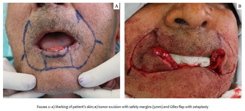
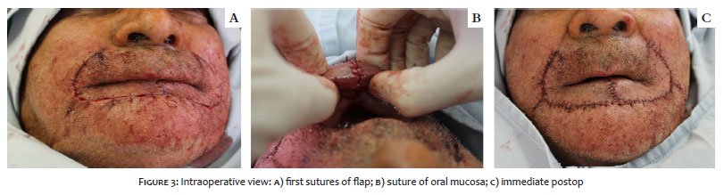
Following exams, excision of the lesion was performed with 5mm safety margins (Figure 2). Since the defect corresponded to more than 60% of the lower lip, the technique chosen for reconstruction was the Gilles fan flap with bilateral zetaplasty (Figures 2 and 3).
PATIENT 2: Male patient, 79 years, photo type III, from Jataizinho, Paraná State, Brazil, a nurse and administrative assistant in the local government, reported an ulcerated lesion on the lower lip for three months. No report of local trauma, but he suspected having been bitten by an insect. Patient presented a psychiatric disorder and was in follow-up with use of haloperidol, fluoxetine, and clonazepam. He was a former smoker with had a history of alcohol consumption.
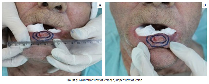
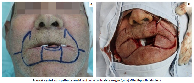
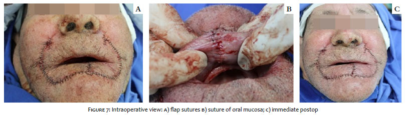
Physical examination showed a poorly demarcated ulcerated lesion on the lower lip. Palpation revealed infiltration of the tissues adjacent to the lesion (Figure 5). Biopsy revealed poorly differentiated SCC. Palpation of cervical lymph nodes was negative, as was computerized tomography of the cervical region. Excision of the tumor was performed with 5mm safety margins. Since the defect also corresponded to more than 60% of the lower lip, the chosen technique was the same as in the previous patient (Figures 6 and 7).
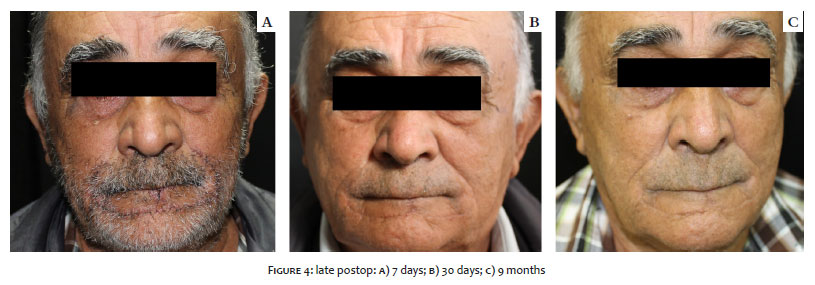
PATIENT 1: Histopathological examination of the surgical specimen showed well-differentiated SCC, without lymph vessel or perineural invasion and with free surgical margins. Patient evolved with good healing and satisfaction, without postoperative complications (Figures 4a, 4b, 4c).
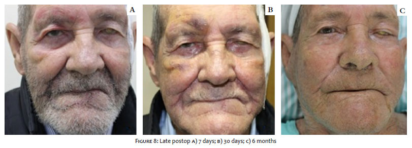
PATIENT 2: Histopathological examination showed moderately differentiated SCC, ulcerated, with infiltration of reticular dermis, moderate lymphocyte infiltrate, without lymph vessel or perineural invasion and free surgical margins. Patient had no postoperative complications and reported good aesthetic satisfaction (Figures 8a, 8b, 8c).
Various techniques for reconstruction of the lower lip have been described in the literature and classified according to the size of the defect: small (up to 30%), medium (30-60%), or large (60% or more). Small defects can be reconstructed with primary closure following excision in V or W. Medium-sized defects can be reconstructed with elliptical excision, edge-to-edge suture, and M-plasty or flaps. However, larger defects need to be reconstructed with more complex techniques, such as Abbé and Estlander flap (repair of the lower lip defect with an upper lip flap), Gilles, or Karapandzic.2
The Gilles flap used in these two patients consists of projection of the lower lip commissure and lateral region to cover the defect left by the lesion on the lower lip, representing a full-thickness flap. To prevent microstomia, we associated zetaplasty with the flap.2
Many studies have indicated a decrease in survival of patients with cervical metastases, since there is a close relationship between tumor size and metastases. This highlights the need for early diagnosis and proper treatment to ensure the patient’s cure.
Both cases showed satisfactory late postop results, tumor treatment with free margins, absence of microstomia, preservation of function, and good aesthetic acceptance, providing better quality of life for the patients.
Vanessa Gheno | 0000-0003-2670-0497
Approval of the final version of the manuscript; conception and planning of the study; elaboration and writing of the manuscript.
Rogério Nabor Kondo | 0000-0003-1848 -3314
Approval of the final version of the manuscript; conception and planning of the study; effective participation in orientation of the research; elaboration and writing of the manuscript; critical review of the literature; critical revision of the manuscript.
Clarissa Patias Lena | 0000-0002-9137-6585
Approval of the final version of the manuscript; conception and planning of the study; elaboration and writing of the manuscript.
1. Pereira SM. Dermatologia Geriátrica. In: Belda Jr W, Di Chiacchio, Criado PR, editors. Tratado de Dermatologia, 2nd ed. São Paulo: Atheneu; 2014. p.867-930.
2. Contin LA, Carvalho MM, Machado Filho CDS, Hayashida ME, Ferraz TS, Gonçalvez Jr BF. Reconstrução do lábio inferior com retalhos de Karapandzic e Gilles após excisão de carcinoma espinocelular. Surg Cosmet Dermatol. 2012;4(2):195-9.
3. Faveret PLS. Reconstrução labial após ressecção de tumores. Rev Bras Cir Plast. 2015;30(2):206-18.
4. Renner JG. Reconstruction of the lip. In: Baker SR, Swanson NA, editors. Local flaps in facial reconstruction. 2nd ed. New York: Elsevier; 2007. p.479-528.
 All content the journal, except where identified, under the Creative Commons Attribution 4.0 International licence - ISSN-e 1984-8773


 Read in Portuguese
 Portuguese PDF
 Print
Print
 Send this article by email
 How to cite this article
 Submit a comment
 Mendeley
 Pocket