


Luiz Gameiro1; Hamilton Ometto Stolf1,2; Giovanni Pellacani3
Received on: 13/04/2020
Approved on: 05/05/2020
Financial support: None
Conflict of interest: None
Research performed at the Department of Dermatology, Faculdade de Medicina da Universidade Estadual Paulista, Botucatu (SP), Brazil
Diagnosing basal cell carcinomas, both clinically and dermoscopically, is part of most dermatologists' daily routine. However, tumors of this lineage, when densely pigmented, can be a challenge for the physician and surgeon. Dermoscopic features typical of melanocytic lesions may be present in these carcinomas, and the similarity with melanoma results in a real dilemma. More in-depth knowledge on this topic can make a difference in the management of these cases.
Keywords: Carcinoma, Basal cell; Dermoscopia; Diagnosis; Melanoma
A relevant success factor for oncologic treatment of pigmented skin lesions is based on precise initial diagnosis. Clinical differentiation between densely pigmented basal cell carcinomas (BCCs) and certain melanomas can be challenging.1-3 Dermoscopic examination allows identifying different skin neoplasms and greatly assists the clinician in making the proper decision. Different dermoscopic structures for diagnosis of BCC have been described.1-4 However, the absence of typical BCC characteristics or the presence of characteristic patterns of melanoma can lead to a mistaken diagnosis. This occurs more frequently in cases of densely pigmented BCCs, as demonstrated by Altamura et al.2 The authors, analyzing 609 BCCs, identified dermoscopic characteristics of melanocytic lesion in 40.6% of these tumors, predominant in those with greater intensity of pigment, and concluded that densely pigmented BCCs were the most difficult type to differentiate from melanoma.2 Since densely pigmented lesions are more common in individuals with higher photo types, careful analysis of these cases is relevant in tropical and subtropical populations. In this case series, we present five patients with densely pigmented lesions, raising the diagnostic challenge of melanoma hidden in four BCCs.
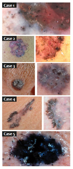
This series of five pigmented lesions shows that the differential diagnosis of pigmented BCC or melanoma is not always immediately evident, even with dermoscopy.1
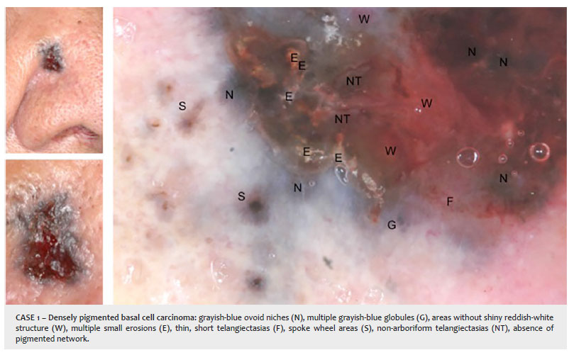
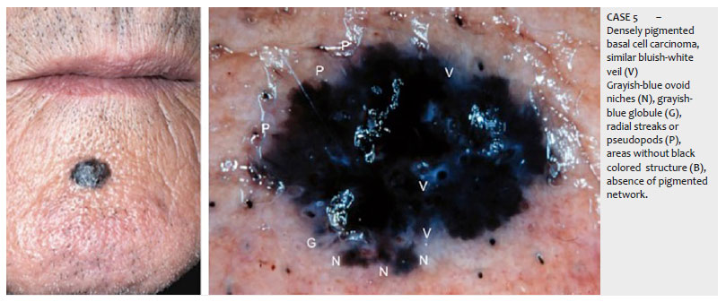
This series includes two cases of BCC (1 and 5) that are reminiscent of nodular melanoma. The dark coloring associated with a bluish veil is typical of melanoma, while the absence of specific characteristics of melanocytic lesions and black color (rule BB negative 5), and presence of spoke wheel and bluish ovoid niches, respectively, were suggestive of BCC.
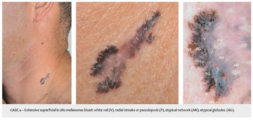
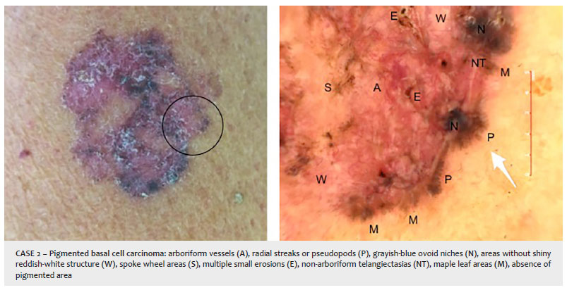
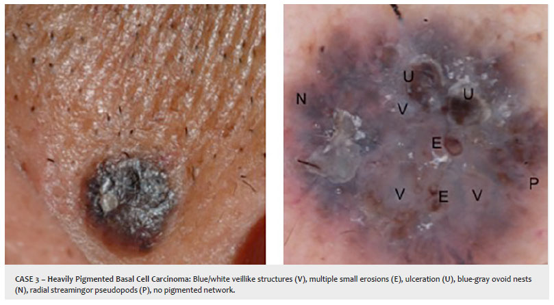
The other three lesions present peripheral structures (similar to radial streaks and pseudopods)6 at first sight. On more careful analysis, peripheral structures of melanoma (case 4) end in a bulbous projection (drumstick-like) compared to the BCCs (cases 2 and 3), which only present radial linear extensions. Besides, in the context of melanoma, some globules may be confused with bluish ovoid niches due to the blue color, but others clearly suggest melanocytic proliferation due to the dark blackish-brown color, resulting from the upward dissemination of melanocytic niches and clusters of Pagetoid cells.7
Differential diagnosis in these cases is obviously not always easy, and patients with high photo type require detailed examination, always with dermoscopy to enhance the differential diagnosis. This difficulty occurs mainly in the differentiation of densely pigmented BCCs.2 Especially in this subtype, the presence of dermoscopic characteristics suggestive of melanocytic lesion can reach 80%.2 Patterns including bluish-white veil and multiple black and blue globules are among the most frequent findings. 2
Thus, in densely pigmented BCCs, careful recognition of the clinical morphology and dermoscopic aspects can increase the diagnostic accuracy. We also propose that other imaging methods, such as confocal reflectance microscopy, which has proven useful in this context,8 can greatly assist the diagnosis of skin cancer in patients with high photo types.
Luiz Gameiro | 0000-0002-0210-8678
Elaboration and writing of the manuscript; critical review of the literature.
Hamilton Ometto Stolf | 0000-0003-4867-0276
Study design and planning.
Giovanni Pellacani | 0000-0002-7222-2951
Critical revision of the manuscript; approval of the final version.
1. Casari A, Pellacani G, Seidenari S, Cesinaro AM, Beretti F, Pepe P, et al. Pigmented nodular basal cell carcinomas in differential diagnosis with nodular melanomas: confocal microscopy as a reliable tool for in vivo histologic diagnosis. J Skin Cancer. 2011. Epub 2010 Oct 14.
2. Altamura D, Menzies SW, Argenziano G, Zalaudek I, Soyer HP, Sera F, et al. Dermatoscopy of basal cell carcinoma: morphologic variability of global and local features and accuracy of diagnosis. J Am Acad Dermatol. 2010;62(1):67-75.
3. Charles CA, Marghoob AA, Busam KJ, Clark-Loeser L, Halpern AC. Melanoma or pigmented basal cell carcinoma: a clinical-pathologic correlation with dermoscopy, in vivo confocal scanning laser microscopy, and routine histology. Skin Res Technol. 2002;8(4):282-287.
4. Menzies SW, Westerhoff K, Rabinovitz H, Kopf AW, McCarthy WH, Katz B. Surface microscopy of pigmented basal cell carcinoma. Arch Dermatol. 2000;136(8):1012-1016.
5. Argenziano G, Longo C, Cameron A, Cavicchini S, Gourhant JY, Lallas A, et al. Blue-black rule: a simple dermoscopic clue to recognize pigmented nodular melanoma. Br J Dermatol. 2011;165(6):1251-5.
6. Menzies SW, Crotty KA, McCarthy WH. The morphologic criteria of the pseudopod in surface microscopy. Arch Dermatol. 1995;131(4):436-40.
7. Longo C, Farnetani F, Moscarella E, Pace B, Ciardo S, Ponti G, et al. Can noninvasive imaging tools potentially predict the risk of ulceration in invasive melanomas showing blue and black colors? Melanoma Res. 2013;23(2):125-31
8. Peccerillo F, Mandel VD, Di Tullio F, Ciardo S, Chester J, Kaleci S, et. al. Lesions mimicking melanoma at dermoscopy confirmed basal cell carcinoma: evaluation with reflectance confocal microscopy. Dermatology 2019;235(1):35-44.
 All content the journal, except where identified, under the Creative Commons Attribution 4.0 International licence - ISSN-e 1984-8773


 Read in Portuguese
 Portuguese PDF
 Print
Print
 Send this article by email
 How to cite this article
 Submit a comment
 Mendeley
 Pocket