


Dian Ardiana1; Nanny Herwanto2; Cita Rosita Sigit Prakoeswa2; Indropo Agusni2
Received on: 18/05/2019
Approved on: 23/09/2019
Financial support: None
Conflict of interests: None
Study conducted at the Department of Dermatology and Venereology, Medical School, Universitas Airlangga, Dr. Soetomo Teaching Hospital, Surabaya, Indonesia
INTRODUCTION: Vitiligo is a pigmentation disorder characterized by white spots on the skin and mucous membranes. Wnt1 protein belongs to the Wnt signaling pathway. Wnt1 induction is linked with melanocyte stem cell differentiation in hair follicles on lentigo solaris, which is a hyperpigmentation skin disorder in sun-exposed area. This assumes the possibility of Wnt1 induction with NB-UVB therapy, which causes perifollicular repigmentation on vitiligo.
OBJECTIVES: To compare Wnt1 expressions in vitiligo repigmentation after NB-UVB treatment and to identify the relationship between Wnt1 expressions and vitiligo repigmentation area.
MATERIALS AND METHODS: The subjects of the study were 18 vitiligo patients. Immunohistochemical examination was conducted on vitiligo skin tissues pre- and post-phototherapy treatment twice a week for two months.
RESULTS: There were significant differences in the vitiligo lesion area between pre and post-therapy. Also, there were significant differences in Wnt1 expressions between pre and post-therapy. There was a correlation between the post-therapy Wnt1 expressions and the post-therapy vitiligo lesion area.
CONCLUSIONS: Wnt1 induction occurs in hair follicles post-NB-UVB therapy, causing pigmentation around hair follicles. Further studies are needed on the Wnt1 protein mechanism resulting in pigmentation around hair follicles.
Keywords: Hair Follicle; Melanocytes; Ultraviolet Therapy; Wnt1 Protein; Phototherapy; Vitiligo
Vitiligo is a skin pigmentation disorder, marked by chalky white patches on the skin and mucosa, and characterized histologically by the lack of melanocytes (melanin pigment formation cell). Vitiligo is detected worldwide, with a prevalence of around 0.1% to 2%.1 Low levels of quality of life are related to disease activity as well as an occurrence at a young age and spots on the hands.2
The narrowband ultraviolet B radiation (NB-UVB) has been a standard treatment for vitiligo. NB-UVB therapy showed statistically and clinically better responses than ultraviolet A (UVA);3-5 however, NB-UVB needs special light, being a time-consuming treatment (from several months to years), and repigmentation may not reach 100%.
Ortonne et al. mention deposits of melanocytes in human hair follicles and the proliferation of these melanocytes with exposure to PUVA, causing repigmentation in vitiligo.6 However, the mechanism of vitiligo perifollicular repigmentation from melanocyte stem cells after treatment with NB-UVB is still unclear.
Wnt is a signaling protein, and its role in human skin and hair pigmentation has not been well explained yet, both during fetal development and in the postnatal period. The Wnt signals are essential in the development of neural crests in the embryology of mice, particularly Wnt1 and Wnt3a, which also play significant roles in the development of the neural crest to form pigment cells; and when depletion of neural crest cells by both proteins, it tends to form neurons instead of pigment cells. Wnt1 transmits signals in the melanoblasts as paracrine to increase the quantity of melanocytes, while the Wnt3a and β-catenin signals act more in determining the neural crest to form melanocytes.7 Wnt stem cells cause the melanocytes stem cells to differentiate and form melanoblasts that will produce melanocytes that, in turn, will provide pigmentation to growing hair follicles.8
A study conducted by Yamada et al. (2013)9 mice showed that melanocyte stem cells in the bulge region induce epidermal pigmentation after treatment with NB-UVB. Melanocyte stem cells differentiate to form melanoblasts by activating the Wnt/β-catenin pathway. The experiment in rats also showed an increase in the expression of Wnt1, stimulating the differentiation of melanocyte stem cells and causing hyperpigmentation of the skin after exposure to UVB.
This study aims to compare the Wnt1 expression of the perifollicular lesion of pre-therapy vitiligo with NB-UVB and the Wnt1 expression in the lesion repigmentation of the post-therapy with NB-UVB and to identify the relationship between the Wnt1 expression and the vitiligo repigmentation area.
A single group, pre-experimental study with a pre-test/ post-test design was conducted. It was approved by the Research Ethics Committee of the Dr. Ramelan Navy Hospital Surabaya. All subjects in this study signed the informed consent form (ICF) before the research process.
The study population comprised patients with vitiligo followed up at the Skin and Venereal Diseases Polyclinic, Dr. Ramelan Navy Hospital Surabaya, Surabaya, Indonesia. The inclusion criteria were patients aged 15 to 60 years with Fitzpatrick skin phototype 4/5, presenting dark hair in the vitiligo lesion. The exclusion criteria were patients with photodermatosis; vitiligo in the mucosa; acrofacial vitiligo; generalized or universal vitiligo; pregnant women; immunodeficient subjects; patients undergoing topical or systemic therapy for vitiligo in the last two months; patients with marked erythema or burn injuries due to radiation; and patients with a history of hypertrophic scarring.
Data were collected and measurements were taken. A punch biopsy with 4 mm was performed initially on the skin with a vitiligo lesion involving hair follicles. At the end of the study, measurements were taken in the areas of the vitiligo lesion and in the repigmentation area adjacent to the initial biopsy. The repigmentation area was considered to be the difference in pigmented area between the pre- and post- NB-UVB therapy period.
The radiation was conducted with DermaPalTM Daavlin NB-UVB at 311-312nm wavelength and 311nm peak (Bryan, OH, USA), with two Philips PL-S 9W/01/2P lamps, radiating in an area of 2.54 x 11.4 cm. The therapy was performed twice a week, on non-consecutive days, for two months. NB-UVB radiation at the initial visit was administered at a dose of 320m J/cm2 and 390m J/cm2 at the subsequent visits.
The immunohistochemical examination of the biopsy was performed at the Laboratory of Pathological Anatomy of the Medical School of Airlangga University Surabaya, with anti-human rat Wnt1 (Novus Biologicals: Wnt-1 Antibody (10C8) NBP1-51575) primary antibody, rabbit anti-rat (TL-012-MHRA MultiVision anti-rabbit/ AP+ anti-rat/HRP polymers) and secondary anti-body.
The results of the area measurement and the calculations of the Wnt1 expression were recorded on a data collection sheet. The normality test was performed using the Shapiro-Wilk test. Differences in the vitiligo area before and after NB-UVB were assessed with the Wilcoxon signed-rank test, as well as the differences in Wnt1 expression before and after NB-UVB therapy. The relationship between the Wnt1 expression and the vitiligo area was analyzed with Spearman's rank correlation coefficient test. The significance < level is 5%.
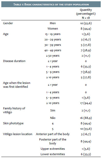
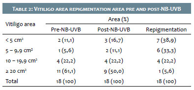
Eighteen vitiligo patients who met the inclusion criteria participated in the study. Table 1 presents the basic characteristic of the study population. The vitiligo before the NB-UVB treatment varied between 2.16 - 283.38 cm2. The mean pre-NB-UVB vitiligo area was 50.83 ± 65.8 cm2. The vitiligo area after NB-UVB varied between 1.98 - 269.17cm2. The mean vitiligo area after the NB-UVB was 43.2 ± 63.97 cm2. The repigmentation area varied between 0.18 - 20.23 cm2. The mean repigmentation was 7.62 ± 6.16cm2 (Table 2).
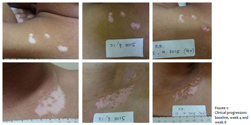
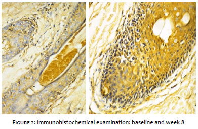
Clinical progression was observed at baseline, at week four, and at week eight (Figure 1), and immunohistochemical examination was performed at baseline, and week four (Figure 2).
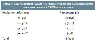
There was a significant difference in the vitiligo area before and after NB-UVB treatment (p=0.000). A comparison between the repigmentation area and the NB-UVB pre-therapy area showed that the lowest percentage of area was 3.2% and the highest was 80.5%. The mean percentage of the repigmentation area was 24.82 ± 21.67%. One patient presented a repigmentation area of 80%, while most of subjects (61.1%) had a repigmentation area <25% (Table 3).
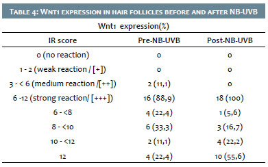
Wnt1 expression was calculated based on two parameters: I. Percentage of positive cells (0: no occurrence of cells expressing Wnt1; 1: occurrence of ≤10% of cells expressing Wnt1; 2: occurrence of 11-50% of cells expressing Wnt1; 3: occurrence of 51-80% of cells expressing Wnt1; 4: occurrence of >80% of cells expressing Wnt1); and II. Color reaction intensity (0: no color reaction; 1: low color intensity; 2: medium color intensity; 3: high color intensity).
The immunoreactivity score (IR) is the multiplication of scores I and II (0: no reaction; 1-2: weak reaction [+]; 3-4: medium reaction [++]; 6-12: strong reaction [+ ++]).
Most of the patients in the study presented a strong Wnt1 expression immunoreactivity reaction score before NB-UVB therapy, whereas, after NB-UVB, all patients demonstrated a strong Wnt1 expression immunoreactivity reaction score (Table 4). There was a significant difference in the immunoreactivity scores of Wnt1 expression in hair follicles before and after NB-UVB (p=0.009). There was a significant correlation between the vitiligo area and the Wnt1 expression after NB-UVB (p = 0.036).
NB-UVB therapy is one of the most used today. NB-UVB provides better statistical and clinical responses than UVA.3,4,5 These studies identified significant differences between the vitiligo area pre and post-NB-UVB (p=0.000). The improvement in vitiligo lesion was marked by the decrease in the area of vitiligo white patches due to pigmentation (repigmentation) with NB-UVB therapy twice a week for two months. Despite the significant difference between the vitiligo area before and after treatment, this study showed that most patients (61.1%) presented less than 25% repigmentation at the end of the study (Table 3). El-Zawahry et al. mentioned that 65% of patients had repigmentation ranging from 40% to over 80% with NB-UVB treatment for three months.5 Kumar et al. demonstrated that only 34% of patients experience repigmentation of less than 25%. In contrast, 48.6% experienced repigmentation of 25% to 75% and 17.4% experience more than 75% of repigmentation with NB-UVB therapy for 12 months.4 The number of patients with repigmentation below 25% in this study still exceeds that of several other studies, which may be because NB-UVB treatment has been implemented twice a week for two months (eight weeks), when still didn't reach a larger area.
Repigmentation begins to occur after the first to the fifth application of phototherapy for almost 35% of patients, and after the sixth to the tenth application for almost 50% of patients. The fastest repigmentation occurs after the third therapy.4,10 The initial repigmentation occurs in weeks 3 to 10, with three treatments per week.11 The variable duration indicates a change in individual NB-UVB responses. Therapeutic responses also depend on the duration of the disease.12 The present study did not show that repigmentation depends on the duration of the disease (p = 0.969).
This study demonstrated a significant difference in the Wnt1 expression in the hair follicle of the vitiligo lesion before NB-UVB treatment and in the Wnt1 expression in the lesion of perifollicular repigmentation after NB-UVB (p=0.009). This shows the occurrence of the induction of Wnt1 expression in hair follicles with NB-UVB during the two-month treatment. The Wnt1 protein can be linked to several types of receptors, including the Frizzled (Fzd) 7-transmembrane receptor. The signal that occurs from the Wnt1 connection to the receiver is still transmitted through three separate pathways: the canonical pathway, which involves Wnt/β-catenin, the non-canonical pathway, which involves Wnt/Ca2+ and Wnt/ polarity pathway (or planar cell polarity pathway).13,14 Wnt1 generally activates the Wnt/β-catenin line.7
The Wnt/β-catenin pathway is known to play a critical role in the differentiation of hair follicle stem cells from melanocytes that cause epidermal pigmentation in mice induced with NB-UV.9 The differentiation of melanocyte stem cells produces melanoblasts, followed by their differentiation and the production of melanocytes in the infundibulum, proliferating and migrating further in the epidermis and causing pigmentation in patients with vitiligo treated with NB-UVB.15 This study demonstrated a significant correlation between the Wnt1 expression in hair follicles and post-NB-UVB vitiligo area (p = 0.036).
The increase in Wnt1 expression in post-NB-UVB hair follicles, as found in this study, is expected to bind to the Fzd receptor to activate intercellular signals via the Wnt/β-catenin pathway, which eventually activates the FTAM gene target. FTAM activation induces melanocyte stem cells to differentiate into melanoblasts. Therefore, the induction of Wnt1 expression in hair follicles with NB-UVB treatment affects the pigmentation of the vitiligo lesion around the hair follicles. Further studies are needed to prove the mechanism.
The present study showed that 94% of the patients showed Wnt1 expressions in the hair follicles with a strong reaction, with an immunoreactivity score of 12 in four patients (22.4%) before therapy. The evaluation of post-NB-UVB treatment showed that all patients presented strong immunoreactivity with a score of 12 in 10 patients (55.65). There are 19 Wnt genes that encode 19 proteins identified in humans. More melanocyte pathways are connected with Wnt1, 3a, 5a, and 10b signals. The Wnt protein in human skin has rarely been studied. Rabbani et al. showed that epithelial stem cells around melanocyte stem cells produce Wnt10b protein, which is capable of activating the Wnt signal in melanocyte stem cells. Then, stem cells differentiate from melanocytes, becoming melanoblasts, and from melanoblasts into melanocytes, which provide pigmentation to growing hair follicles.8
Yamada's study in rats demonstrated that the epithelial pathway expresses the Wnt7a (hair follicle stem cells, hair follicle stem keratinocytes, and epidermal keratinocytes) after exposure to UVB. In contrast, melanocyte stem cells do not express Wnt7a. Wnt7a expressed by the epithelial pathway activates the Wnt/β-catenin signals, which trigger the differentiation of melanocyte stem cells to become melanoblasts. Yamada's additional study in mice showed that the canonical expression of mRNA Wnt1 and Wnt2 experiencing hyperpigmentation increased significantly due to UVB exposure, while the non-canonical expressions of Wnt9a and Wnt5a suffered deregulation.9
This study demonstrated that NB-UVB treatment for two months in vitiligo patients showed changes in the Wnt1 expression in the hair follicles of vitiligo lesions, and this change is related to the occurrence of peripheral re-pigmentation of vitiligo. Additional studies are needed to examine the expression of other Wnt proteins in vitiligo, which plays a more significant role in the differentiation of melanocyte stem cells in vitiligo, and the role of Wnt1 in the epidermal melanocyte.
Dian Ardiana | 0000-0002-8592-1989
Approval of the final version of the manuscript; preparation and writing of the manuscript; data collection, analysis, and interpretation; active participation in research orientation; critical literature review; critical revision of the manuscript.
Nanny Herwanto | 0000-0002-1908-8407
Approval of the final version of the manuscript; preparation and writing of the manuscript; data collection, analysis, and interpretation; active participation in research orientation; critical literature review; critical revision of the manuscript.
Cita Rosita Sigit Prakoeswa | 0000-0001-5325-2963
Approval of the final version of the manuscript; preparation and writing of the manuscript; data collection, analysis, and interpretation; active participation in research orientation; critical literature review; critical revision of the manuscript.
Indropo Agusni | 0000-0001-6729-8831
Approval of the final version of the manuscript; preparation and writing of the manuscript; data collection, analysis, and interpretation; active participation in research orientation; critical literature review; critical revision of the manuscript.
1. Halder R, Taliaferro. Vitiligo. In: Wolff K, Goldsmith L, Katz S, Gilchrest B, editors. Fitzpatrick's dermatology in general medicine. New York: McGraw-Hill; 2008.
2. Karelson M, Silm H, Kingo K. Quality of life and emotional state in vitiligo in an Estonian sample: comparison with psoriasis and healthy controls. Act Derm Venereol. 2013;93(4):446-50.
3. Yones SS, Palmer RA, Garibaldinos TM, Hawk JLM. Randomized Double-blind Trial of Treatment of Vitiligo. Efficacy of Psoralen-UV-A Therapy vs Narrowband-UV-B Therapy. Arch Dermatol. 2007;143(5):578-84.
4. Kumar YHK, Rao GRR, Gopal KVT, Shanti G, Rao KV. Evaluation of narrow-band UVB phototherapy in 150 patients with vitiligo. Indian J Dermatol Venereol Leprol. 2009;75(2);162-6.
5. El-Zawahry BM, Bassiouny DA, Sobhi RM, Abdel-Aziz E, Zaki NS, Habib DF, et al. A comparative study on efficacy of UVA vs. Narrow-band UVB phototherapy in the treatment of vitiligo. Photodermatol Photoimmunol Photomed. 2012;28(2):84-90.
6. Ortonne JP, MacDonald DM, Micoud A, Thivolet J. UVA-induced repigmentation of vitiligo: a histochemical (spilt-DOPA) and ultrastructural study. Br J Dermatol. 1979;101(1):1-12.
7. O'Connell MP, Weeraratna AT. Hear the Wnt Ror: how melanoma cells adjust to changes in Wnt. Pigment Cell Melanoma Res. 2009;22(6):724-39.
8. Rabbani P, Takeo M, Chou W, Myung P, BoseNBerg M, Chin L, et al. Coordinated activation of Wnt in epithelial and melanocyte stem cells initiates pigmented hair regeneration. Cell. 2011;145(6):941-55.
9. Yamada T, Hasegawa S, Inoue Y, Date Y, Yamamoto N, Mizutani H, et al. Wnt/beta-catenin and kit signaling sequentially regulate melanocyte stem cell differentiation in UVB-induced epidermal pigmentation. J Invest Dermatol. 2013;133(12): 2753-62.
10. Girish PN, Narendra JS, Vinma HS, Sandhya I, Umananda M, Ranjith KSB, et al. Evaluation of narrow-band UV B phototherapy for vitiligo. J Evolut Med Dental Sci. 2013; 2(44):8591-8.
11. Khullar G, Kanwar AJ, Singh S, Parsad D. Comparison of efficacy and safety profile of topical calcipotriol ointment in combination with NB-UVB vs. NB-UVB alone in the treatment of vitiligo: a 24-week prospective right-left comparative clinical trial. J Eur Acad Dermato Venereol. 2015;29(5):925-32.
12. Hallaji Z, Ghiasi M, Eisazadeh A, Damavandi MR. Evaluation of the effect of disease duration in generalized vitiligo on its clinical response to narrowband ultraviolet B phototherapy. Photodermatol Photoimmunol Photomed. 2012; 28(3):115-9.
13. Clevers H. Wnt/beta-catenin signaling in development and disease. Cell. 2006;127(3):469-80.
14. Van Camp JK, Beckers S, Zegers D, Van Hul W. Wnt signaling and the control of human stem cell fate. Stem Cell Rev. 2014;10(2):207-29.
15. Goldstein NB, Koster MI, Hoaglin LG, Spoelstra NS. Narrow Band Ultraviolet B Treatment for Human Vitiligo Is Associated with Proliferation, Migration, and Differentiation of Melanocyte Precursors. J Invest Dermatol. 2015;135(8):2068-76.
 All content the journal, except where identified, under the Creative Commons Attribution 4.0 International licence - ISSN-e 1984-8773


 Read in Portuguese
 Portuguese PDF
 Print
Print
 Send this article by email
 How to cite this article
 Submit a comment
 Mendeley
 Pocket