


Helena Reich Camasmie; Antonio Macedo D'Acri
Received on: 13/01/2018
Approved on: 08/03/2019
This study was performed at the Department of Dermatology, Dermatology Sector, Hospital Universitário Gaffrée & Guinle, Universidade Federal do Estado do Rio de Janeiro (UNIRIO) - Rio de Janeiro (RJ), Brazil. Financial support: None
Conflict of interestss: None
Milia are keratin cysts of 1-3mm in diameter that occur due to the obstruction of eccrine sweat glands or hair follicles. We describe the case of a female patient with multiple white-yellow papules over a tattoo made six months prior to the consultation. Conservative treatment is an option, since there is the possibility of the lesion being transient and that it will spontaneously resolve. We opted for a conservative treatment with excellent final cosmetic outcome.
Keywords: Primary Treatment; Tattooing; Ink; Therapeutics
Milia are keratin cysts that measure 1-3mm in diameter and occur due to obstruction of eccrine sweat glands or hair follicles. 1 The origin of these cysts is matter of debate and it has been suggested that they might originate from the inferior portion of the vellus hair's infundibulum, however their histogenesis remains uncertain. Visible on the face as multiple whitish papules, milia are usually treated by manual extraction. 2 They can be classified into primary, spontaneous and secondary, and might occur after minor trauma, use of topical or systemic drugs, and in association with inflammatory skin conditions. 1
Considering the increasing number of people who decide to have tattoos on their skin, it is believed that this habit might pose a significant risk for the public health. The most frequent skin reactions to tattoos include allergic, infectious and granulomatous dermatoses. 3 The act of tattooing leads to damage of the cutaneous barrier, which may facilitate the hematogenous spread of several pathogens, since the needles reach the vessels of the dermis. Therefore, infections in the bloodstream may also occur. 4
Tattoos are associated with increased risk of inflammatory conditions such as eczema, psoriasis and neoplasms. 5 The description of milia on tattoos is rare, with few cases having been described. In fact it is not clear whether the low frequency of this condition is due to the absence of reports or the low incidence of cases.
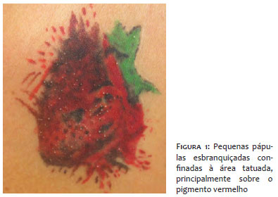
The authors of this paper describe a case of a female patient with multiple white-yellowish papules scattered over a tattoo performed six months before the medical appointment. The papules emerged approximately one month after the procedure, being clinically diagnosed as milia. The lesions were restricted to the tattooed area, mainly on the red pigment, but there were also some on the green pigment (Figure 1).
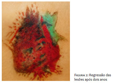
The authors of the present paper decided for adopting an expectant approach to the case. The lesions disappeared spontaneously after two years of follow-up (Figure 2).
Allergic reactions to red ink are the second most common complication after having a tattoo performed on the skin and occur due to the haptenization process that the red pigment undergoes. Although not exclusively, most of our patient's lesions were located over the region tattooed in red. Such lesions may occur at any time after the procedure and are generally asymptomatic.3 The precise cause that could explain the emergence of milia after following tattooing procedures is not well explained in the literature and the authors of the present paper believe it is due to the trauma process and anomalous healing.
Conservative treatment is an option, since there is a possibility that the lesion may be transient and disappear spontaneously. Manual extraction with needles and dermabrasion are valid approaches, however can damage the original drawing.
The number of tattoos in the population is increasing significantly, and dermatologists should be aware of the possible complications as well as of the available therapeutic options.
Helena Reich Camasmie | ORCID 0000-0003-0231-3003
Discussion and planning of the theme; data and bibliographic references analysis; drafting and final approval of the manuscript.
Antonio Macedo D’Acri | ORCID 0000-0002-2682-525X
Discussion and planning of the theme; data and bibliographic references analysis; drafting and final approval of the manuscript.
1. Avhad G, Ghate S, Dhurat R. Milia en plaque. Indian Dermatol Online J. 2014; 5(4):550-1.
2. Kurokawa I, Kakuno A, Tsubura A. Milia may originate from the outermost layers of the hair bulge of the outer root sheath: A case report. Oncol Lett. 2016;12(6):5190-2.
3. Ross N, Farber M, Sahu J. Eruptive Milia within a Tattoo: A Case Report and Review of the Literature. J Drugs Dermatol. 2017;16(6):621-4.
4. Dieckmann R, Boone I, Brockmann SO, Hammerl JA, Kolb-Mäurer A, Goebeler M, et al. Risk of bacterial infection after tattooing. Dtsch Arztebl Int. 2016;113(40):665-71.
5. Duan L, Kim S, Watsky K, Narayan D. Systemic allergic reaction to red tattoo ink requiring excision. Plast Reconst Surg Glob Open. 2016;4(11):e1111.
 All content the journal, except where identified, under the Creative Commons Attribution 4.0 International licence - ISSN-e 1984-8773


 Read in Portuguese
 Portuguese PDF
 Print
Print
 Send this article by email
 How to cite this article
 Submit a comment
 Mendeley
 Pocket