


Felipe Siqueira Ramos; Elisangela Manfredini Andraus de Lima; Flávia Regina Ferreira; Samuel Henrique Mandelbaum
Received on: 10/12/2018
Approved on: 22/01/2019
Study conducted at Taubaté University Hospital, Universidade de Taubaté - Taubaté (SP), Brazil.
Financial support: The products used, Epifactor® e cold cream, were given by the company Infinity Pharma, Campinas (SP)
Conflict of interests: None
INTRODUCTION: Healing is a phenomenon that occurs after tissue injury and involves complex cellular and molecular mechanisms. Growth factors seem to be an effective and safe complement for the treatment of wounds.
OBJECTIVE: To evaluate wound healing after electrocoagulation, comparing the vehicle in isolation and its association with epidermal growth factor.
METHODS: Double-blind clinical trial in a Dermatology service between 2016 and 2018. Patients of both genders, older than 18 years of age, submitted to electrocoagulation of two lesions and subsequent application of the vehicle (cold cream) on one and epidermal growth factor in cold cream on the other were included. Evaluations after 7, 14 and 28 days, analysed erythema, edema, crusting, discharge and healing. Analyzed: edema, edema, crusting, discharge and healing. The binomial test was used for two ratios and Fisher’s exact test was used for dichotomic data.
RESULTS: Variable results were found regarding erythema, edema, crusting and discharge, sometimes favoring the vehicle, sometimes the growth factor, however with no statistical significance. Regarding healing, epithelialization was quicker with with epidermal growth factor (p<0.05).
CONCLUSIONS: This study evaluated the impact of epidermal growth factor in the healing process, and its results reinforce scarce data of the current literature and are a foundation for future studies.
Keywords: Epidermal growth factor; Evaluation; Wound healing
Healing is a phenomenon that occurs after tissue injury of any nature and involves complex cellular and molecular mechanisms. Inflammation, proliferation, angiogenesis, reepithelization, tissue regeneration and remodeling are part of this biological process.1 Thermal burns (accidental or intentional) generate areas of necrosis that extend beyond the wound, even causing obstruction of blood and lymphatic vessels.2
The process of tissue repair is modulated by growth factors, that are produced by epidermal and epithelial cells, such as macrophages, fibroblasts and keratinocytes. Growth factors are biologically active molecules and act directly from within the cell, regulating the cell cycle.3 However, the availability of these growth factors can be insufficient in the bed of the wound resulting from burns due to their excessive degradation or reduced production. Therefore, treatment with growth factors seems to be an effective and safe complement to the treatment of wounds.4,5 The objective of this study was to evaluate wound healing after electrocoagulation comparing the vehicle alone (cold cream) to its association with epidermal growth factor.
This is a comparative, double-blind clinical trial, conducted at a Service of Dermatology between June 2016 and July 2018. Patients from both genders and older than 18 years of age were included, with two lesions similar in nature and size, on the same body area. Both lesions were submitted to electrocoagulation, with subsequent daily application of the vehicle (cold cream) on one and 5% epidermal growth factor in cold cream (Epifactor®) on the other, for 28 days. Evaluations were performed after 7, 14 and 28 days, and the wounds photographed according to the standardization. These photos were then evaluated by an independent researcher (dermatologist). The variables analyzed were: erythema, edema the use of the vehicle alone. However, on the 28th day, both groups had a coinciding end result for this variable.(presence or absence and intensity), crust, discharge (presence or absence) and healing (crust, ulceration and epithelization). Tables were made. For the comparison between two independent samples, the binomial test was used for two of significance adopted was alpha = 5%, and the statistical program used was BioEstat 5.0. The study was approved by the Committee ratios and Fisher's exact test for dichotomic data. The level of Ethics in Research of the institution under the number 1.861.616.
Thirteen patients were included (seven men and six women), with minimum and maximum ages of 36 and 86 years, respectively.
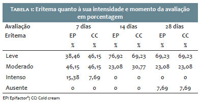
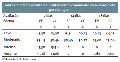
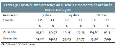
Table 1 demonstrates the progression of erythema along the 28 days. After 14 days, there were similar percentages between the two groups for this variable, predominating mild erythema. In table 2, we can evaluate data regarding edema. There was an apparent lower initial edema (7th and 14th day) with regarding the variable 'crust formation', data can be seen on table 3. Most wounds (in both groups) showed crust after seven days. The elimination of the crust was faster with the use of the vehicle alone (69.23% of the lesions had no crust on the 14th day).
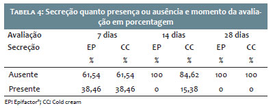
Table 4 illustrates the variable 'discharge'. In it, we can observe a faster resolution of the discharge in the wounds where Epifactor® was used (100% on the 14th day).
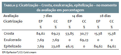
Regarding healing, only 7.69% of the wounds (from both groups) had ulceration on the 7th day (Table 5). The epithelization occurred faster in the wounds where Epifactor® was used (46.15% versus 0% - 14th day), p<0.05.
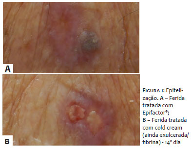
Figure 1 illustrates epithelization of the wound treated with Epifactor® and with vehicle on the 14th day.
This original study, evaluating the impact of epidermal growth factor in the healing process, supports the literature where studies on this subject are still scarce, what made difficult the discussion of the results found. Regarding the variables erythema, edema, crust and discharge, there was a mild predominance of a better result sometimes for one group, other times for the other, however, with no statistical significance. Healing (epithelization) was faster in the group using epidermal growth factor, supporting the findings by Zhang et al, who demonstrated that the topical use of growth factors significantly reduced healing time for partial thickness burn wounds.4 Limitation: Factors not taken into account in this study, such as gender, age, site, comorbidities, could have interfered with our findings.
This study allowed for the evaluation of the healing of wounds after electrocoagulation, comparing the vehicle alone to its association with epidermal growth factor and concluded that the topical use of epidermal growth factor accelerated wound epithelialization, significantly reducing healing time.
To all staff of the Service of Dermatology at University Hospital of Taubaté and to Prof. Dr. Luiz Carlos Laureano da Rosa for the help with the statistical analysis of this study.
Felipe Siqueira Ramos | ORCID 0000-0002-3109-4359
Statistical analysis, approval of the final version of the manuscript, design and planning of the study, preparation and writing of the manuscript, data collection, analysis and interpretation, active participation in mentoring the research, intellectual participation in propaedeutics and/or therapeutics of the cases studied, critical review of the literature, critical review of the manuscript.
Elisangela Manfredini Andraus De Lima | ORCID 0000-0002-2390-0410
Approval of the final version of the manuscript, design and planning of the study, preparation and writing of the manuscript, data collection, analysis and interpretation, active participation in mentoring the research, intellectual participation in propaedeutics and/or therapeutics of the cases studied, critical review of the manuscript.
Flávia Regina Ferreira | ORCID 0000-0001-5679-4282
Approval of the final version of the manuscript, design and planning of the study, data collection, analysis and interpretation, active participation in mentoring the research, intellectual participation in propaedeutics and/or therapeutics of the cases studied, critical review of the literature, critical review of the manuscript.
Samuel Henrique Mandelbaum | ORCID 0000-0002-4631-4828
Approval of the final version of the manuscript, design and planning of the study.
1. Velnar T, Bailey T, Smrkolj V. The Wound Healing Process: an Overview of the Cellular and Molecular Mechanisms. J Int Med Res. 2009;37(5):1528-42.
2. Lee JH, Bae IH, Choi JK, Park JW. Evaluation of a Highly Skin Permeable Low-Molecular-Weight Protamine Conjugated Epidermal Growth Factor for Novel Burn Wound Healing Therapy. J Pharm Sci. 2013;102(11):4109-20.
3. Vieira ACQM, Medeiros LA, Palácio SB, Lyra MAM, Alves LDS, Rolim LA, et al. Fatores de crescimento: uma nova abordagem cosmecêutica para o cuidado antienvelhecimento. Rev Bras Farm. 2011;92(3):80-9.
4. Zhang Y, Wang T, He J, Dong J. Growth factor therapy in patients with partial-thickness burns: a systematic review and meta-analysis. Int Wound J. 2016;13(3):354-66.
5. Marchese C, Chedid M, Dirsc OR, Csaky KG, Santanelli F, Latini C, et al. Modulation of Keratinocyte Growth Factor and its Receptor in Reepithelializing Human Skin. J Exp Med. 1995;182(5):1369-76.
 All content the journal, except where identified, under the Creative Commons Attribution 4.0 International licence - ISSN-e 1984-8773


 Read in Portuguese
 Portuguese PDF
 Print
Print
 Send this article by email
 How to cite this article
 Submit a comment
 Mendeley
 Pocket