


Samara Eberlin1; Gustavo Facchini2; Samir Eberlin3; Ana Lúcia Tabarini Alves Pinheiro4; Michelle Sabrina da Silva5; Adriano da Silva Pinheiro6; Adilson Costa7
Introduction: Infrared radiation (IR-A) causes structural changes in the skin, similar to those caused by prolonged exposure to ultraviolet radiation. Evaluation of efficacy and safety of cosmetic products concentrates in in vitro tests and clinical trials. A promising alternative is the use of fragments of human skin from elective cosmetic surgery, to evaluate the actual clinical benefits of a product applied topically.
Objective: The objective of this study was to correlate IR-A radiation effects in biopsies and in ex vivo skin fragments and in human fibroblasts culture by quantifying MMP-1, TIMP-1 and GADD45a mediators.
Methods: Collection of biopsies from 15 volunteers after IR-A applications for 5 consecutive days. Exposure to IR-A radiation of human skin fragments from elective cosmetic surgery, and human fibroblasts culture. Measurement of MMP-1, TIMP-1 and GADD45a mediators for further comparison of results.
Results: In the three models used, the IR-A radiation induced an increase in MMP-1, inhibited the synthesis of GADD45a, and did not changed TIMP-1 values.
Conclusion: Due to the positive correlation of the models studied, it may be suggested the use of ex vivo skin as plausible and sustainable tool to overcome differences between knowledge generated from in vitro and clinical experiments.
Keywords: SKIN AGING; MATRIX METALLOPROTEINASE 1; SOLAR RADIATION; IN VITRO TECHNIQUES
The electromagnetic spectrum emitted by solar radiation is composed of a wide range of wavelengths. Nevertheless, only a few fractions of these lengths reach the Earth's surface, including ultraviolet radiation (UV 280-400nm), the visible light (VL 400-760nm) and infrared radiation (IR 760nm-1mm). 1
For many years, photoaging and cutaneous damage were attributed almost exclusively to UV radiation, which represents only 6.8% of solar radiation as compared with the infrared and visible radiations, which correspond 54.3% and 38.9% of incident solar energy, respectively. 1 Currently, however, it is known that IR radiation also induces histological alterations similar to those induced by chronic exposure to UV. 2
Infrared radiation (IRR) is classified into IR-A (760 – 1,400nm), IR-B (1,400-3,000nm) and IR-C (3,000nm-1mm), according to the wavelength and its penetration into the skin layers. 1-3 Infrared radiation has two effects: thermal (which can be beneficial or harmful, depending on the dose) and oxidative damage (which arises from the range close to IR-A, 760 - 1,500nm). Infrared radiation-A reaches deeper layers of the skin, with 35% of the radiation being dispersed in the epidermis, 48% in the dermis and 17% in the subcutaneous tissue.2, 4 Although not yet completely understood, the mechanism by which IR-A radiation causes harmful effects involves disturbances in the transportation of mitochondrial electrons, leading to a decrease in energy production and an increase in the formation of reactive oxygen species. 5-7 With the loss of mitochondrial homeostasis, there is oxidative stress and changes in gene expression and dermal metabolism translated into increased expression of metalloproteinase 1 (MMP-1), decreased collagen synthesis, development of solar elastosis and skin hyperpigmentation. 8-11
In addition, DNA damage, cytotoxicity induction and generation of oxidative stress, with a decrease in antioxidant activity have been reported after acute exposure to IR-A radiation.2, 9, 12-15 Excessive and repeated exposures to IR-A has also been shown to cause chronic damage as erythema ab igne and squamous cell carcinoma,5, 16 probably as a result of the reduction in the DNA repair process.17-18
With the advent of the 3R policy (Replace, Refine and Reduce), which supports the use of alternative tests to replace, refine and reduce the use of animals in research, safety and efficacy assessment of cosmetics became restricted to in vitro and clinical tests. In vitro trials predict possible toxic effects and determine probable biological mechanisms of action responsible for the clinical benefit of the cosmetic product, complementing the in vivo results. Nonetheless, direct inference from the results requires caution due to the fact that not always the mechanisms observed in cell cultures or equivalent skin models can be extrapolated to the real condition of use. Likewise, although clinical results offer an undeniable contribution to the assessment of safety and efficacy of cosmetic products, they do not provide data regarding the mechanisms of action such as those obtained by in vitro techniques.
The evaluation of the biological mechanisms of action using skin biopsies obtained from healthy human volunteers as test-systems19-21 constitutes a model for understanding the real damage that an aggressor agent can trigger, as well as for the genuine clinical benefits generated by a cosmetic or dermatological treatment. However, although frequently reported in the literature, this procedure can be considered invasive when used as an everyday research tool.
Thus, a plausible and sustainable alternative to bridge this gap between the in vitro and the clinical is the use of skin fragments obtained from elective plastic surgery (ex vivo study), which is characterized as the most suitable model for approximating the actual effect responsible for the clinical benefits of a product applied topically.
The objective of the present study was to correlate the effects of IR-A radiation, both in biopsies and in ex vivo skin fragments and cultured human fibroblasts, through the quantification of MMP-1 mediators (matrix metalloproteinases), TIMP-1 (tissue inhibitor of metalloproteinase 1) and GADD45a (growth interruption protein and DNA damage).
Human Fibroblasts HFF-1 (BCRJ, Rio de Janeiro, Brazil) were seeded in 75cm2 bottles (Nunc, Denmark), cultured and expanded in an incubator at 37°C in the presence of 5% CO2, using specific culture medium. On reaching confluency, cells were seeded in 24-well plates (Nunc, Denmark).
The skin fragments used in the present study were obtained from a 54-year old healthy, skin phototype III 22 individual who had undergone elective plastic surgery in the abdominal region (abdominoplasty). After the surgical procedure, the skin fragments were fractionated into pieces of approximately 1.5cm2, weighed and kept in 24-well plates.
The cultures of HFF-1 and skin fragments underwent a 360 J/cm2 dose of IR-A radiation using the Hydrosun 750 and HBM1 devices (Hydrosun Medizintechnik GmbH, Müllheim, Germany). After radiation, the test-systems were incubated in fresh culture medium and maintained for 24 hours for collection of the supernatant, cell lysate and homogenized tissue.
The clinical trial for efficacy evaluation was characterized as open, single-center and prospective, involving 15 volunteers aged between 35 and 45 years, with skin phototypes II and III. Two areas were demarcated in the paravertebral region of all participants included in the study – one area served as a control and did not undergo application of IR-A radiation, while the other was exposed to IR-A radiation. The application of IR-A radiation in the study participants was performed with the 750T Hydrosun IRA device. A dose of 360 J/cm2 was applied daily for five consecutive days. This radiation dose is physiologically relevant given that the human skin is exposed to significant amounts of solar radiation type IR-A, with an average dose of 108 J/cm2/hr (summer, Campinas, SP, Brazil).
The study involving the participation of human volunteers and the use of human skin fragments obtained in elective surgeries was conducted after the approval of the Research Ethics Committee of the Universidade São Franciso - SP, Brazil.
The concentrations of MMP-1, TIMP-1 and GADD45a were measured by an immunoenzymatic trial, using commercially available kits (R&D Systems, Minneapolis, MN, USA; Uscn Life Science Inc., Houston, TX, USA). The absorbance reading was performed on monochromator Multiskan GO (Thermo Fisher Scientific Oy, Vantaa, Finland). The mediators' levels were calculated based on the reference values obtained by the standard curve, which was built with known concentrations of recombinant proteins.
The paired t-test with a 95% confidence interval (Graph-Pad Prism v6) was used for the statistical evaluation.
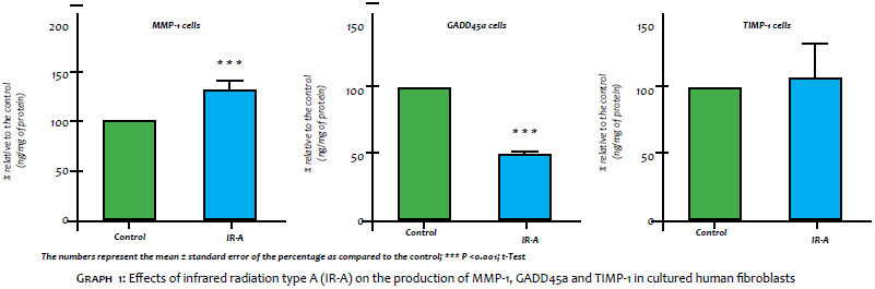
Graph 1 depicts the effects of IR-A radiation on the production of MMP-1, TIMP-1 and GADD45a in cultured human fibroblasts. As can be seen, the IR-A radiation produced a significant increase (31.2%) in the production of MMP-1 as compared to the non-irradiated baseline control. Regarding the GADD45a, the IR-A radiation led to a reduction of 50.5%, however it did not alter the TIMP-1's values.
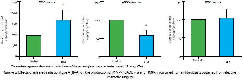
In Graph 2, it is possible to observe the results obtained after the exposure of human skin fragments to IR-A irradiation, which promoted a statistically significant increase (65.5%) in the production of MMP-1 in addition to a significant reduction in the synthesis of GADD45a (41.6%). TIMP-1 levels did not change compared to the non-irradiated control.
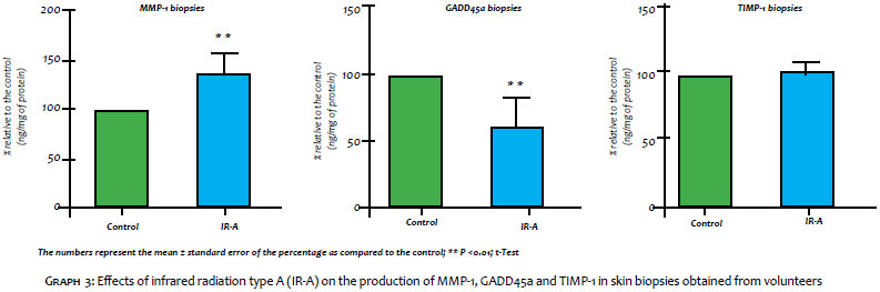
The results obtained in the homogenized tissue of the biopsies harvested after exposure of the volunteers to the IR-A radiation are in Graph 3. The radiation was able to promote a significant increase (33.9%) in the synthesis of MMP-1 and a reduction of 37.9% in the GADD45a protein – however it did not change the levels of TIMP-1.
A fairly common occurrence after exposure to IR-A radiation is the decrease in the synthesis of the main dermal proteins – collagen and elastin – essential for providing structural support for tissues.9, 23 This change occurs as a result of oxidative stress induced by reactive oxygen species generated upon exposure to radiation and leads to increased proteolytic enzymes, such as matrix metalloproteinase-1 (MMP-1).12, 24 This proteinase in turn triggers a collapse in the extracellular matrix and therefore the premature onset of signs of aging skin.9, 23 The MMPs' activity can be controlled by tissue inhibitors of metalloproteinases (TIMPs), which are synthesized by the fibroblasts located in the dermis and act locally, with the specific function of blocking the activity of MMPs, in this manner preventing the degradation of the matrix extracellular. 25
Another aspect of the oxidative response induced by IR-A radiation is the damage inflicted to cellular DNA. The ability to promptly repair that type of damage is an important cellular mechanism that protects cells and maintains genomic stability, preventing early oncogenesis.26-27 Animal cells have a complex defense mechanism to preserve genomic integrity and prevent that damage resulting from the genotoxic stress becomes permanent.25 Among these mechanisms are the disruption of cell cycle progression or direct activation of apoptosis, depending on the extent of damage and cell type.26-29 In this context, GADD45a protein plays a crucial role as a cellular stress sensor through the interaction with other proteins, promoting control of cell cycle regulation, DNA repair, epigenetic changes, apoptosis, survival and senescence.26-29
In the present study, the authors evaluated markers involved in skin aging using three human test-system models: fibroblast culture, ex vivo skin fragments and skin biopsies after exposure to IR-A.
The purpose of this comparative analysis was to validate the use of human skin obtained from elective cosmetic surgery as an alternative tool for assessing the effectiveness of cosmetic ingredients and products, in light of the fact that biological trials in animal models with this product category were practically banned and replaced by in vitro and clinical tests.
Despite the innovation of cell culture techniques and the development of increasingly complex three-dimensional skin equivalent models, there is still a gap in the extrapolation of the results for the clinical benefits that a cosmetic is able to promote. Furthermore, considering that the cutaneous tissue interacts structurally and functionally with the entire body and plays a vital role in the maintenance and regulation of the immune, endocrine and nervous systems, 30 biological effects obtained from in vitro studies may not precisely convey the observations, results and conclusions that are likely to occur clinically.
The in vivo (clinical) evaluation using skin biopsies from volunteers who undergo aesthetic treatments 19-21 constitutes a methodology that allows investigating the pharmacodynamics of molecules or products for, unlike other models, it does not exclude hormonal, nutritional or even immune individual variability. However, due to the fact that it is an invasive procedure, it might in some cases be deemed an aggressive method for proving the effectiveness of cosmetic and dermatological products, being consequently precluded as a day-to-day research tool.
According to a report by the International Society of Aesthetic Plastic Surgery (ISAPS), 31 Brazil ranked first in number of surgical procedures performed in 2013, in special liposuction, breast implants placement and abdominoplasty. The survey also shows that Brazil has nearly doubled the number of cosmetic surgeries performed in the past four years, with a growth of 97.2%.
While fragments of excess skin removed during elective plastic surgery are routinely discarded as infectious waste, its use is a feasible and sustainable experimental alternative that bridges the gap between the in vitro and the clinical, leading to almost similar outcomes to those of a product topically applied in a real situation.
The outcomes obtained in the present study confirm the deleterious effects that IR-A radiation is able to promote in the skin tissue, such as accelerated aging and weakening of the mechanisms involved in tissular repair. The production of MMP1 increased after exposure of the three test-systems – fibroblast culture, ex vivo fragments of skin and skin biopsies – to a dose of 360 J/cm2 of radiation IR-A. Similarly, the IR-A radiation led to a significant reduction in the production of GADD45a when compared to the unirradiated baseline control. One possible explanation for this effect is the increase of consumption and degradation of this protein as a result of genotoxic stress, which could result in a transient reduction in the levels of GADD45a in the cultures. As already mentioned, the absence of this protein can lead to genomic instability and impairment in the capacity to repair DNA damage.28-29, 32 Regarding the TIMP-1 levels, there was absence of significant alterations after exposure of the test-systems to IR-A radiation.
The results obtained in the present study clearly show that the ex vivo skin model is effective in mimicking the effects of IR radiation on the skin, proving that the use of human skin fragments obtained from elective plastic surgery is currently the safest option and most promising noninvasive option for the study of new active principles and formulations in the cosmetic/ dermatological industry.
Due to the positive correlation of results among the three assessed models, the authors can suggest that the ex vivo trial of skin fragments obtained from elective plastic surgery is an alternative approach to the use of human biopsies, given that it has been proven as a credible and sustainable tool to address differences between the knowledge generated from in vitro and clinical experiments.
1. Krutmann J. Skin Aging. In: Krutmann J, Humbert P, editors. Nutrition for Healthy Skin. Berlin Heidelberg: Springer-Verlag; 2011. p .15-24.
2. Schieke SM, Schroeder P, Krutmann J. Cutaneous effects of infrared radiation: from clinical observations to molecular response mechanisms. Photodermatol Photoimmunol Photomed. 2003;19(5):228-34.
3. Yoon HS, Kim YK, Matsui M, Chung JH. Possible role of infrared or heat in sun-induced changes of dermis of human skin in vivo. J Dermatol Sci. 2012;66(1):76-8.
4. Sklar LR, Almutawa F, Lim HW, Hamzavi I. Effects of ultraviolet radiation, visible light, and infrared radiation on erythema and pigmentation: a review. Photochem Photobiol Sci. 2013;12(1):54-64.
5. Schroeder P, Pohl C, Calles C, Marks C, Wild S, Krutmann J. Cellular response to infrared radiation involves retrograde mitochondrial signaling. Free Radic Biol Med. 2007; 43(1):128-35.
6. Karu T. Primary and secondary mechanisms of action of visible to near-IR radiation on cells. J Photochem Photobiol B. 1999;49(1):1-17.
7. Butow RA, Avadhani NG. Mitochondrial signaling: the retrograde response. Mol Cell. 2004;14(1):1-15.
8. Schroeder P, Calles C, Benesova T, Macaluso F, Krutmann J. Photoprotection beyond ultraviolet radiation-effective sun protection has to include protection against infrared A radiation-induced skin damage. Skin Pharmacol Physiol. 2010;23(1):15-7.
9. Kim MS, Kim YK, Cho KH, Chung JH. Regulation of type I procollagen and MMP-1 expression after single or repeated exposure to infrared radiation in human skin. Mech Ageing Dev. 2006;127(12):875-82.
10. Costa A, Eberlin S, Clerici SP, Abdalla BM. In vitro effects of infrared A radiation on the synthesis of MMP-1, catalase, superoxide dismutase and GADD45 alpha protein. Inflamm Allergy Drug Targets. 2015;14(1):53-9.
11. Ayres EL, Costa A, Eberlin S, Clerici SP. Estudo ex vivo para avaliação da atividade clareadora do Pycnogenol® após exposição à radiação ultravioleta, infravermelha e luz visível. Surg Cosm Dermatol. 2015;7(4):303-7.
12. Schroeder P, Lademann J, Darvin ME, Stege H, Marks C, Bruhnke S, et al. Infrared radiation-induced matrix metalloproteinase in human skin: implications for protection. J Invest Dermatol. 2008;128(10):2491-7.
13. Zastrow L, Groth N, Klein F, Kockott D, Lademann J, Renneberg R, et al. The missing link-light-induced (280-1,600 nm) free radical formation in human skin. Skin Pharmacol Physiol. 2009;22(1):31-44.
14. Darvin ME, Haag SF, Lademann J, Zastrow L, Sterry W, Meinke MC. Formation of free radicals in human skin during irradiation with infrared light. J Invest Dermatol. 2010;130(2):629-31.
15. Jung T, Höhn A, Piazena H, Grune T. Effects of water-filtered infrared A irradiation on human fibroblasts. Free Radical Biol Med. 2010;48(1):153-60.
16. Piazena H, Kelleher DK. Effects of infrared-A irradiation on skin: discrepancies in published data highlight the need for an exact consideration of physical and photobiological laws and appropriate experimental settings. Photochem Photobiol. 2010;86(3),687-705.
17. Calles C, Schneider M, Macaluso F, Benesova T, Krutmann J, Schroeder P. Infrared A radiation influences the skin fibroblast transcriptome: mechanisms and consequences. J Invest Dermatol. 2010;130(6):1524-36.
18. Leatherbarrow EL, Jenner TJ, O'Neill P, Botchway SW, Conein E, Gaur V, et al. [Internet]. Characterization of DNA damage induced by near infrared multiphoton absorption, Central Laser Facility Annual Report; 2004/2005, 151-154. [updated 2016 May. 24]. Available from: http://www.clf.stfc.ac.uk/resources/PDF/ar04-05_s6_characterisation_dna.pdf
19. Treiber N, Maity P, Singh K, Ferchiu F, Wlaschek M, Scharffetter-Kochanek K. The role of manganese superoxide dismutase in skin aging. Dermatoendocrinol. 2012;4(3):232-5.
20. Ramos SM, Carneiro SCS. Elderly skin and its rejuvenation: products and procedures for the aging skin. J Cosmet Dermatol. 2007;6(1):40-50.
21. Bolognia JL. Aging Skin. Am J Med. 1995;98(1A):99S-103S.
22. Fitzpatrick TB, Pathak M, Parrish JA. Protection of human skin against the effects of the sunburn ultraviolet (290-320nm). In: Fitzpatrick TB, editor. Sunlight and Man, normal and abnormal photobiological responses. Tokyo: University of Tokyo Press; 1994. p .751.
23. Cho S, Lee MJ, Kim MS, Lee S, Kim YK, Lee DH, et al. Infrared plus visible light and heat from natural sunlight participate in the expression of MMPs and type I procollagen as well as infiltration of inflammatory cell in human skin in vivo. J Dermatol Sci. 2008;50(2):123-33.
24. Schroeder P, Krutmann J. Infrared A-induced skin aging. In: Farage MA, Miller KW, Maibach HI, editors. Textbook of Aging Skin. Berlin Heidelberg: Springer-Verlag; 2010. p .421-5.
25. Wang XY, Bi Z. UVB-irradiated human keratinocytes and interleukin-1alpha indirectly increase MAP Kinase/AP-1 activation and MMP-1 production in UVA irradiated dermal fibroblasts. Chin Med J (Engl). 2006;119(10):827-31.
26. Liebermann DA, Hoffman B. Gadd45 in stress signaling. J Mol Signal. 2008;3:15.
27. Maeda T, Espino RA, Chomey EG, Luong L, Bano A, Meakins D, et al. Loss of p21WAF1/Cip1 in Gadd45-deficient keratinocytes restores DNA repair capacity. Carcinogenesis. 2005;26(10):1804-10.
28. Smith ML, Ford JM, Hollander MC, Bortnick RA, Amundson SA, Seo YR, et al. p53-mediated DNA repair responses to UV radiation: studies of mouse cells lacking p53, p21, and/or gadd45 genes. Mol Cel Biol. 2000;20(10):3705-14.
29. Cretu A, Sha X, Tront J, Hoffman B, Liebermann DA. Stress sensor Gadd45 genes as therapeutic targets in cancer. Cancer Ther. 2009;7(A):268-76.
30. Slominski A, Wortsman J. Neuroendocrinology of the skin. Endocr Rev. 2000;21(5):457-87.
31. Sociedade Brasileira de Cirurgia Plástica [Internet]. Brasil lidera ranking de cirurgias plásticas no mundo. São Paulo [updated jul, 2014]. Disponível em: //www2.cirurgiaplastica.org.br/de-acordo-com-a-isaps-brasil-lidera-ranking-de-cirurgias-plasticas-no-mundo/. Acesso em 24/05/2016.
32. Hildesheim J, Bulavin DV, Anver MR , Alvord WG, Hollander MC, Vardanian L, et al. Gadd45a protects against UV irradiation-induced skin tumors, and promotes apoptosis and stress signaling via MAPK and p53. Cancer Res. 2002;62(24):7305-15.
This study was carried out at Kolderma Instituto de Pesquisa Clínica Eireli, Grupo Kosmoscience – Valinhos (SP), Brazil
 All content the journal, except where identified, is under a Creative Commons Attribution-NonCommercial 4.0 International license - ISSN-e 1984-8773


 Read in Portuguese
 Portuguese PDF
 Print
Print
 Send this article by email
 How to cite this article
 Submit a comment
 Mendeley
 Pocket