


Natacha Quezada Gaón1; Cristian Vera-Kellet2; Alvaro Abarzúa3
The present paper describes the case of a 47 year-old patient with facial Lupus Panniculitis, with absence of disease activity in excess of one year. The large malar and temporomandibular atrophy caused by the pathology has become a great problem for the patient, with impacts on her quality of life. A cutaneous filling procedure was carried out with hyaluronic acid using microcannulas, compensating the defect and with aesthetically appropriate results.
Keywords: CHYALURONIC ACID; PANNICULITIS, LUPUS ERYTHEMATOSUS; QUALITY OF LIFE
Chronic cutaneous lupus is an autoimmune disease with an incidence of 4.3/100,000 per year in the population and prevalence of 73/100,000. Of these cases, a percentage that varies from 2% to 18% can develop into systemic lupus within a period of 8.2 years. 1
Lupus panniculitis is an unusual manifestation, comprising less than 3% of chronic cutaneous lupus cases. 2 When not diagnosed and treated early, it can lead to large deformations that compromise the facial appearance and cause an important impact on the patient's quality of life. 3
The authors present the case of a 47 year-old female patient with history of lupus panniculitis. She sought care at the service aiming at aesthetically improving the lupus-induced facial defect she had.
The physical examination revealed well-defined areas of skin atrophy in the malar and temporomandibular areas, with histology compatible with lupus panniculitis.
The patient reported a history of clinical stability of existing lesions, with no new lesions arising for more than 18 months, with normal blood count and biochemical profile, negative ANA, normal C2 and C3, negative double helix anti-DNA and ENA profile, having been under clinical control with 200mg/day hydroxychloroquine.
After stabilization of the clinical picture, the facial lesions have become an actual cosmetic problem for the patient, causing great impact on her quality of life. 3, 4 Deep lupus is an uncommon presentation, with absence of reports in the literature that absolutely contraindicate cutaneous filling with hyaluronic acid in collagenopathies once the picture is deemed stable. 5
It is worth to note that the use of volumizers in collagenopathies is reported in the literature, having been described in several studies, in special related to lupus panniculitis and Parry-Romberg syndrome.6, 7 Bearing in mind that hyaluronic acid is an innocuous filler, the authors planned the restoration of the volume lost due to the disease using 3ml of hyaluronic acid (Emervel® Volume, Galderma, Santiago, Chile) in the malar and temporal regions with a n. 21G microcannula in the supraperiosteal plan using the retroinjection technique. 8_10 (Figure 1).
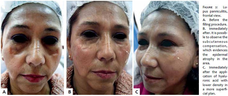
In face of the improvement in the subcutaneous volume, the epidermal atrophy caused by lupus became evident and was treated with 1ml of hyaluronic acid (Emervel® Touch, Galderma, Santiago, Chile) using a 30G cannula and very superficial fanlike retroinjections , compensating the defect in a aesthetically adequate manner (Figure 2).
The authors present a lupus panniculitis case, which is a rare form of cutaneous lupus that, when located on the face, has great psychological and cosmetic relevance to the patient. Treating the resulting defect greatly improves the patient's quality of life and, after considering the evident stability of the base clinical picture, cutaneous filling with hyaluronic acid was chosen, due to its excellent biocompatibility and versatility regarding its viscosity.
1. Durosaro O, Davis M, Reed K, Rohlinger A. Incidence of Cutaneous Lupus Erythematosus,1965-2005: A Population-Based Study. Arch Dermatol. 2009;145(3):249-53.
2. Hawilo A, Mebazaa A, Trojjet S, Zribi H, Cheikh Rouhou R, Zaraa I, et al. Acquired unilateral Facial lipoatrophy: presentation suggestive of lupus panniculitis. Tunis Med. 2012;90(6):499-501.
3. Verma SM, Okawa J, Propertr KJ, Werth PJ. The impact of skin damage due to cutaneous lupus on quality of life. Br J Dermatol. 2014;170(2):315-21.
4. Massone C, Kodama K, Salmhofer W, Abe R, Shimizu H, Parodi A, et al. Lupus erythematosus panniculitis (lupus profundus): Clinical, histopathological, and molecular analysis of nine cases. J Cutan Pathol. 2005;32(6):396-40.
5. Monteiro MR. Doenças autoinmunes, diabetes e cosmiatría. Mateus A, Palermo E. Cosmiatria e Laser. São Paulo:AC Farmacéutica; 2012. p.102-7.
6. Eastham B, Liang C, Femia A, Lee T, Vleugels R, Merola J. Lupus erythematosus panniculitis-induced facial atrophy: Effective treatment with poly-L-lactic acid and hyaluronic acid dermal fillers. J Am Acad Dermatol. 2013;69(5):e260-2.
7. Thareja S, Sadhwani D, Fenske ND. En coup de sabre morphea treated with hyaluronic acid filler. Report of a case and review of the literature. Int J Dermatol. 2015; 54(7):823-6.
8. Braz A, Sakuma T. Región malar y cigomática. Lesqueves Sandoval MH, Leis Ayres E. Rellenos 1º Ed. São Paulo: AC Farmacéutica; 2014. p. 199-203.
9. Carruthers JD, Carruthers A. Facial sculpting and tissue augmentation. Dermatol Surg. 2005;31(11 pt 2):1604-12.
10. Morley AMS, Malhotra R. Use of Hyaluronic acid filler for tear trough rejuvenation as analternative to lower eyelid surgey. Opththal Plast Reconstr Surg. 2011:27(2);69-73.
The present study was conducted at the Instituto Lauro de Souza Lima (ILSL) - Bauru (SP), Brazil.
 All content the journal, except where identified, under the Creative Commons Attribution 4.0 International licence - ISSN-e 1984-8773


 Read in Portuguese
 Portuguese PDF
 Print
Print
 Send this article by email
 How to cite this article
 Submit a comment
 Mendeley
 Pocket