


Clarissa Prieto Herman Reinehr1; Juliana Cattucci Boza2; Roberta Horn3
Post-inflammatory hyperchromia is an important sequel of inflammatory dermatoses. In addition to depigmenting topical treatments, chemical peels can be effective in treating it. The present report describes a case of post-inflammatory hyperchromia after treatment of Lichen Planus handled with thioglycolic acid peels. Thirty percent thioglycolic acid applied in the form of a peel is described as a therapeutic option with discrete results and can be considered an ally in managing this dermatologic condition.
Keywords: CHEMEXFOLIATION; HYPERPIGMENTATION; THIOGLYCOLATES
Post-inflammatory hyperpigmentation (PIH) is an important sequel of inflammatory dermatoses.1 It can ensue several skin diseases, and its treatment is challenging.1 The authors describe a case of PIH that ensued a picture of extensive lichen planus treated with thioglycolic acid peels (TA).
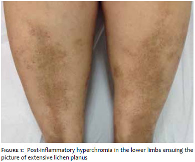
A 54 year-old female patient sought treatment due to asymptomatic oral lesions, and pruriginous cutaneous lesions with seven months of development. Physical examination revealed lichenoid purplish papules on the upper limbs, trunk and neck, with absence of oral lesions. Biopsy of one of the lesions was consistent with lichen planus, and treatment with 20mg/day prednisone was instituted. Three months after the end of the treatment, the patient presented PIH in the lower limbs and wanted to treat it (Figure 1). She began using 4% hydroquinone and 10% glycolic acid cream for four months without success. A triple formulation (4% hydroquinone + 0.05% tretinoin + 0.1% fluocinolone) was then introduced with little response after three months of use. Due to the ineffectiveness of the treatments, he authors decided to perform 30% TA gel peelings with fortnightly intervals. The peelings were performed starting with the application of 30% TA gel for two minutes. The time of exposure of the skin to TA was increased by three minutes on each session, up until a maximum of 15 minutes was reached. In each session, once the time of exposure to the peeling was reached, neutralization of the TA was carried out by cleansing with gauze moistened with distilled water and saline solution. A total of 6 sessions were conducted. The daily use of a depigmentation formula (1.5% TA + 3% tranexamic acid) at night was associated in order to enhance the peeling treatment. The patient was instructed about the importance of applying daily photoprotection, using SPF 50 sunscreen, as well as physical protection.
There was partial improvement of the PIH with the whitening of the lesions, as seen in the progression picture (Figure 2).
Post-inflammatory hyperpigmentation (PIH) is an important sequel of inflammatory dermatoses, and its management is a challenge for dermatologists. 1
PIH may result from a number of dermatoses, including lichen planus. 1, 2 The pathogenesis is not completely understood, however it is known that it results from increased production of melanin or from its irregular deposit after dispersion caused by the action of mediators of the inflammatory process. 1 The intensity of the PIH is greater in dermatoses in which inflammation is chronic, recurrent, and when there is damage to the basal layer. 2
When PIH is confined to the epidermis, it is possible to observe a brownish hue and there is an increase in the production and transfer of melanin to keratinocytes. The presence of hemosiderotic component in lesions is also described. If not treated, it tends to resolve spontaneously after months or years. 1 In the dermis, it is possible to observe a blue-grayish color and damage to the basal layer, with deposition of melanin in the upper layers of the dermis. These deposits are phagocytized, forming melanophages. 1, 2 The removal of melanin deposited in the dermis is a very slow process. 1
The treatment of PIH begins with the approach of the underlying condition, however caution is needed for the management of the basic dermatosis can exacerbate PIH. 1 After controlling the underlying dermatosis, the PIH is treated with topical depigmenting agents and procedures, such as chemical peels and laser applications. 1 Photoprotection is essential in any therapeutic modality and should not be underestimated. 1 It must be carried out through the daily use of sunscreen containing a combination of physical and chemicals agents, in addition to physical external protection against ultraviolet radiation. 1
It is crucial to perform photographic records of the PIH before and during the procedure aimed at assessing the results.
The first choice therapy in PIH is the topical application of 4% hydroquinone associated with photoprotection, being more effective and well tolerated if associated with tretinoin and topical corticosteroids, as in triple whitening formulations (4% hydroquinone + 0.05% tretinoin + 0.1% fluocinolone acetonide), for the combination allows greater penetration by reducing the irritation potential. 1 In the present case, the patient had no therapeutic response with the use of the Kligman formula. Hydroquinone has limited effect when the pigment is deposited in the skin. 2 In resistant cases, other topical depigmenting agents (azelaic or kojic acid, tretinoin, licorice extract, soybean derived depigmenting) can be associated after 8 to 12 weeks of treatment, and the therapy with chemical peels is also indicated (isolatedly or associated with topical treatments, as was done in the present case). Therapies with light/laser (photodynamic therapy with blue light, selective laser photothermolysis, and Nd:YAG laser) can also be employed. 1, 2
Dyschromias - among them PIH - are indications for peelings. 1 Superficial chemical peels tend to be effective in the treatment of PIH when properly applied, with 20-70% glycolic acid, 20-30% salicylic acid, 10-25% trichloroacetic acid or the Jessner's solution being possible options. Preparing the skin with a formula containing 4% hydroquinone for 15 days enhances the results.
Thioglycolic acid (TA) - or mercaptoacetic acid - belongs to the class of thioglycolates, which are substances that solubilize hemosiderotic deposits. 3 Thioglycolic acid has sulfur in its composition, is highly soluble in water, alcohol and ether, and is easily oxidizable. 4 The use of this substance as a depigmenting agent in concentrations ranging between 0.5 and 30% was reported by Izzo and Verzella in 1998. 3 Its topical use in the form of peels usually does not cause erythema or pain. 5 Sensitization occurs rarely, and the resulting desquamation is mild and transient. 5 In the literature, the weekly or biweekly application in gel vehicle is recommended, with a total of five to six sessions.4, 5 The treatment begins with the application of TA gel for two minutes, with three minutes being added on each session, until a maximum of 15 minutes of application is reached. 4
In the present case, as the PIH did not showed satisfactory response to common whitening treatments, the authors decided to carry out 30% TA peelings with sessions at fortnightly intervals, associated with topical depigmenting substance, thus enabling the synergism between the adopted techniques, with good results.
For periocular hyperchromia, the literature mentions a concentration of 10% in gel. 4 The same concentration was used to treat Schamberg disease in the lower limbs. 5 As the authors did not find data in the literature describing the use of 30% TA in PIH, they chose to use this concentration and observe the patient's tolerance to each application, with special attention to any signs of local irritation that required the suspension of the treatment.
Some considerations are relevant in the discussion of this case. First, as the patient used association of the depigmenting topical treatment with TA peelings, it was not possible to define the extent of the improvement achieved resulted from the isolated action of the peelings, as compared with that caused by the use of topical depigmenting substances. Further studies evaluating the isolated action of TA peelings, and even comparing them to the isolated use of topical depigmenters may elucidate this issue. Furthermore, the slight improvement observed may have been spontaneous, since this occurs during the natural development of PIH.
In light of the present study's findings, the authors believe that the TA peeling can be considered a therapeutically in the treatment of resistant PIH that does not show improvement with first-line treatments, and can be used in combination with topical depigmenting treatments for improving results. Moreover, since the results obtained were discrete and obtained from only one patient, the authors deem that studies reproducing this technique in isolation and with more patients are necessary.
1. Davis EC, Callender VD. Postinflammatory hyperpigmentation: a review of the epidemiology, clinical features, and treatment options in skin of color. J Clin Aesthetic Dermatol. 2010;3(7):20-31.
2. Cestari TF, Dantas LP, Boza JC. Acquired hyperpigmentations. An Bras Dermatol. 2014;89(1):11-25.
3. Izzo, Marcello.Verzella, Gianfranco. The use of thioglycolic acid as depigmenting agent. Eur Pat EP0975321 [Internet]. [cited 2015 Jan 13]; Available from: http://www.freepatentsonline.com/EP0975321B1.html
4. Costa A, Basile AVD, Medeiros VLS, Moisés TA, Ota FS, Palandi JAC. 10% thioglycolic acid gel peels: a safe and efficient option in the treatment of constitutional infraorbital hyperpigmentation. Surg Cosmet Dermatol. 2010;2(1):29-33.
5. Hammerschmidt M, Gentili AC, Hepp T, Mukai MM. Peeling de ácido tioglicólico na doença de Schamberg. Surg Cosmet Dermatol. 2013;5(2):165-8.
The present study was performed at the Dermatology Service of the Hospital de Clínicas de Porto Alegre - Porto Alegre (RS), Brazil.
 All content the journal, except where identified, under the Creative Commons Attribution 4.0 International licence - ISSN-e 1984-8773


 Read in Portuguese
 Portuguese PDF
 Print
Print
 Send this article by email
 How to cite this article
 Submit a comment
 Mendeley
 Pocket