


Eloisa Leis Ayres1; Adilson Costa2; Samara Eberlin3; Stefano Piatto Clerici4
Introdução: Hyperpigmentation is a common dermatological condition, usually associated with exposure to the sun and relative difficulty of treatment. Pycnogenol® is an extract obtained from the French pine tree Pinus pinaster, with known antioxidant and anti-inflammatory activity, having been proven to inhibit melanin synthesis in previous studies. Objective: To evaluate the whitening activity of Pycnogenol® in an ex vivo experimental model after exposure to ultraviolet A and B, infrared-A radiations and visible light. Method: Human skin fragments obtained from elective plastic surgery were treated with Pycnogenol ® and then irradiated with ultraviolet A and B, and infrared-A radiations, and visible light, having been sent for histological evaluation of melanin pigmentation by the Fontana-Masson staining technique. Results: The histological evaluation demonstrated an increase in melanin deposition in all irradiated groups. However, fragments pre-incubated with Pycnogenol® showed a reduction in the deposition of this pigment after irradiation. Conclusions: This study demonstrates the reduced melanin deposition in skin culture treated with Pycnogenol® after irradiation with ultraviolet A and B, and infrared-A radiations, and visible light, concluding that this substance may have lightening properties.
Keywords: HYPERPIGMENTATION; SKIN LIGHTENING PREPARATIONS; SOLAR RADIATION
When it comes to skin care, clinical signs evidenced by chronic exposure to solar radiation, such as the presence of wrinkles and hyperpigmentation, are among the main aesthetic concerns that affect human beings, given the amount of active principles and cosmetic products marketed based on their alleged action in the mitigation of such effects. 1-6
Cutaneous pigmentation depends both on the number of melanocytes and the activity of melanogenic enzymes, which culminates in the production of melanin. Melanin plays an important role in protecting the skin from the harmful effects of solar radiation, however an excessive amount of this pigment can become an aesthetic problem. 7
Exposure to sunlight, artificial tanning, the use of medications and other chemical agents, as well as chronic inflammatory processes and hormonal influences, can increase the production of mediators, such as the melanocyte stimulating hormone (a-MSH) and endothelin-1, which trigger a melanization process in the skin.7-9 Studies show that, in addition to the exposure to ultraviolet light, infrared-A radiation and visible light also promote changes in skin pigmentation.10-12
The availability of active principles and more effective and lower irritating power products with whitening or depigmenting properties is steadily sought after in the cosmetic marketplace. Aligned with this, the pursuit of substances that promote the homeostasis of the skin and are capable of delaying or reversing the clinical signs of cutaneous photoaging is a research and development tool in the cosmetic and dermatology fields.
Pycnogenol® is a standardized extract sourced from the bark of French maritime pine Pinus pinaster, which is rich in procyanidins, which in turn has demonstrated antioxidant and anti-inflammatory activity. 13 Several possible effects of Pycnogenol have been investigated, including its action in reducing pigmentation and melanogenesis, in addition to its anti-edematous and vascularization actions. 14
The present study is aimed at evaluating the whitening activity of Pycnogenol® based on the histological evaluation of melanin pigmentation using the Fontana-Masson technique in human skin fragments subjected to ultraviolet A and B (UVA/ UVB) and infrared-A (IRA) radiations, visible light (VL), and the association of UVA/UVB, VAT and VL.
Fragments of human skin harvested in an elective surgery were incubated in culture medium and treated with Pycnogenol® (Flebon®, Pinus pinaster, FQM Farmoquímica S/A, Brazil) at concentrations of 0.0316, 0.0100 and 0.00316 mg/ml, defined after performing a cell viability test for determining non-cytotoxic concentrations for the efficacy trial. After 48 hours of incubation, all fragments were underwent irradiation as described below.
For exposure of the fragments to UVA/UVB radiations (10 J/cm2 UVA) the following devices were used: UVA/UVB Cube 400, SOL 500 H2 filter and UV Meter (Hönle UV America Inc., MA, USA). Regarding the infrared-A radiation (360 J/cm2 IRA radiation), the Hydrosun 750 and HBM1 devices (Hydrosun Medizintechnik GmbH, Müllheim, Germany) were used. For the irradiation of visible light (480 J/cm2) the devices used were UVA/UVB Cube 400, SOL 500 H1 filter for filtering the UVA radiation; Rosco Cinegel 3114 filter for filtering UVB and VLP-471RAD Radiometric Sensor (Deta Ohm, Caselle di Selvazzano, Italy).
The fragments were maintained for 48 additional hours and fixed in 10% buffered formalin, embedded in paraffin blocks, and subjected to serial sectioning on a microtome to about 5µm thick. The sections were stained with hematoxylin-eosin (H/E) associated with the Fontana-Masson technique. The slides were photographed under an optical microscope (Nikon Eclipse) at 10X magnification with the aid of the Image-Pro software.
The use of human skin fragments harvested in elective surgeries for carrying out the present study was approved by the Research Ethics Committee of the Universidade San Francisco (SP), Brazil.
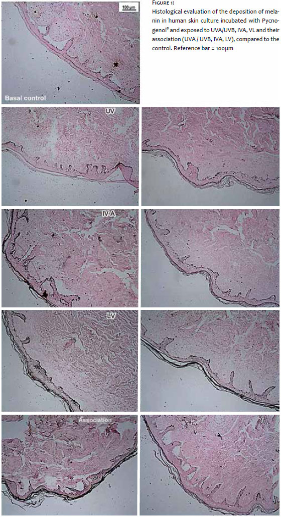
As can be seen in Figure 1, the histological evaluation showed that cultures of human skin fragments exposed to UVA/UVB, IRA, VL and to the association of the radiations showed higher density of melanin pigmentation by Fontana-Masson technique as compared to the control group (fragments maintained at baseline).
Conversely, all fragments treated with Pycnogenol® showed noticeable reductions in melanin density as compared to the fragments that were only photoexposed.
It is important to note that the results observed in this ex vivo experiment corroborate the in vitro trials obtained in our laboratory using cultured human melanocytes (in progress; unpublished data) in which the radiation promote, isolatedly, increased production of melanin and tyrosinase enzyme activity. An interesting observation obtained from the in vitro trials indicates that the increase verified in the production of melanin is significantly higher in cell groups exposed to the association of the radiations than that observed in the groups irradiated isolatedly.
Several studies have attempted to relate the use of antioxidants to skin pigmentation disorders and justify its use as a possible therapeutic alternative in these cases.
Procyanidins have demonstrated a significant free radicals sweeping action, promoting anti-edematous and anti-inflammatory action in conditions that occur with capillary fragility. Its topical and oral use in experimental studies with mice has been shown capable to inhibit erythema induced by UV radiation and to increase vascular permeability. Grape seed extract rich in proanthocyanidins, was proven to be able to suppress the formation of melanin in pig skin, as well as to have a whitening effect on hyperpigmentation induced by UV radiation. Later on it was observed in Japanese women that grape seed extract could inhibit melanogenesis or the proliferation of melanocytes in the skin of the face affected by melasma. 13
Handog et al. have shown that the association of procyanidins, vitamin A, C and E could be effective as an adjuvant in the clinical treatment of melasma. In this study, the use of 48mg/day Pycnogenol® was combined with 6mg of beta-carotene, 60mg ascorbic acid and 15UI D-alpha-tocopherol, and compared to the administration of placebo for eight weeks. The results evaluated with the assistance of a mexameter showed that there was a decrease in pigmentation and also a significant improvement in the Masi score, leading to the conclusion that this combination is safe and effective in the treatment of melasma. 15
In 2002, a study by Ni et al. investigated the efficacy of Pycnogenol® in the treatment of melasma. Twenty-five milligrams (25mg) were administered three times a day (75mg/day) in 30 women with melasma. At the end of 30-day period there was a decrease in the melasma area, in the intensity of pigmentation, with 80% effectiveness rate, according to the authors. 14
Pycnogenol® is a plant extract obtained from the bark of French maritime pine Pinus pinaster, a source of flavonoids. It has attracted attention due to its powerful antioxidant action, with demonstrated ability to modulate melanogenesis, UV radiation induced erythema, and the expression of kappa-B nuclear transcription factor (NFkB). 13 In vitro studies have demonstrated that Pycnogenol® is more potent than vitamins E and C, having the ability to recycle vitamin C, regenerate vitamin E and increase the endogenous antioxidant system. 16
The improvement of techniques for its extraction allowed to obtain extracts with higher antioxidant activity, favoring the chromatographic identification of the main pharmacological components of Pycnogenol®, such as caffeic acid, ferulic acid, catechin and taxifolin. 17
Studies have shown that, in addition to the antioxidant action, Pycnogenol® has anti-inflammatory activity, also being capable to stimulate the synthesis of Enos (endothelial nitric oxide synthase).18, 19
The use of this active principle in cardiovascular disorders has also been reported with promising results. 20 In the skin, in addition to studies involving the reduction in melanin production, the inhibition of metalloproteinases type 1, 2 and 9 (MMP-1, MMP-2 and MMP-9) has also been observed, recognizing its role in improving skin hydration and elasticity. 21
In the study by Kim et al. it was possible to demonstrate that Pycnogenol® is able to suppress reactive oxygen species (ROS) and has a strong antityrosinase action, leading to suppression of melanin biosynthesis, corroborating its antimelanogenic potential. Its antioxidant capacity was assessed based on the suppression of the activity of peroxynitril, superoxide, nitric oxide and hydroxyl radicals, with a positive regulation in the ratio reduced glutathione/oxidized glutathione in B16 cells also taking place. Its inhibitory action on tyrosinase activity was compared to that of other inhibitors; kojic acid was used as the reference inhibitor, with the results showing a significantly greater potency for Pycnogenol®. The treatment with the product promoted a reduction of the melanin content in B16 cells in a concentration-dependent way, ranging from 22.2% (50mg/ml) to 58.9% (5mg/ml) as compared to the control group treated with a-MSH, demonstrating its action in controlling melanin synthesis.22, 23
Using human skin fragments incubated with Pycnogenol®, the present study demonstrated that the deposition of melanin pigment was less intense after exposure to UVA/UVB radiation, IVA, VL and their association, as compared with untreated fragments, confirming the literature data, which demonstrate its antimelanogenic activity. However, the treatment of pigmentary disorders remains in the focus of new promising studies. Plant extracts rich in flavonoids have been investigated due to their ability to modulate cutaneous pigmentation, and Pycnogenol® is a substance that is gaining prominence.
The present study demonstrates that ultraviolet A and B, IRA radiations and visible light, in addition to the association of all these radiations, are capable of increasing the melanin's pigmentary density. It also demonstrates that cultures that have been previously treated with Pycnogenol® showed a noticeable decrease in the deposition of melanin, leading to the conclusion that this substance may have whitening properties.
Recent studies have shown that due to its antimelanogenic action, Pycnogenol® can be considered an option in the treatment of disorders like melasma and can have great importance in the maintenance and control of recurrences, although further studies may be required.
1. Thomas JR, Dixon TK, Bhattacharyya T. Effects of topicals on the aging skin process. Facial Plast Surg Clin N Am. 2013;21(1):55-60.
2. Ertel K, Wyborski R, Zheng Q. Futuro dos cosmecêuticos. In: Costa A. Tratado Internacional de Cosmecêuticos. Rio de Janeiro: Guanabara Koogan; 2012. p.680-91.
3. Tsatsou F, Trakatelli M, Patsatsi A, Kalokasidis K, Sotiriadis D. Extrinsic aging UV-mediated skin carcinogenesis. Dermato-Endocrinol 2012;4(3):285-97.
4. Gkogkolou P, Böhm M. Advanced glycation end products key player in skin aging? Dermato-Endocrinol 2012;4(3):259-70.
5. Shibao J, Bastos DHM. Produtos da reação de Maillard em alimentos: implicações para a saúde. Rev Nutr 2011;24(6):895-904.
6. Glaser DA. Anti-aging products and cosmeceuticals. Facial Plast Surg Clin N Am. 2003;11(2):219-27.
7. Solano F, Briganti S, Picardo M, Ghanem G. Hypopigmenting agents: an updated review on biological, chemical and clinical aspects. Pigment Cell Res. 2006;19(6):550-71.
8. Bolognia JL, Orlow SJ. 2003. Pigmentary disorders. In: Bolognia JL, Jorizzo J, Rapini R, editors. Dermatology. Philadelphia: Mosby; 43-52.
9. Slominski A, Tobin DJ, Shibahara S, Wortsman J. Melanin pigmentation in mammalian skin and its hormonal regulation. Physiol Rev. 2004;84(4):1155-228.
10. Sklar LR, Almutawa F, Lim HW, Hamzavi I. Effects of ultraviolet radiation, visible light, and infrared radiation on erythema and pigmentation: a review. Photochem Photobiol Sci. 2013;12(1):54-64.
11. Ju Hee Lee, Mi Ryung Roh, and Kwang Hoon Lee. Effects of Infrared Radiation on Skin Photo-Aging and Pigmentation. Yonsei Medical J. 2006;47(4):485- 90.
12. Kollias N, Baqer A. An experimental study of the changes in pigmentation in human skin in vivo with visible and near infrared light. Photochem Photobiol. 1984;39(5):651-9.
13. D'Andrea G. Pycnogenol: A blend of procyanidins with multifaceted therapeutic applications? Fitoterapia. 2010;81(7):724-36.
14. Ni Z, Mu Y, Gulati O. Treatment of melasma with pycnogenol. Phytother Res. 2002;16(6):567-71.
15. Handog EB, Galang DAVF, Leon-Godinez MA, Chan GP. A randomized, double-blind, placebo-controlled trial of oral procyanidin with vitamins A, C, E for melasma among Filipino women. Int J Dermatol. 2009,48(8):896-901.
16. Maimoonaa A, Naeema I, Saddiqea Z, Jameelb K. A review on biological, nutraceutical and clinical aspects of French maritime pine bark extract. J Ethnopharm. 2011;133(2):261-77.
17. 1Rohdewald P. A review of the French mari- time pine bark extract (Pycnogenol ®), a herbal medication with a diverse clinical pharmacology. Int J Clin Pharmacol Ther. 2002;40(4):158-68.
18. Blazso G, Gabor M , Rohdewald P. Antiinflammatory activities and superoxide radical scavenging activities of a procyanidin containing extract from the bark of Pinus pinaster Sol and its fractions. Pharm Pharmacol Lett. 1994;3:217-20.
19. Nishioka K, Hidaka T, Nakamura S, Umemura T, Jitsuiki D, Soga J, et al. French maritime pine bark extract. Augments endothelium-dependent vasodilation in humans. Hypertension Research. 2007;30(9):775-80.
20. Gulati OP. The neutraceutical pycnogenol: its role in cardiovascular health and blood glucose control. Biomedical Reviews. 2005;16:49-57.
21. Marinia A, Grether-Becka S, Jaenickea T, Webera M, Burkib C, Formanna P, et al. Pycnogenol® Effects on Skin Elasticity and Hydration Coincide with Increased Gene Expressions of Collagen Type I and Hyaluronic Acid Synthase in Women. Skin Pharmacol Physiol. 2012;25(2):86-92.
22. Kim YJ, Kang KS, Yokozawa T. The anti-melanogenic effect of pycnogenol by its anti-oxidative actions. Food Chem Toxicol. 2008;46(7):2466-2471.
23. Kim YM, Yun J, Lee CK, Lee H, Min KR, Kim Y. Oxyresveratrol and hydroxystilbene compounds: inhibitory effect on tyrosinase and mechanism of action. J Biol Chem. 2002;277(18):16340-4.
The present study was conducted at the Kolderma Instituto de Pesquisa Clínica Eireli EPP, Campinas (SP), Brazil.
 All content the journal, except where identified, under the Creative Commons Attribution 4.0 International licence - ISSN-e 1984-8773


 Read in Portuguese
 Portuguese PDF
 Print
Print
 Send this article by email
 How to cite this article
 Submit a comment
 Mendeley
 Pocket