


Célia Luiza Petersen Vitello Kalil1; Valéria Barreto Campos2; Christine Rachelle Prescendo Chaves3; Luiza Helena Urso Pitassi4; Stela Cignachi5
Introduction: Photodamaged skin of the anterior thorax region is characterized by flacidity, wrinkles, hyperpigmentation, erythema, telangiectasia, and atrophy. Microneedling has been used for the transdermal drug delivery of active agents to the skin through microchannels. Objective: To clinically evaluate the rejuvenation of the skin of the anterior thorax region resulting from the use of microneedling associated with drug delivery. Methods: Double-blind randomized, placebo-controlled study conducted with 22 women who underwent three microneedling sessions followed by the topical application of a test product containing hyaluronic acid and other active principles or placebo. The evaluation was performed through photographic comparison carried out by a dermatologist oblivious to the study and the application of self-assessment questionnaires to patients. Results: Clinical evaluation showed improvement in the overall skin rejuvenation of the anterior thorax region in 100% of the patients. The statistical analysis showed a 28% improvement (p <0.05) with use of the test product as compared to the placebo. The responses to the questionnaires demonstrated an improvement of 30% in the patients treated with microneedling and the test product. Conclusions: The microneedling technique associated with the transdermal administration of drugs used for rejuvenation provides improved overall appearance of the skin of the anterior thorax region, with a high degree of tolerability and satisfaction.
Keywords: REJUVENATION; COLLAGEN; ADMINISTRATION; CUTANEOUS
Skin aging results from the action of both individual genetically determined factors and external factors, such as smoking, pollution, chronic exposure to sunlight, and other adjuvant factors, such as stress, use of drugs, the impact of cutaneous and systemic diseases, in addition to hormonal factors.1-3
The anterior region of the thorax is an area that shows the extent of skin aging, since it is usually exposed to sunlight and often not treated at all.4, 5 There are few published studies on the treatment of the skin in this region. The aging of the anterior thorax region manifests as muscle and skin sagging, brownish and irregular diffuse pigmentation, xerosis, superficial and deep rhytids, ephelides, poikiloderma,6, 7 hypomelanosis and pre-neoplastic and neoplastic lesions.8, 9 Moreover, it is known that the healing process becomes impaired, causing tissue repair of spontaneous injuries or those caused by surgical procedures in this region to happen more slowly10, 11 due to the reduced number of adnexa.
In order to correct these alterations, diverse procedures can be employed, namely chemical peels, injectable hyaluronic acid, botulinum toxin, radio frequency, infrared radiation, intense pulsed light, photodynamic therapy and fractional ablative and non-ablative lasers.12, 13 A promising technique known as microneedling is currently used in the successful treatment of facial cutaneous aging. Nevertheless, there is lack of studies describing the use of this technique for rejuvenating the skin of the anterior thorax region.14, 15
Furthermore, microneedling can be associated with a procedure that allows the transdermal delivery of selected active principles (drug delivery), and can optimize the results sought. This technique uses the transport of drugs through the skin, with the advantage of being easily obtainable, non-invasive, safe, and effective. However, its clinical application is limited by the stratum corneum, which is the barrier of the main skin. Due to its hydrophobic characteristic and negative electrical charge, the transportation of hydrophilic and ionized molecules is a major challenge.
In this manner, the present study aims at evaluating the clinical efficacy and safety of a new and original approach to the treatment of the anterior surface of the chest area using microneedling associated with the drug delivery process.
This is an original, prospective, multicenter, comparative, double-blind, randomized (by drawing lots), and placebo controlled study aimed at evaluating the clinical efficacy of a test product applied on the skin of the anterior chest region, in association with the microneedling procedure. There is an absence of similar publications. It was conducted at two research centers located within dermatologic private practices - one in Porto Alegre (RS, Brazil) and the other in Jundiaí (SP, Brazil) - in 2014.
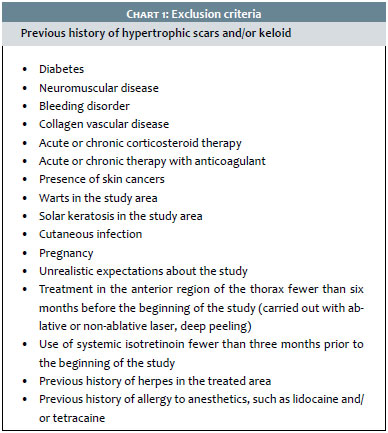
Twenty-two women treated for rejuvenation of the anterior surface of the skin of the thorax were included (average age = 55, Fitzpatrick skin phototype I to IV16 and Glogau classification of aging 2 to 4. 17 The exclusion criteria are listed in Chart 1.
The evaluation of the results of the treatment was carried out before (D0) and 30 days after the three monthly procedures (D90) of microneedling associated with the application of the test product or placebo (D0, D30, and D60), according to randomization by drawing lots. The study was conducted after the signing of the Free and Informed Term of Consent, and according to the ethical guidelines of the Helsinki Declaration.
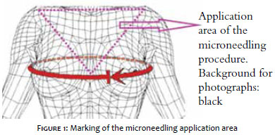
The microneedling procedure was performed after the application of topical anesthetic cream and after the marking of the region (Figure 1).
The microneedling device used in the study was a roller (Dr. Roller®, Moohan Enterprise, South Korea) consisting of 192 micro needles made of surgical steel measuring 0.07 mm in thickness and 1.5 mm in length (registered at ANVISA/MS under the number 80669600001, and imported by the company MTO Importadora e Distribuidora Ltda., São Leopoldo (RS), Brazil). To perform the procedure, the device was positioned in one hand with minimum pressure at an angle of 45º on the area to be treated. Ten movements with passes in four directions (horizontal, vertical, diagonal right and left) were carried out, causing uniform micro bleeding points. Cleansing with gauze moistened with thermal water was carried out immediately after, with the test product or placebo being subsequently applied. This application was directed and performed by the technician responsible for the study at the research center. Three similar sessions were performed at monthly intervals (D0, D30, and D60).
The test product was a compounded formulation comprising: 2% Juvenile®; 0.5% PhytoCellTec Malus Domestica®; 2% Cell to Cell®; 5% Homeostatine®; 2.5% Hyaluronic Acid; 30 ml anhydrous fluid serum QS. The association of these substances of cosmetic use was used with the main purpose of increasing the duration of the opening of the pores, and stimulating the production of collagen and elastin, relying on anti-inflammatory action to avoid discomfort during the application.
After the first procedure (D0), the patients were instructed to carry out the application of the same product or placebo at home, gently massaging with the fingertips, until it was completely absorbed, once a day, at night, in the area being treated. The procedure should be repeated up until the following sessions and evaluations (D30, D60 and D90). Each volunteer used only one of the treatments up until the end of the study.
The following evaluations were performed: clinical efficacy (through photographic assessment of the overall skin rejuvenation in the anterior chest, comparing D0 with D90, performed by a dermatologist blinded to which patients had applied the test formulation or placebo); subjective evaluation through the application of patient questionnaires on the improvement of the following factors related to the rejuvenation of anterior chest region's skin: wrinkles, texture, luminosity, smoothness, tone, firmness and overall appearance. Assessment was also performed through Visia® digital photography system (Canfield Imaging Systems, USA) in some patients. The safety assessment of the procedure and the product was based on the patient's acceptance and tolerance as well as on the adverse effects observed.
Statistical analysis of the clinical efficacy
The results were expressed as mean ± standard error of the mean value (SEM) and statistically evaluated through the analysis of the Student t test. A significance limit of p <0.05 was used in all results. The data represent the mean ± SEM. *P<0.05 versus placebo within the same group.
The 22 patients treated completed the study. The duration of the follow-up was three months. The results obtained are described below.
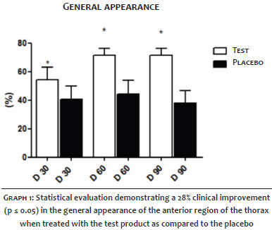
The clinical efficacy assessment demonstrated a significant improvement in 100% of the treated patients. The statistical analysis suggested there was 28% of improvement in the overall skin rejuvenation of the anterior thorax (p <0.05) when the test product was used as compared to the placebo, after three months of treatment (Graph 1).
The patients' self-assessment suggested an improvement of 30% in the patients treated with the combined use of microneedling and the test product, when considering the following variables: skin's texture, smoothness and firmness.
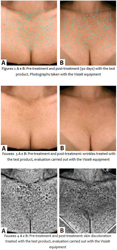
Photographs were taken of a group of patients with the Visia® device for the comparative quantitative and qualitative study of the same body region on D0 and on D90, regarding the variables wrinkles and pigmentation. Although there was no statistical significance between the groups, clinical improvement with the use of test product was observed in the above items (Figures 2-4). No adverse effects were seen in the treated patients.
The skin has an excellent barrier property, protecting the body from physical and chemical agents, thus being able to restrict the transdermal access of a large number of drugs - even those with low molecular weight or lipophilic ones.18 The topical application of compounds has the potential to be a safe and ideal alternative route for administration into the tissue.19, 20 The use of techniques that facilitate the permeation of active principles increases the potential of the latter for action.21, 22 The combined use of microneedling and drug delivery proves to be essential for the positive clinical results obtained.
Microneedling has been described as a virtually painless, simple, and minimally invasive technique.23, 24 The use of microneedling allows the creation of a means of transport accessible to macromolecules and other hydrophilic substances into the skin.25 Thus, microneedling is a fundamental tool for the product used in the study to act in the dermis through an essential and necessary amount for achieving the results effectively and quickly, with only three treatment sessions.
The technique promotes the disruption of the stratum corneum (this is microscopically proven through the visualization of channels and the increased transepidermal water loss - TEWL).26, 27 As a consequence, there is increased permeation of the formulations' hydrophilic molecules and macromolecules when it is applied once the microneedling's perforations have been performed.28, 29 In this way, the length of the needles was long enough to pierce the more superficial layers of the epidermis and short enough not to excite the nerve endings in the skin, with good patient tolerability and acceptance. All patients in the present study continued the treatment up until its end.
There are few therapeutic resources described for a treatment aimed at a general rejuvenation of the skin of the anterior chest region. According to Peterson et al. 30 the main techniques described in the literature are: the use of injectable polylactic and hyaluronic acids, botulinum toxin, sclerotherapy, chemical peels, and more recently lasers and light-based therapies, such as intense pulsed light (IPL), photodynamic therapy (PDT), fractional ablative and non-ablative lasers. These are minimally invasive options for rejuvenating the skin of the anterior thorax region, which are well tolerated by patients and present a low incidence of side effects. However, the use of fractional ablative laser can increase recovery time, since the skin is thinner and has smaller concentrations of pilosebaceous units. Consequently, if not performed carefully, some of these procedures can cause local scarring and hyperpigmentation alterations. According to the results obtained in the present study, microneedling appears to be a safer and less invasive technique for the treatment of the anterior surface of the thorax than fractional ablative laser. As a result, the authors aimed at developing a new and original approach to the treatment of the skin of the anterior thorax region using the microneedling technique combined with drug-delivery.
The choice of the formulation's components and characteristics of the drugs used in the drug delivery may influence the permeation and degree of skin irritation. The use of hyaluronic acid, for instance, has been indicated to increase the duration of pore opening. 31 Controlled release systems can help to increase the depth reached by the active principles, such as liposomes, which increase the bioavailable concentration of the active principle crossing the stratum corneum, for enhanced bioavailability in the skin.32, 33 The choice of the ideal vehicle for the formulation is also another determining factor for good performance.28, 31 Fluid anhydrous serum is safe and effective, without causing burning or discomfort to the patient at the moment of application, and also has the advantage of forming a film on the skin, causing occlusion, which is of the utmost importance for significantly increasing the duration of the opening of pores and reducing the transepidermal water loss (TEWL).
The present study sought to use a topical product that facilitated the permeation in a safe manner and allowed for the association with rejuvenating active principles that were crucial for the positive results obtained with the combined treatment.
The active principles used in the present study's test formulation achieved the primary goal of a general rejuvenation of the skin of the anterior thorax region. The active principles used prevent the rupture of collagen fibers, have an anti-inflammatory action and inhibit metalloproteinases (MMPs), and activate the cutaneous cell metabolism.34, 35 Furthermore, the set of active principles used - Cell to Cell®, Juvenile®, apple stem cells and Homeostatine® - lends synergy of action to the formulation, with an absence of signs of irritation or burning in the skin, even immediately after the procedure.36
Applying the test formula at home after the procedure was key to improved healing, moisturizing, and lower TEWL, with better clinical responses.
During the study it was possible to observe significant changes in the progressive improvement of the treated area in the skin of the anterior thorax region (the differences in the results of patients became evident from 60 days of treatment), in the alignment with the subjective and clinical evaluations, and in the statistical analysis, which showed a significant improvement of 28% when comparing the pre-treatment experimental time point with 90 days after.
This study has demonstrated that the use of formulations during and after the procedure not only enhances the outcome of the technique but also minimizes the potential for side effects, such as pigmentary alterations. With the statistical analysis, it was possible to prove correctness of the subjective data, with the significant improvement in patients treated with the test product as compared to the placebo.
The multicenter study used a standardized sample that presented the greatest possible similarity to the routine patient in a dermatologist's practice. In addition, a cohesive sample is associated with the adequate comparative response of the data, therefore allowing the assertion that the data found are due to the efficiency of the active principles and not to idiosyncratic responses to the treatment. The microneedling technique is considered safe for any skin phototype (especially higher ones).
Our findings demonstrate the general clinical improvement in the rejuvenation of the skin of the anterior thorax region with microneedling techniques in association with drug-delivery. However, further studies are necessary to estimate the histological improvement with the application of the technique.
The microneedling technique is firmly established and safe, nevertheless in order to obtain an advanced degree of general rejuvenation, it is necessary to combine specific active principles for drug delivery. In this manner, it is possible to infer that the combined technique of microneedling and drug delivery for rejuvenating the skin of the anterior thorax region provided a significant improvement in the overall appearance, arising as a well tolerated procedure, with minimal adverse effects and high rates of patient satisfaction.
In addition, there is a scarcity of scientific studies published on this treatment modality, which strengthens the innovative and original character of the present study.
1. Beynet D, Neuhaus IM, Yu SS. Abordagem do paciente estético. In: Alam M, Gladstone HB, tung RC. Dermatologia Cosmética.Rio de Janeiro:Elsevier; 2010. p.1-6.
2. Addor FAZ. Abordagem nutricional do envelhecimento cutâneo: correlação entre os efeitos em fibroblastos e os resultados clínicos. Surg Cosmet dermatol 2011;3(1):13-6.
3. Montagner S, Costa A. Bases biomoleculares do fotoenvelhecimento. Ann Bras Dermatol 2009;84(3):263-9.
4. Montagna W, Carllise K. Structural changes in aging skin. Br J Dermatol 1990;122s;(35): 61-72.
5. Lessa S, Nanei M, Flores E. Histologic study of the structural changes in fine palpebral skin following selective photothermolysis with CO2 laser. Aesthetc Plast Surg 2009:33(1):66-71.
6. Bernstein EF. Aging skin: intrisic aging and chronic photoaging. In: Lasers and non Surgical Rejuvenation. DibernardoEB, Pozner JN. Elsevier; 2009.p:1-9.
7. Do Nascimento LV. Tipos de envelhecimento. In: Kede MPV, Sabatovich O. Dermatologia Estética. São Paulo: segunda edição, Atheneu, 2009. p.53-6.
8. Kadunc BV. Cirurgia dermatológica. In: Azulay RD, Azulay DR, Abulafia LA, editores. dermatologia. rio de janeiro: Guanabara Koogan; 2008. p.773-7.
9. Bezerra SMC, Lima EA, Jardim MML. Eletrocirurgia.In: Tratado de cirurgia dermatologica, cosmiatria e laser da sociedade brasileira de dermatologia. Rio de janeiro:Elsevier; 2013. p.535-40.
10. Papaléo Netto M, Pontes JR. Envelhecimento: desafio na transição do século. In: Papaléo Netto M. Gerontologia. São Paulo, Atheneu, 1996.
11. Abulafia LA, Montagner S, Costa A. Envelhecimento cutâneo: bases fisiopatológicas. In: Kadunc B, Palermo E, Addor F, Metsavaht L. Tratado de cirurgia dermatológica, cosmiatria e laser da Sociedade Brasileira de dermatologia; Ed Elsevier;2013.p:237-40.
12. Reilly MJ, Cohen M, Hokugo A, Keller GS. Molecular Effects of Fractional Carbon Dioxide Laser Resurfacing on Photodamaged Human Skin. Arch Facial Plast Surg. 2010;12(5):321-5.
13. Gabay MA, Curi MP. Peelings e procedimentos combinados para o rejuvenescimento do pescoço e do face anterior do tórax. In: Mateus A, Palermo E. Cosmiatria e laser: Editora Gen; 2012. p:193-205.
14. Hoesly FJ, Borovicka J, Gordon J, Nardone B, Holbrook JS, Pace N, Ibrahim O, et al. Safety of a novel microneedle device applied to facial skin. Arch Dermatol 2012;148(6):711-17.
15. Wermeling DP, Banks SL, Hudson DA. Et al. Microneedles permit transdermal delivery of skin-impermeant medication to humans. PNAS; 2008;12(105):2058-63.
16. Glogau RG. Aesthetic and anatomic analysis of the aging skin. Semin Cutan Med Surg. 1996;15(3):134-8.
17. Fitzpatrick TB. Editorial: the validity and practicality of sun-reactive skin types I through VI. Arch Dermatol. 1998;124(6):869-71.
18. Li G, Badkar A, Kalluri H, Banga AK. Microchannels Created by Sugar and Metal Microneedles:Characterization by Microscopy, Macromolecular Fluxand Other Techniques. J Pharm Sci. 2010;99(4):1931-43.
19. Aust MC, Fernandes D, Kolokythas P, Kaplan HM, Vogt PM. Percutaneous collagen induction thrapy: an alternative treatment for scars, wrinkles, and skin laxity. Plast Recosntr Surg.2008;121(4):1421-9.
20. Kumar R, Philip A. Modified transdermal technologies: Breaking the barriers of drug permeation via the skin. Trop J Pharm Res. 6(1):633-44.
21. Brogden NK, Milewski M, Ghosh P, Hardi L, Crofford l J, Stinchcomb A. Diclofenac delays micropore closure following microneedle treatment in human subjects. J Control Realese. 2002;163(2):220-9.
22. Luo S, Zheng Y, Ni H, Liu, Y, Liu Y, Li X, et al. Effects of topical application of growth factors followed by microneedle therapy in women with female pattern hair loss: a pilot study. J Dermatol. 2012;40(1): 81-3.
23. Benson HA, Namjoshi S. Proteins and peptides:Strategies for delivery to and across the skin. J Pharm Sci. 2008;97(9):3591-610.
24. Al-Qallaf B, Das DB. Optimizing Microneedle Arrays to Increase Skin Permeability for Transdermal Drug Delivery. Ann New York Acad Sci. 2009;1161: 83-94.
25. Schuetz YB, Naik A, Guy RH, Kalia YN. Emerging strategies for the transdermal delivery of peptide and protein drugs. Expert Opin Drug Deliv. 20052(3):533-48.
26. Harvinder S. Gill , Mark R. Prausnitz. Coated microneedles for transdermal delivery. J Control Release. 2007;117(2):227-37.
27. Gill HS, Prausnitz MR. Pocketed Microneedles for Drug Delivery to the skin. J Phys Chem Solids. 2008;69(5-6):1537-41.
28. Paudel K S, Milewski M, Swadley C L, Brogden N K, Ghos, P, Stinchcom, A L. Challenges and opportunities in dermal/ transdermal delivery. Ther Deliv. 2011;1(1):109-131.
29. Gupta J, Gill HS, Andrews SN, Prausnitz MR.Kinetics of Skin Resealing After Insertion of Microneedles in Human Subjects. J Control Release. 2011;154(2):148-55.
30. Petersen J D, Goldman M P. Rejuvenation of the Aging Chest: A review and our experience. Dermatol Surg. 2011;37(5):555-71.
31. Donnelly RF, Singh TR, Garland MJ, Migalska K, Majithiya R, McCrudden CM. Hydrogel- Forming Microneedle Arrays for Enhanced Transdermal Drug Delivery. Adv Funct Mater. 2012;22(23):4879-90.
32. Barrak Al-Qallaf, Diganta Bhusan Das. Optimizing Microneedle Arrays to Increase Skin Permeability for Transdermal Drug Delivery. Interdisciplinary Transport Phenomena: Ann N.Y. Acad Sci. 2009;1161(1):83-94.
33. Mark R. Prausnitz. Microneedles for transdermal drug delivery. Adv Drug Deliv Rev. 2004;56(5):581-7.
34. Badran, M M, Kuntsche, J, Fahr, A. Skin penetration enhancement by a microneedle device (Dermaroller®) in vitro: dependency on needle size and applied formulation. Eur J Pharm Sci. 2009;36(4-5):511-23.
35. Fabbrocini G, De Vita V, Fardella N, Pastore F, Annunziata MC, Mauriello MC, et al.Skin Needling to Enhance Depigmenting Serum Penetration in the Treatment of melasma. Plast Surg Int. 2011;2011:158241.
36. McAllister, D V, Ping, M W, Davis, S P, Park, J-H, Canatella, P J, Allen, M G, Prausnitz, M R. Microfabricated needles for transdermal delivery of macromolecules and nanoparticles: fabrication methods and transport studies. Proc Natl Acad Sci U S A. 2003;100(24):13755-60.
The present study was conducted at the Dermatology Departments of the Santa Casa de Misericórdia de Porto Alegre - Porto Alegre (RS); of the Universidade de Jundiaí - Junidiaí (SP); and of the Universidade de Campinas (Unicamp) - Campinas (SP), Brazil.
 All content the journal, except where identified, under the Creative Commons Attribution 4.0 International licence - ISSN-e 1984-8773


 Read in Portuguese
 Portuguese PDF
 Print
Print
 Send this article by email
 How to cite this article
 Submit a comment
 Mendeley
 Pocket