


Flávia Alvim Sant’Anna Addor
Keywords: COLLAGEN; DERMIS; NUTRIENTS; SKIN AGING
Cutaneous aging is a progressive degenerative process resulting from a decline in the physiological functions of the cutaneous tissue, both at the epidermal and dermal levels.
In the dermis, there is a decrease of collagen synthesis, as well as the components of the other extracellular matrices, characteristic of chronological (intrinsic) aging. This decrease can be exacerbated by metalloproteinase, which increases in expression due to photodamage, and leading to its fragmentation. 1
Menopause is also an accelerating factor of degenerative changes to the collagen and thickness of the dermis through the progressive loss of collagen, which reaches its peak in the first five years (with a loss of up to 30%) and then stabilizes at loss rate of 1% to 2% per year. Hormone replacement therapy allows for partial recovery, however not all patients can use it. 2
The use of oral supplements containing collagen as a means to improve the signs of aging is not a recent practice; nevertheless the scarcity of studies and publications on this subject means there have always been doubts about its actual value.
Nonetheless, the topic is once again being discussed with renewed interest due to the development of technologies that allow for the isolation of peptides for oral consumption and the emergence of a new generation of collagen supplementation-specific peptides capable of enhancing the expression of certain molecules linked to the synthesis of collagen and association with other substances, such as vitamins and Phytoextracts, which act synergistically enhancing that effect.
The objective of the present study was to demonstrate the effects of a nutritional supplement in improving dermal structure, evaluating its thickness, and the clinical properties of cutaneous firmness, elasticity, and hydration.
A monocentric, open, blind, non-comparative study was carried out at a private research center between August and November 2014.
Thirty female patients (35 to 65 years of age) complaining of some degree of facial sagging were invited and subsequently included in the study. All patients stopped using any cosmiatric treatment four weeks before the beginning of the study. Patients using corticosteroids or immunosuppressants, bearing active endocrine diseases or any clinical condition that could interfere with the evaluations, were excluded.
After signing a term of free and informed consent and undergoing a dermatologic evaluation in order to rule out any dermatosis in the body site to be assessed, a multichannel color Doppler ultrasonography with a frequency transducer of up to 15MHz (Voluson E device - GE Healthcare) was performed in two body sites (submental and malar areas), for standardization. The ultrasound evaluation was carried out with an aim at calculating the dermal thickness of the selected area and capturing any differences between each experimental occasion.
Subsequently, the patients received the dietary supplement containing collagen peptides, vitamin C, and Hibiscus sabdariffa in sachet form, under evaluation and with instructions to consume 2 sachets diluted in 200 ml of cold or hot water once a day for 12 weeks.
Lastly, the patients were asked to return monthly for clinical safety reasons, subjective and ultrasonographic assessments, or if at any time there was a case of doubt or complication. At all visits a questionnaire was given to evaluate the firmness, elasticity, hydration, and overall appearance of the skin. The rating system used in the questionnaire was conceptualized as follows, in order to obtain accuracy in the answers:
Worsened: there has been visible deterioration as compared to the previous state for this location; Unchanged: there was no improvement or worsening as compared to the previous state for this location; Partial improvement: there was some degree of perceived improvement; Total improvement: the perceived improvement is significant, easily noticeable, and the best possible that the patient could expect with this type of treatment.
The features of the product were also evaluated regarding their solubility and taste, with ratings of very bad, bad, good, or very good.
Ethical aspects
The study protocol was approved by the Independent Ethics Committee and conducted in accordance with the Good Clinical Practice standards.
Statistical evaluation
In order to assess the normality of the data's distribution, the authors used the Shapiro-Wilk test. To compare the effects in between the experimental occasions, the Wilcoxon test was performed using the medians, with a 95% significance level.
Of the 30 patients who started the study, 28 completed the assessments. The average age was 48.5 years. Two adverse events were reported: one case of headache and another of migraine. Both were classified as not serious and unrelated to the product. The two patients who experienced adverse events were excluded from the study, since they discontinued the use of the product.
None of the patients developed any digestive symptoms, such as nausea, vomiting, or abdominal discomfort during the study. No further complaint or adverse effect was recorded during the study.
The patients showed good adherence to the treatment.
Subjective assessment
At each visit, all patients completed a questionnaire evaluating the degree of improvement or worsening regarding the studied parameter.
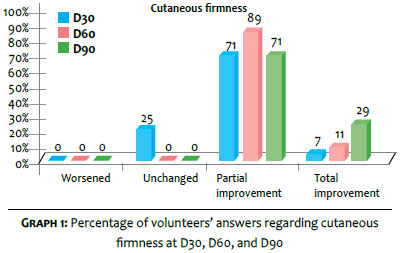
Graph 1 depicts the data collected for the evaluation of cutaneous firmness. Firmness was defined as having a higher resistance to traction or digital pressure.
Graph 1 allows us to observe that after 30 days of supplement use there was a perceived improvement of 75%; in 60 days, the perceived improve reached 100% of the sample. At the end of the study, 100% of the sample had noticed partial or total improvement in the cutaneous firmness of the face.
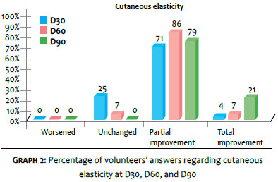
Graph 2 shows the results obtained for the item regarding cutaneous elasticity. This variable was defined as the ability to return to its original state after traction or digital pressure.
Graph 2 shows that after 30 days of supplement use there was a perceived improvement of 75%; in 60 days, the perceived improvement reached 93% of the sample. At the end of the study, 100% of the sample noticed partial or total improvement in facial cutaneous elasticity.
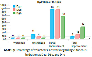
Another parameter evaluated was cutaneous hydration, defined as skin with a healthy, homogenous, bright appearance and smooth to the touch. Graph 3 shows the time results for that variable.
Graph 3 shows that after 30 days of supplement use there was a perceived improvement of 89% in the perception of cutaneous hydration; in 60 days, the perceived improvement reached 96% of the sample. At the end of the study, 100% of the sample noticed partial or total improvement of the facial cutaneous hydration.
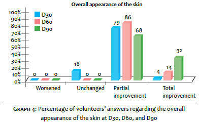
Finally, the overall appearance of the skin was defined as the perceived appearance of the skin during observation in the mirror without taking into account any specific parameter, except for the impression of vitality and smoothness of features/ signs of aging.
Graph 4 shows the results obtained for this variable:
Chart 4 shows that after 30 days of supplement use there was a perceived improvement in the overall appearance of the skin of 83%; in 60 days, an improvement of 100% could be noticed. At the end of the study, 100% of the sample noticed partial (68%) or total (32%) improvement in the hydration of facial skin
Evaluation of the supplement: characteristics of patient self-administration
The product's solubility, defined as the degree of difficulty of dissolution in 200 ml of cold or hot water, was also evaluated. After 90 days of treatment, 96% of patients reported that the solubility was easy or very easy.
Regarding the taste, 100% of the patients considered it good or very good.
Ultrasonographic evaluation
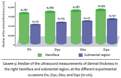
Based on the data obtained about the dermal thickness on each visit, the group's median was calculated for each experimental event: D0, D30, D60, and D90 (D = Day). It was possible to observe a progressive and significant increase in the median thickness in both evaluated areas, with increases of 17.0% and 18.8%, in the malar and in the submental areas respectively, at the end of the study. Both results were statistically significant according to the Wilcoxon test (p <0.001). Graph 5 details the measurements obtained for the evaluated areas at each experimental occasion:
Comparisons made on D60 relative to D30 and D90 also showed a statistically significant (p<0.001) increase in the thickness of the malar and the dermis of the submental regions, characterizing progress in the effect seen over time.
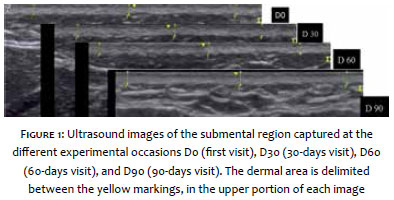
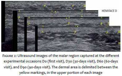
Figures 1 and 2 illustrate the ultrasonographic findings for the study areas.
The use of collagen as an oral supplement is not a recent development. Given that collagen is a nutrient with structural value, cases of malnutrition, rigorous diets, or malabsorption syndromes would naturally and evidently benefit from protein supplementation.
As the studies aimed at delaying the signs of the aging progresses, the current discussion focuses on the possibility of collagen supplementation in offering a beneficial and significant effect on dermal loss related to skin aging.
Few studies have focused on evaluating the impact of collagen supplementation with gelatin and other enriched foods (such as shakes and even capsules) on the parameters of aging.
Gelatin is a polypeptide derived from collagen with a high molecular weight, however it is deficient in essential amino acids. Despite the fact that its nutritional value is indisputable, it does not have specific properties. 3
Finally, the study by Nishimoto et al. on Wistar rats, using a model to histologically demonstrate the synthesis of collagen based on the hydroxyproline index, has demonstrated that isolated peptides would have a significant effect on the collagen synthesis, when compared to that obtained with gelatin, whose levels were not superior to those of the placebo. 4
Based on findings like this, hydrolyzed collagen (HC) has once again attracted attention.
Hydrolyzed collagen is digested and absorbed in the digestive tract -identified in the blood by its constituent peptides - and reaches the skin in up to four days. 5
Due to its similarity to collagen - particularly type I dermal collagen - its effect would be not only that of restoration, but also of promoting type I collagen synthesis, playing a positive role in aging and even in other disorders with dermal involvement, like tissue repair. 6,7
Hydrolyzed collagen is characterized by a relatively low molecular weight (<6 kDa), which facilitates its absorption and bioavailability. Nonetheless, there are many types of hydrolyzed collagen according to the protein source, and the synthesis and supply processes. 8,9
Each hydrolyzed collagen type can have a set of effects, and during the study their effects were evaluated in vitro, often using models that are not comparable. For these reasons, it is very difficult to gather comparable data on the subject, since the term "hydrolyzed collagen" refers to a large group of peptides associations.
Therefore, results obtained for a particular HC cannot be extrapolated to others.
The HC that has been studied in the present article is a polypeptide derived from the biotechnological enzymatic process of animal skin, devoid of impurities and other molecules such as lipids and carbohydrates, and therefore easily digestible, susceptible to the action of proteolytic enzymes, and with an absorption rate of more than 90%. 10
The collagen peptide evaluated in the present study consists of 18 type-I collagen peptides, 8 of which are essential. It is characterized by higher concentrations of glycine, proline, and hydroxyproline - amino acids that represent about 50% of the total content of amino acids in the composition. This composition was demonstrated to be capable of promoting collagenesis from fibroblasts, in addition to the fact that the synthesized collagen is firmer. 3
The specific peptides generated from the digestion of collagen peptides - Gly-Glu and Pro-Hypro - are chemoattracted by dermal fibroblasts as a signal of "destruction" of collagen, thus activating fibroblasts. 11
There is also the activation of the enzyme Hyaluronan synthase 2, with an increased synthesis of hyaluronic acid and glycosaminoglycans. 12,13
The collagen peptide has also been shown to stimulate the production of Decorin, a proteoglycan, which is a connective tissue component that binds to type-1 collagen and plays a role in the organization of the extracellular matrix, and by regulating the aggregation of bundles leads to the production of collagen fibers. 14
Another trend that induces a greater diversity of supplements in this area is the association of other molecules, with protective or beneficial effects in the collagen synthesis, thus favoring the effects of the peptides. The action of vitamin C in stimulating fibroblast proliferation is widely known. 15
Studies by Pinnel in the 1980s demonstrated that the L-ascorbic acid is capable of inducing procollagen synthesis in cultured dermal fibroblasts. 16 These studies demonstrated that this increased synthesis occurred even in older fibroblasts, which becomes especially relevant considering that there is a decrease of physiological vitamin C reserves in the dermis with age. 17
The combination of substances such as phytoextracts can also increase the effects of the peptides associations of hydrolyzed collagen, enabling a better performance through the neutralization of free radicals, for example.
The plant Hibiscus sabdariffa, traditionally used in food preparation due to its taste, demonstrated antioxidant, lipolytic, hypoglycemic, and diuretic properties, among others, in in vitro models. Its use for reducing resistance to insulin was demonstrated based on its polyphenols, for it contains high concentrations of phenolic acid and proanthocyanidins.18,19
Its broad antioxidant mechanism, however, is based on the combination of additional compounds that are not present in that extract. The Hibiscus sabdariffa's powerful antioxidant action mechanism has already been studied in neoplasias, having been more recently evidenced with the isolation of its main antioxidant compounds: chlorogenic and neochlorogenic, cryptochlorogenic acids, rutin, and isoquercetin. 20, 21
The present study was aimed at evaluating the effects of a nutritional supplement that brings together the synergistic action of these three molecules (collagen peptide, Vitamin C, and Hibiscus sabdariffa) on the human dermis. A non-invasive study was carried out with the assistance of ultrasonographic evaluation, which allowed for the confirmation of the signs perceived by patients. In addition to the progressive and significant improvement in the cutaneous firmness, elasticity, and hydration, there was a real and measurable increase in the dermal thickness in two facial areas: the malar region (where there was a 17.0% increase of the thickness) and the submental region (where there was an increase of 18.8%). The latter finding is of particular interest, since this area offers more complexity to treat sagging.
The evaluation of these patients allowed for the observation of safety levels in the use of the product, which did not cause any systemic reaction during the three months of consumption.
The daily use of the supplement was demonstrated to be palatable, a fundamental factor for adherence to prolonged treatments. Regarding the taste, it is important to highlight a finding: even during continued use (three months), there was no reduction of tolerance (i.e. patients did suffer from "taste fatigue" when ingesting the supplement over time) - which is a major factor in treatment adherence.
The daily use of a nutritional supplement containing collagen peptides, Vitamin C, and Hibiscus sabdariffa promoted a significant increase in the dermal thickness of the studied areas of the face (malar and submental), with a positive impact on the evaluation of cutaneous firmness, elasticity, and hydration. The safety of its use combined with its proven effects allows for the conclusion that this association is beneficial in the approach to facial aging, as well as dermal sagging and atrophy.
1. Fisher GJ, Kang S, Varani J, Bata-Csorgo Z, Wan Y, Datta S, et al. Mechanisms of photoaging and cronological skin aging. Arch Dermatol. 2002;138(11):1462-70.
2. Brincat M, Mouniz CF, Studd JW, Cooper d. Sex hormones and skin collagen content in post menopausal women. Br Med J (Clin Res Ed). 1983;287(6402):1337-8.
3. Zague V. A new view concerning the effects of collagen hydrolysate intake on skin properties. Arch Dermatol Res. 2008;300(9):479-83.
4. Hiura, N, Sato R, Suzuki K, Asano R. Effect of oral administration of gelatin and collagen peptides on the hydroxyproline content of rats skin. Journal of the Japanese Society for Food Science and Technology. 2002;49;3 [acesso em 30 de janeiro de 2015]. Disponível em: http://agris.fao.org/agris-search/search.do?recordID=JP2002003318
5. Ohara H, Matsumoto H, Ito K, Iwai K, Sato K. Comparison of quantity and structures of hydroxyproline-containing peptides in human blood after oral ingestion of gelatin hydrolysates from different sources. J Agric Food Chem. 2007;55:1532-35.
6. Zague V. Collagen hydrolysate intake increases skin collagen expression and suppresses matrix metalloproteinase 2 activity. J Med Food. 2011;14(6):618-24.
7. Atamas SP, Luzina IG, Ingels J, Choi J, Wong WK, Furst DE, et al. Stimulation with type I collagen induces changes in gene expression in peripheral blood mononuclear cells from patients with diffuse cutaneous systemic sclerosis (scleroderma). Clin Exp Immunol. 2010;161(3):426-35.
8. Zhuang, Yongliang, Zhao X, Zhang Z, Li B. Effects of collagen and collagen hydrolysate from jellyfish (Rhopilema esculentum) on mice skin photoaging induced by UV irradiation. J Food Sci. 2009;74(6):H183-8.
9. Postlethwaite AE, Wong WK, Clements P, Chatterjee S, Fessler BJ, Kang AH, et al. A multicenter, randomized, double-blind, placebo-controlled trial of oral type I collagen treatment in patients with diffuse cutaneous systemic sclerosis: I. oral type I collagen does not improve skin in all patients, but may improve skin in late-phase disease. Arthritis Rheum. 2008;58(6):1810-22.
10. Oesser S, Adam M, Babel W, Seifert J. Oral administration of (14)C labeled gelatin hydrolysate leads to an accumulation of radioactivity in cartilage of mice (C57/BL). J Nutr. 1999;129(10):1891-5.
11. Postlethwaite AE, Seyer JM, Kang AH. Chemotactic attraction of human fibroblasts to type I, II, and III collagens and collagen-derived peptides. Proc Natl Acad Sci U S A. 1978;75(2):871-5.
12. Matsuda N, Koyama Y, Hosaka Y, Ueda H, Watanabe T, Araya T, et al. Effects of ingestion of collagen peptide on collagen fibrils and glycosaminoglycans in the dermis. J Nutr Sci Vitaminol (Tokyo). 2006;52(3):211-5.
13. Minaguchi J, Koyama Y, Meguri N, Hosaka Y, Ueda H, Kusubata M, et al. Effects of ingestion of collagen peptide on collagen fibrils and glycosaminoglycans in Achilles tendon. J Nutr Sci Vitaminol (Tokyo). 2005;51(3):169-74.
14. Pulkkinen L, Alitalo T, Krusius T, Peltonen L. Expression of decorin in human tissues and cell lines and defined chromosomal assignment of the gene locus (DCN). Cytogenet Cell Genet. 1992;60(2):107-11.
15. Hata R, Senoo H. L-ascorbic acid 2-phosphate stimulates collagen accumulation, cell proliferation, and formation of a three-dimensional tissuelike substance by skin fibroblasts. J Cell Physiol. 1989;138(1):8-16.
16. Pinnel SR, Murad S, Darr D. Induction of collagen synthesis by ascorbic acid. A possible mechanism. Arch Dermatol. 1987;123(12):1684-6.
17. Patnaik BK, Kanungo MS. Ascorbic acid and aging in the rat. Uptake of ascorbic acid by teeth and concentration of various forms of ascorbic acid in different organs. Biochem J. 1966;100(1):59-62.
18. Da-Costa-Rocha I, Bonnlaender B, Sievers H, Pischel I, Heinrich M. Hibiscus sabdariffa L. - a phytochemical and pharmacological review. Food Chem. 2014;165:424-43.
19. Peng CH, Yang YS, Chan KC, Wang CJ, Chen ML, Huang CN. Hibiscus sabdariffa polyphenols alleviate insulin resistance and renal epithelial to mesenchymal transition: a novel action mechanism mediated by type 4 dipeptidyl peptidase. J Agric Food Chem. 2014;62(40):9736-43.
20. Worawattananutai P, Itharat A, Ruangnoo S. In vitro antioxidant, anti-inflammatory, cytotoxic activities against prostate cancer of extracts from Hibiscus sabdariffa leaves. J Med Assoc Thai. 2014;97 Suppl 8:S81-7.
21. Wang J, Cao X, Jiang H, Qi Y, Chin KL, Yue Y. Antioxidant activity of leaf extracts from different Hibiscus sabdariffa accessions and simultaneous determination five major antioxidant compounds by LC-Q-TOF-MS. Molecules. 2014;17;19(12):21226-38.
This study was performed at a private practice - São Paulo (SP), Brazil.
 All content the journal, except where identified, under the Creative Commons Attribution 4.0 International licence - ISSN-e 1984-8773


 Read in Portuguese
 Portuguese PDF
 Print
Print
 Send this article by email
 How to cite this article
 Submit a comment
 Mendeley
 Pocket