


Adilson Costa1; Samara Eberlin2; Stefano Piatto Clerici3; Beatrice Martinez Zugaib Abdalla4
Keywords: ANTI-INFLAMMATORY AGENTS/ADVERSE EFFECTS; DERMATITIS, ATOPIC; SOAPS
Like any body organ, the skin may present degeneration, metabolic abnormalities, malformations, dysfunction, and inflammation, leading to histological alterations and resulting in elementary lesions.1
Depending on the anatomical location and environmental influences, the skin has exceptional structural and functional diversity, as it is continually exposed to external aggressions such as solar radiation, mechanical stimuli, climate change and/or chemical and biological damage.2 The maintenance of the skin's structural integrity is therefore critical and, when affected, it should be able to count on mechanisms for the rapid restoration of the epidermal barrier.3-5
Hydration and the integrity of the stratum corneum (SC) are crucial to the skin's appearance, metabolism, mechanical properties and cutaneous barrier function.6 During the epidermal differentiation, several proteins are involved in the formation of the stratum corneum, including loricrin, involucrin, filaggrin, and keratins. 7-9
The variation of the genes of the stratum corneum results in the pathogenesis of three major skin disorders: ichthyosis vulgaris, psoriasis, and atopic dermatitis (AD). 10-11 AD is a chronic inflammation characterized by pruritus and one in which hereditary, environmental, and immunological components play a key role. 12
AD fluctuates between two phases: the acute phase, with a predominance of Th2 type response and IgE production, and the chronic, with strong Th1 type response, with the production of interferon-γ (IFN-γ) and interleukin-12 (IL-12). 11
Keratin is a tough, waterproof protein responsible for the skin's protection.13 The keratins 5 and 14 are expressed in the basal keratinocytes while keratins 1 and 10 participate in the differentiation of suprabasal keratinocytes.14 Mutations in keratin genes can result in human diseases characterized by an alteration in epidermal homeostasis, including the impairment of the barrier function. 14 The rupture of the permeable barrier of the epidermis - both acute and chronic - results in changes in the synthesis of structural proteins in keratinocytes, relating to increased suprabasal cell proliferation and differentiation, in an attempt to repair the skin barrier. 14
The local production of inflammatory mediators is correlated to the integrity of the barrier, as a result of being activated following the release of cytokines, histamine, and eicosanoids. 15 These, in turn, increase the sensitivity of the nociceptors, such as TRPV-1 (V1 transitional channel vanilloid receptor), a nonselective calcium channel widely expressed in the cutaneous tissue, including keratinocytes and peripheral sensory nerve fibers.16 TRPV-1 has its sensitivity increased in AD lesions, which translates into hyperalgesia and additional production of inflammation mediators. 17-18
The availability of cosmetic products that minimize the occurrence of unwanted side effects such as skin xerosis and cutaneous hypersensitivity, can be a key differential to ensure adherence to and success of the treatment. The present study was aimed at evaluating whether some cosmetic products in liquid soap marketed in Brazil provide in vitro anti-inflammatory and protective efficacy to the skin barrier.
Four commercial liquid soaps were evaluated in the present in vitro study: Test-substance A (Dermacyd Infantil, Sanofi-Aventis Farmacêutica Ltda., São Paulo, SP, Brazil, batch 246782); Test-substance B (Sabonete de Glicerina Granado Bebê, Casa Granado Laboratórios, Farmácias e Drogarias S.A., Belém, PA, Brazil, batch R1446); Test-substance C (Huggies Turma da Mônica Recém-Nascido, Kimberly-Clark Brasil Indústria e Comércio de Produtos de Higiene Ltda, Mogi das Cruzes, SP, Brazil, batch Lkg2354); and Test-substance D (Johnson's Baby Sabonete Líquido Da Cabeça aos Pés, Johnson & Johnson Industrial Ltda, São José dos Campos, SP, Brazil, batch 3013b09).
To that end, human fibroblast cultures (Human Foreskin Fibroblasts-1, "HFF-1"; ATCC SCRC-1041 (Banco de Células do Rio de Janeiro, Rio de Janeiro/RJ, Brazil, Catalogue number 0275) were seeded in 75 cm3 bottles (Nunc, Roskilde, Denmark), cultured and expanded in an incubator at 37ºC in the presence of 5% CO2, using a specific culture medium for the determination of cell viability through the XTT method.
Cell viability was determined by a colorimetric method using the XTT dye (2, 3-bis [2-methoxy-4-nitro-5-sulfopheny]-2H-tetrazolium-5-carboxyanilide inner salt), which is converted in water-soluble orange formazan by the mitochondrial enzyme succinate dehydrogenase in viable cells (Xenometrix AG, Switzerland). The fibroblasts were seeded and incubated with the Test-substances in eight concentrations using decimal geometric dilution. 13 After 48 hours of incubation the Test-substances were removed and the culture medium was replaced. Then, the XTT dye was added to the culture, and the plate was incubated for a further three hours. The absorbance (optical density - OD) of each well was determined at 480nm with the assistance of a Multiskan GO monochromator (Thermo Scientific, Finland). The percentage of cell viability was calculated according to the following equation:
Viability % = (DOST / DOCN) x 100
Where:
- DOST is the optical density of the Test-substance
- DOCN is the optical density of the negative control.
HaCat human keratinocytes (Banco de Células do Rio de Janeiro, Rio de Janeiro/RJ, Brazil, Catalogue number 0341) were seeded and cultured in the same way that the HFF-1 human fibroblasts were. Upon reaching confluence, the cells were seeded in plates for later incubation with the Test-substance and quantification of the proposed mediators.
The keratinocyte cultures were incubated with three non-cytotoxic concentrations of the Test-substances determined by the XTT method. Cells were maintained in contact with the test-substances for 48 hours. After this period, the cell culture's supernatant was collected for measurement of the mediators related to the cutaneous barrier protective activity. In order to evaluate the anti-inflammatory and reductive activity of the dermal hypersensitivity, the inflammatory stress was mimicked through the addition of interleukin-1 alpha (IL-1α, 10ng/mL) to the keratinocyte cultures, concurrently with the treatment using the Test-substance, for 48 hours.
Using commercially available kits for Sandwich ELISA, the concentration of the following mediators were assessed in the supernatants obtained from cell cultures of human keratinocytes: Keratin 10 (USCN Life Science Inc., Wuhan, Hubei, China); keratin 14 (USCN Life Science Inc., Wuhan, Hubei, China); loricrin (USCN Life Science Inc., Wuhan, Hubei, China); interleukin-12 (IL-12; BD Biosciences, San Diego, CA, USA); interferon-γ (IFN-γ, BD Biosciences, San Diego, CA, USA); and transient channel vanilloid receptor V-1 (TRPV-1; USCN Life Science Inc., Wuhan, Hubei, China). The absorbance reading was performed using a Multiskan GO monochromator (Thermo Scientific, Finland).
For the statistical evaluation of the mediators Keratin 10, Keratin 14, loricrin, IL-12, IFN-γ, and TRPV-1, the one-way ANOVA test was used (GraphPad Prism 4 Software Inc. San Diego, CA, USA), which allows for the measuring of the variation in results by comparing the data from the Test-substances, evidencing the differences among them. As the F-test was significant, the authors applied the nonparametric Tukey test. A 5% significance level was adopted.
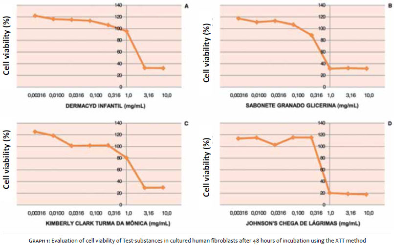
Graph 1 represents the Test-substances' concentration-cell viability curve. As can be seen, Test-substances A, C, and D showed non-cytotoxic concentrations starting with the 0.316mg/mL dilution. The Test-substance B showed non-cytotoxic concentration starting with the dilution 0.100 mg/mL.
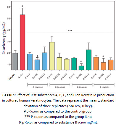
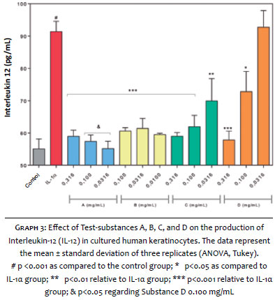
From a methodological point of view, the incubation of cultured human keratinocytes with pro-inflammatory cytokine interleukin-1 alpha (IL-1α) promoted an increase of 2.7 times and 1.7 times the production of IFN-γ and IL-12, respectively. Aimed at assessing the anti-inflammatory activity of each Test-substance, Graphs 2 and 3 depict the effect of the Test-substances in the IFN-γ and IL-12 synthesis in cultured human keratinocytes. All Test-substances evaluated were able to significantly reduce the synthesis of both inflammatory cytokines.
Regarding IFN-γ, Graph 2 suggests that the Test-substances promoted significant decreases in all concentrations evaluated. At a a concentration of 0.100 mg/mL, Test-substances A, C, and D have shown a slightly stronger anti-inflammatory response (p<0.05) as compared to substance B at the same concentration.
The response obtained in the synthesis of cytokine IL-12 was similar to that of IFN-γ. From Graph 3 it can be seen that Test-substance A at a concentration of 0.100 mg/mL and 0.0316 mg/mL, was able to reduce IL-12 production to levels significantly lower (p<0.05) than those observed with Test-substance D at 0.100 mg/mL concentration.
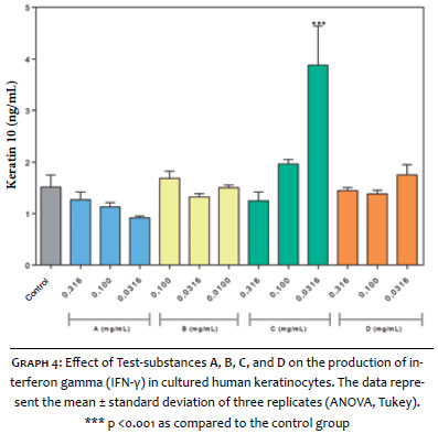
Regarding the protective activity of the skin barrier, Graph 4 represents the effects of the Test-substances in the synthesis of Keratin 10 in cultured human keratinocytes. As can be seen, only substance C in the concentration of 0.0316 mg/mL produced a significant increase in the synthesis of Keratin 10 (p <0.001) when compared to the control.
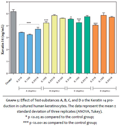
The response observed for Keratin 14 (Graph 5) was contrary to that observed with Keratin 10. All Test-substances - either in one or more concentrations evaluated - caused significant reduction in the synthesis of Keratin 14 as compared to the control group.
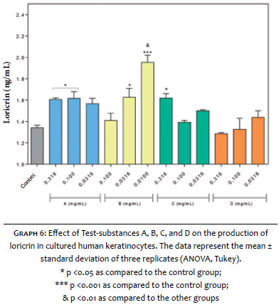
Graph 6 depicts the effects of Test-substances on loricrin synthesis in cultured human keratinocytes. Test-substances A, B, and C, in one or more tested concentrations promoted significant increases in loricrin synthesis. The greatest increase observed was obtained with Test-substance B in a concentration of 0.0100 mg/mL (p <0.01), as compared to other groups evaluated.
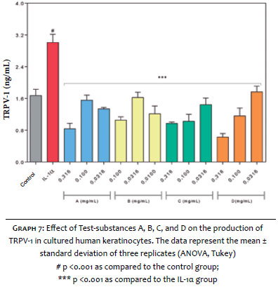
Regarding the dermal hypersensitivity activity, Graph 7 shows the effect of Test-substances on TRPV-1 receptor synthesis in cultured human keratinocytes. The addition of IL-1α to the cell cultures caused a significant increase of 79.7% in the synthesis of TRPV-1 receptor. In contrast, all Test-substances reduced the production of this receptor when compared to the group that was stimulated with IL-1α.
The primary function of the epidermis is that of protection. 2 The barrier function is performed by a semipermeable stratum corneum composed of lipid lamellae.2
The regulation of the lipid barrier's synthesis has been studied in a variety of models. Genetic alterations in lipid metabolism or in the protein content components of the stratum corneum produce desquamation or ichthyosiform skin with abnormal structure and function of the lipid barrier. 2
The concept of skin hygiene began with the dawn of human history and is related to the removal of impurities. 14 Nowadays, the act of cleansing the skin is related to an individual's appearance and skin health. 14
It is known that the use of water alone is not sufficient for skin hygiene; therefore, in order to remove finer particles it is necessary to use emulsifiers, which reduce the skin's surface tension and remove dirt, sebum, microorganisms, and cells of the stratum corneum.15 An ideal soap must have these characteristics, however without damaging or irritating the skin and attempting to maintain the hydration of the skin's surface, 16 providing a good balance of the two. 15
This in vitro study compared four soaps regarding the activity of their anti-inflammatory, barrier protective, and cutaneous hypersensitivity reduction. In general, from the biochemical, pre-clinical standpoint, all Test-substances showed satisfactory and very similar results, which can be useful for the clinical indication in patients with dermatoses that compromise the skin barrier.
Disorders with a purely inflammatory and chronic character - such as psoriasis and atopic dermatitis - lead to a decreased barrier function. 17 It has been known for a long time that soaps and products containing surfactants play an important role in the alteration of the cutaneous barrier, triggering AD outbreaks. 18 For this reason, the indication of commercial products that have little effect in triggering such crises is mandatory in dermatological clinical practice; therefore, it is desirable to know which ones may be useful in this indication.
AD typically demonstrates a decreased irritability threshold of the skin barrier; 19 therefore soaps that are less irritating and demonstrate the ability to maintain good hydration of the stratum corneum represent a benefit for the atopic xerodermic skin, influencing the treatment's outcome and the development of the disease, with an improvement in the overall management of the condition. 19
Based on the results obtained it is possible to infer that the four Test-substances promoted significant reductions in the synthesis of IFN-γ (Test-substances A, C, and D showed marked reductions as compared to B) and IL-12 (Test-substance A was able to reduce IL-12 production to levels significantly below those observed with Test-substance D in the concentration of 0.100 mg/mL). Test-substance C triggered a significant increase in the synthesis of Keratin 10, a phenomenon not seen with other Test-substances, although all have shown significant reductions in the synthesis of Keratin 14. All Test-substances promoted significant increases in loricrin synthesis and significant reductions in TRPV-1 synthesis. Based on these findings, it is possible to conclude that all Test-substances have anti-inflammatory, hypersensitivity reduction, and epidermal barrier restorative activities.
1. Sampaio AP, Rivitti EA. Dermatologia. 2 ed. São Paulo: Artes Médicas; 2001. p. 39-40.
2. Elias P, Feingold K, Fluhr J. The skin as an organ of protection. In: Friedberg IM, Eisen AZ, Wolff K, Austen KF, Goldsmith LA, Katz SI, editors. Dermatology in General Medicine. New York: McGraw-Hill; 2003. p .07-118.
3. Martin P. Wound healing-aiming for perfect skin regeneration. Science. 1997;276(5309):75-81.
4. Gurtner GC, Werner S, Barrandon Y, Longaker MT. Wound repair and regeneration. Nature. 2008;453(7193):314-21.
5. Feingold KR. Thematic review series: skin lipids. The role of epidermal lipids in cutaneous permeability barrier homeostasis. J Lipid Res. 2007;48(12):2531-46.
6. Madison KC. Barrier function of: "la raison d'etre" of the epidermis. J Invest Dermatol. 2003;121(2):231-41.
7. Hoffjan S, Stemmler S. On the role of the epidermal differentiation complex in ichthyosis vulgaris, atopic dermatitis and psoriasis. Br J Dermatol. 2007;157(3):441-9.
8. Kalinin A, Marekov LN, Steinert PM. Assembly of the epidermal cornified cell envelope. J Cell Sci. 2001;114(Pt 17):3069-70.
9. Ishida-Yamamoto A, Iizuka H. Structural organization of cornified cell envelopes and alterations in inherited skin disorders. Exp Dermatol. 1998;7(1):1-10.
10. Smith FJ, Irvine AD, Terron-Kwiatkowski A, Sandilands A, Campbell LE, Zhao Y, et al. Loss of function mutations in the gene encoding filaggrin cause ichtyosis vulgaris. Nat Genet. 2006;38(3):337-42.
11. O'Driscoll J, Muston GC, McGrath JA, Lam HM, Ashworth J, Christiano AM. A recurrent mutation in the loricrin gene underlies the ichthyotic variant of Vohwinkel syndrome. Clin Exp Dermatol. 2002;27(3):243-6.
12. Varothai S, Nitayavardhana S, Kulthanan K. Moisturizers for patients with atopic dermititis. Asian Pac J Allergy Immunol. 2013;31(2):91-8.
13. Chen J, Cheng X, Merched-Sauvage M, Caulin C, Roop DR, Koch PJ. An unexpected role for keratin 10 end domains in susceptibility to skin cancer. J Cell Sci. 2006;199(Pt 24):5067-76.
14. Ekanayake-Mudiyanselage S, Aschauer H, Schmook FP, Jensen JM, Meingassner JG, Proksch E. Expression of epidermal keratins and the cornified envelope protein involucrin is influenced by permeability barrier disruption. J Invest Dermatol. 1998;111(3):517-23.
15. Luger TA. Neuromediators - a crucial component of the skin immune system. J Dermatol Sci 2002;30(2):87-93.
16. Riol-Blanco L, Ordovas-Montanes J, Perro M,Naval E, Thiriot A, Alvarez D, et al. Nociceptive Sensory Neurons Drive Interleukin-23 Mediated Psoriasiform Skin Inflammation. Nature. 2014;510(7503):157-161.
17. Imamachi N, Park GH, Lee H, Anderson DJ, Simon MI, Basbaum AI, et al. TRPV1-expressing primary afferents generate behavioral responses to pruritogens via multiple mechanisms. Proc Natl Acad Sci USA. 2009;106(27):11330-5.
18. Sadofsky LR, Ramachandran R, Crow C, Cowen M, Compton SJ, Morice AH. Inflammatory stimuli up-regulate transient receptor potential vanilloid-1 expression in human bronchial fibroblasts. Exp Lung Res. 2012;38(2):75-81.
19. ECVAM-Dbalm. 3T3 Neutral Red Uptake (NRU) Phototoxicity Assay. DB-ALM Protocol nº 78. [atualizado 2008; acesso 16 de fevereiro de 2015]. Disponível em: http://ecvam-dbalm.jrc.ec.europa.eu/public_view_doc2.cfm?id=736F27E9E9F7A9D869FB4808
7878D2497180BB0BC12CB10496CDA74B54630A05A3291B895581F634]
This study was performed at the Kolderma Instituto de Pesquisa Clínica and at the Dermatology Service of the Pontifícia Universidade Católica de Campinas - Campinas (SP), Brazil.
 All content the journal, except where identified, under the Creative Commons Attribution 4.0 International licence - ISSN-e 1984-8773


 Read in Portuguese
 Portuguese PDF
 Print
Print
 Send this article by email
 How to cite this article
 Submit a comment
 Mendeley
 Pocket