


Daniele Bani1; Alessandro Quattrini Li2; Giulia Lo Russo3
Keywords: ADIPOCYTES, WHITE; HIGH-INTENSITY FOCUSED ULTRASOUND ABLATION; SUBCUTANEOUS FAT
To fulfill the increasing demand for non-invasive fat reduction methods, as an alternative to liposuction, 1 numerous physical treatments--namely mechanical and electric stimulation, radio frequency, and low-level laser--have been investigated. However, most of them have not met with expectations, 1-4 and some have also raised safety issues.5 Of the most promising non-invasive approaches to lipo-reduction, ultrasound holds a pivotal place. 6-9 In spite of its accelerating use in aesthetic medicine (with satisfactory results), the mechanisms of action on adipose cells remain to be fully elucidated and may likely vary according to the mode of ultrasound delivery. In fact, ultrasonic energy can be transmitted to the skin in non-focused or focused modes. In the non-focused mode, energy attenuates with depth: thus, to deliver enough energy to the subcutaneous fat, the superficial skin is exposed to maximum energy intensity and may undergo injury. Instead, focused ultrasound can be concentrated in a defined subcutaneous area to produce clinically relevant fat lysis while limiting damage to the upper tissues. 10,11 However, focused ultrasounds can induce marked heating, thereby causing adipocyte necrosis in the treatment area.12
The most recent lipo-reductive ultrasound devices are specifically designed to prevent tissue injury. Contour ITM (UltraShape, Yoqneam, Israel), a focused ultrasound emitter, was first demonstrated to achieve selective adipocyte lysis and clinically relevant reduction of the volume of subcutaneous fat pad, in the absence of significant adverse reactions.7,13 Med2ContourTM (General Project, Montespertoli, Italy) takes advantage of two angled non-focused transducers that create a weakly focused ultrasound field where the two beams overlap, i.e. within the subcutaneous fat pad.14,15
The cellular mechanisms underlying the lipo-reductive effects of ultrasound are not fully understood and are thus a matter for investigation. It has been shown that the impact of ultrasounds on adipose cells can induce transitory pore opening at the plasma membrane, allowing triglyceride leakage.14,15
The current study aims at providing further evidence for the efficacy of weakly focused high-frequency ultrasounds, delivered by the Med2ContourTM, for non-invasive liporeduction and to search for possible morphological clues that can help understand the mechanism of action on adipose cells.
This study complied with the guidelines of the Declaration of Helsinki, as amended in Edinburgh, 2008. It was approved by the Ethical Committee of the Faculty of Medicine, University of Florence, Italy. All subjects gave written informed consent to their participation in the study.
Study on human skin explants
Full-thickness biopsies of normal skin, about 15 mm thick, were taken at a surgery from three patients undergoing abdominoplasty (1 male, 2 females, aged 40-65 years). Each biopsy was cut in half, and each half placed in a Petri dish on ice, the subcutaneous tissue facing downwards, and mixed with 2 ml of pre-oxygenated incubation medium (Dulbecco's modified Eagle medium, DMEM; Gibco Invitrogen, Milan, Italy). A first sample was treated with non-focused ultrasound using the Med2ContourTM device (General Project, Montespertoli, Italy) set at: 3 W power output, 20 kHz frequency, pulsed mode (2 pulses, 6 s. each, separated by a 10 s. pause). A single transducer of the Med2ContourTM was placed in direct contact with the epidermis through a thin layer of Aquasonic ClearTM ultrasound gel (Parker, Fairfield, USA). The above power and frequency settings were adopted because they were similar to those yielding the best clinical performance,7,14,15 while the timing protocol was chosen to avoid tissue overheating, considering that the skin explants lacked blood flow-related temperature homeostasis. Tissue temperature was continuously monitored with a digital thermometer and found not to exceed 38º C. The other specimen was sham-treated (i.e. subjected to the same handling procedure but with no ultrasound emission) and used as control. At the end of the experiments, fragments of adipose tissue were taken from the central part of the treated and control specimens, fixed in isotonic 4% glutaraldehyde and 1% OsO4, dehydrated and embedded in Epon epoxy resin (Fluka, Buchs, Switzerland) for light and electron microscopic studies.
Adipocyte size was measured by computer-aided morphometry on digital photomicrographs of semi-thin sections, 2 mm thick. The surface area of adipocyte lipid vacuoles was measured on 10 randomly chosen micrographs (test area: 65,700 mm2) from each specimen using ImageJ 1.33 image analysis program (http://rsb.info.nih.gov/ij), upon setting an appropriate threshold to only include the osmiophilic lipid vacuoles of the adipocytes. Vacuolar profiles < 1000 mm2, consistent with polar cross-sections, were excluded. Data were reported as mean values (± SEM) of the control and treated groups. For transmission electron microscopy, ultrathin sections were stained with uranyl acetate and alkaline bismuth subnitrate, viewed and photographed under a JEM 1010 transmission electron microscope (Jeol, Tokyo, Japan).
In vivo study
This was performed on three obese volunteers (2 males, 1 female, aged 34-53 years) scheduled for abdominal lipo-reductive surgery, who were subjected to weakly focused high-frequency ultrasounds using the Med 2ContourTMset at: 2 W power output, pulsed mode, 20 kHz frequency, 15 minute treatment. In each patient, the right hypogastrium was the test area whereas the left hypogastrium was the sham-treated area. Two patients received four treatments at 27, 20, 12, and 1 day before surgery. The remaining patient received three treatments at 27, 20, and 12 days before surgery: the aim of this protocol being to study whether the effects of the ultrasound were maintained over time. During the surgery, fragments of subcutaneous adipose tissue were taken from the central part of the test and control areas and processed for ultrastructural examination, as described above. Similarly, during the experiment on skin explants, morphometry of adipocyte size was performed on semi-thin sections and ultrastructural analysis on ultra-thin sections of the Epon-embedded specimens.
Further, three overweight patients (2 males, 1 female, aged 30-33 years) were enrolled for assessment of subcutaneous adipose tissue mass in control and ultrasound-treated abdominal skin. They were subjected to four weekly ultrasound treatments, similarly to the above-described protocol. Before each treatment and one week after the last treatment, the thickness of subcutaneous fat pads in the control and test areas were assessed by measuring the depth of skin folds with a Harpenden caliper (FitnessAssist, Wrexham, UK). To normalize individual differences, the values were expressed as percent changes over the initial measures.
Statistical analysis
The assayed quantitative parameters were statistically analyzed with Graph Pad Prism 4.03 statistical software (GraphPad, San Diego, CA, USA), assuming the individual patients as sample units (n=3). Statistical significance of differences between groups was assessed by unpaired Student's t test; p < 0.05 was considered significant.
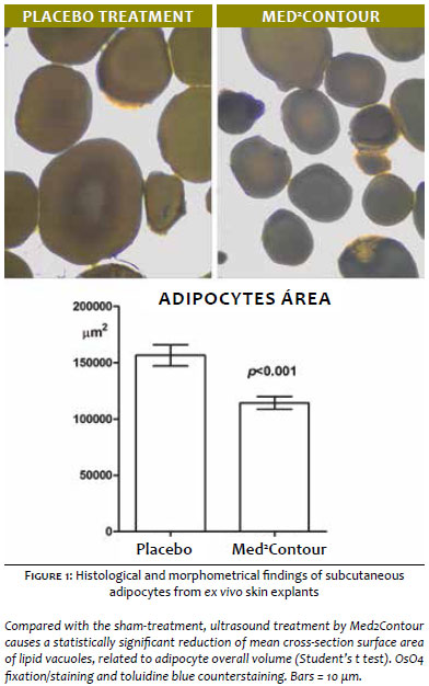
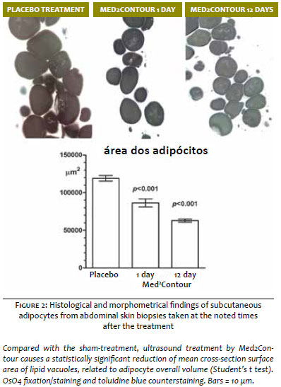
Light microscopic and morphometric analysis of semi-thin sections of subcutaneous adipose tissue of the ex vivo skin explants (n=3) showed that ultrasound treatment by Med2ContourTM induced a marked, statistically significant decrease (-23%) in the size of adipocyte lipid vacuoles (Figure 1). Similar findings were observed in the subcutaneous fat biopsies taken at surgery from sham- or Med2ContourTM-pretreated abdominal skin (Figure 2). In the biopsies collected one day after the last ultrasound application (n=2), we found a significant reduction (-26%) of the size of adipocyte lipid vacuoles. In the biopsies taken 12 days after the last ultrasound application (n=1), the treatment induced an even more marked reduction (-47%) of adipocyte lipid vacuoles. No differences were observed among the sham-treated biopsies from the three patients (data not shown).
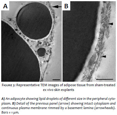
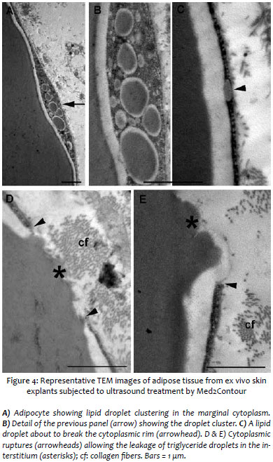
Ultrastructural analysis of adipose tissue from sham-treated ex vivo skin explants demonstrated normal adipocytes, showing a large, osmiophilic lipid vacuole with a peripheral electron-lucent rim contiguous to a thin cytoplasmic layer containing scanty organelles, pinocytosis micro vesicles and small lipid droplets (Figure 3). Cells were surrounded by a continuous basement membrane. Blood micro vessels, mainly capillaries, and interstitial connective tissue composed of a loose matrix containing thin collagen fibers, showed a normal appearance (data not shown). Normal features of adipocytes and stromal components were also observed in the adipose tissue biopsies taken from sham-treated areas of the three patients enrolled in the study (data not shown). Conversely, the subcutaneous adipose tissue of ultrasound-treated ex vivo skin explants displayed well-appreciable differences compared with the sham-treated specimens. In particular, many adipocytes showed peculiar abnormalities, consisting in lipid droplet clustering and focal ruptures of the peripheral cytoplasmic rim (Figure 4). Such ruptures were usually restricted to small areas of the cell surface, approx. 0.5-1.5 mm in diameter, but large enough to allow leakage of triglyceride droplets from the inner cytoplasmic vacuole to the extracellular space. Of note, no signs of adipocyte demise or cell remnants were observed. Of note, the cellular and intercellular stromal components showed a normal appearance, with no signs of damage (data not shown).
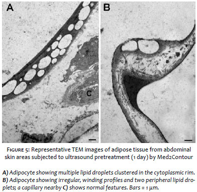
Ultrastructural examination of subcutaneous fat biopsies taken at surgery from ultrasound-pretreated abdominal skin areas showed different features from those of the ex vivo specimens. In all the patients examined, regardless of whether the biopsies were taken one or 12 days after the last ultrasound application, triglyceride leakage from adipocytes was not detected in the images. The interstitial stromal components showed a normal appearance (data not shown). However, adipocytes that had been exposed to ultrasounds consistently showed irregular, winding profiles and multiple lipid droplets clustered in the cytoplasmic rim (Figure 5). These features were never found in the sham-treated adipocytes and are consistent with marked reduction of cell volume, conceivably related to triglyceride discharge.
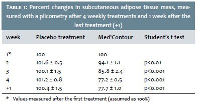
Assessment of subcutaneous adipose tissue mass by plicometry showed a time-related decrease of the measured values in the ultrasound-treated abdominal skin regions as compared with the corresponding sham-treated areas (Table 1). Of note, the decreasing trend in the Med2ContourTM-treated regions continued one week after the last treatment (longer times not assayed).
The present findings indicate that weakly focused high-frequency ultrasounds delivered by the Med2ContourTM device on human skin can yield a substantial reduction of subcutaneous fat and adipose cell size, confirming the previous clinical and histological observations of a marked lipo-reductive effect of this technique.7,14,15 This study provides additional morphological clues to better understand the mechanism of action of ultrasounds on adipocytes. In fact, exposure of full-thickness skin explants to two short ultrasound cycles (6 sec. each), with a similar energy output to that used for clinical purposes, resulted in a statistically significant shrinkage of subcutaneous adipocytes. By electron microscopy, the ultrasound treatment appeared to cause destabilization of adipocyte cytoplasm and plasma membrane enveloping the lipid vacuole, possibly by coalescence of lipid droplets. In turn, this phenomenon causes focal ruptures of the adipocyte cytoplasm, approx. 0.5-1.5 mm in diameter, which allow leakage of lipids from the inner vacuole to the extracellular space. Similar findings were observed in adipose tissue biopsies taken from patients who had been treated with Med2ContourTM. In particular, as compared with the sham-treated areas, the mean size of subcutaneous adipocytes was markedly reduced one day after the last treatment and remained well appreciable after 12 days. Ultrastructurally, images of triglyceride leakage from adipocytes were no longer observed in any of the studied patients, although the adipocytes still showed ultrastructural features consistent with triglyceride emptying.
Of note, in both the ex vivo and in vivo experiments, lipid discharge was not accompanied by any morphological signs of adipocyte damage or interstitial inflammation. Moreover, the effects of ultrasound treatment appear to be restricted to adipocytes, while blood vessels and interstitial stroma showed normal features, as observed in the sham-treated controls. This is in keeping with previous in vivo porcine and human studies with both Contour ITM and Med2ContourTM, in which ultrasound treatment was shown to cause selective adipose cell reduction without injury to skin, vessels, nerves, or connective tissue.7,14,15 The present findings suggest that the treatment with weakly focused high-frequency ultrasound, at appropriate settings and timing, does not create local adverse conditions which may favor tissue injury and subsequent inflammatory/fibrotic reaction. On the other hand, the integrity of the vascular components of the adipose tissue can favor the removal of interstitial fat droplets and putative pro-inflammatory mediators released from adipocytes, conceivably by lymphatic drainage.14,15
The observation that ultrasound treatment induces triglyceride leakage from adipocytes to the interstitial stroma poses the issue of their fate. It is conceivable that triglycerides can then be absorbed and metabolized by endogenous lipases to glycerol and free fatty acids, as well as incorporated in the total lipoprotein pool. Of note, serum lipids were unchanged7,11,14 or slightly elevated, but still within the normal range,13 in experimental animals and in patients subjected to lipo-reductive ultrasound treatments, accounting for substantial safety of this procedure from a metabolic viewpoint. At variance with a previous report,16 we did not observe any signs of disarrangement of adipose tissue collagen network or induction of adipocyte apoptosis, but this discrepancy is reasonably due to the far longer (10 min.) exposure of the skin samples to ultrasounds adopted in that study.16
In conclusion, this study further strengthens the current view that non-invasive trans-cutaneous high-frequency ultrasound, one of the most sought-after plastic and aesthetic surgical procedures, is a promising technology for localized reduction of fat. Generalization of the meaning of our study is hampered by the fact that we enrolled a limited number of patients: however, the consistency of the observed findings provides support to the notion that Med2ContourTM, owing to its unique design yielding a weakly focused ultrasound field within the subcutaneous fat pad, can be an effective and safe tool for lipo-reductive purposes.
Acknowledgments:
The authors gratefully acknowledge Dr. Moreno Naldoni, MSEE, CEO of General Project Ltd., for kindly providing the Med2ContourTM device used in this study.
1. Coleman KM, Coleman WP 3rd, Benchetrit A. Non-invasive, external ultrasonic lipolysis. Semin Cutan Med Surg. 2009;28(4):263-7.
2. Neira R, Arroyave J, Ramirez H, Ortiz CL, Solarte E, Sequeda F, et al. Fat liquefaction: effect of low-level laser energy on adipose tissue. Plast Reconstr Surg. 2002;110(3):912-22.
3. Jackson RF, Dedo DD, Roche GC, Turok DI, Maloney RJ. Low-level laser therapy as a non-invasive approach for body contouring: a randomized controlled study. Lasers Surg Med. 2009;41(10):799-809.
4. Manuskiatti W, Wachirakaphan C, Lektrakul N, Varothai S. Circumference reduction and cellulite treatment with a Tri-Polar radiofrequency device: a pilot study. J Eur Acad Dermatol Venereol. 2009;23(7):820-7.
5. de Felipe I, Del Cueto SR, Pérez E, Redondo P. Adverse reactions after nonablative radiofrequency: Follow-up of 290 patients. J Cosmet Dermatol. 2007;6(3):163-6.
6. Zocchi ML. Clinical aspects of ultrasonic liposculpture. Perspect Plast Surg. 1993;7:153-174.
7. Teitelbaum SA, Burns JL, Kubota J, Matsuda H, Otto MJ, Shirakabe Y, et al. Noninvasive body contouring by focused ultrasound: safety and efficacy of the Contour I device in a multicenter, controlled, clinical study. Plast Reconstr Surg. 2007;120(3):779-89.
8. Brown SA, Greenbaum L, Shtukmaster S, Zadok Y, Ben-Ezra S, Kushkuley L. Characterization of nonthermal focused ultrasound for noninvasive selective fat cell disruption (lysis): Technical and preclinical assessment. Plast Reconstr Surg. 2009;124(1):92-101.
9. Ascher B. Safety and efficacy of UltraShape Contour I treatments to improve the appearance of body contours: multiple treatments in shorter intervals. Aesthetic Surg J. 2010;30(2):217-24.
10. Fatemi A, Kane MA. High-intensity focused ultrasound effectively reduces waist circumference by ablating adipose tissue from the abdomen and flanks: a retrospective case series. Aesthetic Plast Surg. 2010;34(5):577-82.
11. Jewell ML, Baxter RA, Cox SE, Donofrio LM, Dover JS, Glogau RG, et al. Randomized sham-controlled trial to evaluate the safety and effectivenessof a high-intensity focused ultrasound device for noninvasive body contouring. Plast Reconstr Surg. 2011;128(1):253-62.
12. Kennedy JE, ter Haar GR, Cranston D. High intensity focused ultrasound: surgery of the future? Br J Radiol. 2003;76(909):590-9.
13. Moreno-Moraga J, Valero-Altés T, Riquelme AM, Isarria-Marcosy MI, de la Torre JR. Body contouring by non-invasive transdermal focused ultrasound. Lasers Surg Med. 2007;39(4):315-23.
14. Garcia O Jr, Schafer M. The effects of nonfocused external ultrasound on tissue temperature and adipocyte morphology. Aesthetic Surg J. 2013;33(1):117-127.
15. Bani D, Quattrini Li A, Freschi G, Russo GL. Histological and ultrastructural effects of ultrasound-induced cavitation on human skin adipose tissue. Plast Reconstr Surg Glob Open. 2013;1(6):e41.
16. Palumbo P, Cinque B, Miconi G, La Torre C, Zoccali G, Vrentzos N, et al. Biological effects of low frequency high intensity ultrasound application on ex vivo human adipose tissue. Int J Immunopathol Pharmacol. 2011;24(2):411-22.
This study was performed at the Department of Experimental & Clinical Medicine, Research Unit of Histology & Embryology Dept. Surgery & Translational Medicine, Plastic Surgery Unit, University of Florence - Florence, Italy
 All content the journal, except where identified, under the Creative Commons Attribution 4.0 International licence - ISSN-e 1984-8773


 Read in Portuguese
 Portuguese PDF
 Print
Print
 Send this article by email
 How to cite this article
 Submit a comment
 Mendeley
 Pocket