


Daniela Tiemi Sano1; Jeane Jeong Hoon Yang2; Cristiano Luiz Horta de Lima Júnior3; José Roberto Pereira Pegas4
Keywords: POROMA; SWEAT GLAND NEOPLASMS; MELANOMA.
Eccrine poroma is a benign tumor of the eccrine or apocrine sweat gland that is composed of cells similar to those of the acrosyringium. It is characterized by a monochrome skin lesion, usually located on the palms and soles and possibly affecting other body areas.1-3 There are clinical variants, which include poromatosis, linear eccrine poroma and pigmented eccrine poroma. 4 The pigmented variant is rare and can have a possible clinical resemblance to nodular malignant melanoma, due to the pigmentation of the lesion. Due to the fact that the clinical features of the poroma are not specific, it is important to perform a differential diagnosis against pyogenic granuloma, pigmented basal cell carcinoma, hemangioma, and melanoma, with the definitive diagnosis established by histology1, 5 The present study reports the clinical case of a pigmented eccrine poroma in an unusual location for the tumor, simulating a malignant melanoma.
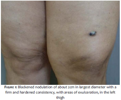
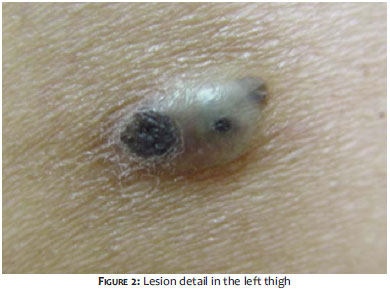
A 53-year-old female patient with brown pigmented skin, referred for the appearance of a nodule on the anterior region of the left thigh, beginning eight years before. Initially, it emerged as a skin color nodule that evolved into a reddish color, becoming darker later on. The patient described a progressive enlargement of the lesion, associated with mild local pain. During this period, the patient sought the care of a dermatologist due to the presence of the blackened nodulationon the left thigh. Diagnoses of a skin tumor (pigmented basal cell carcinoma or malignant melanoma) and of a nodule caused by a thrombosed vein have been suggested. The patient had a history of hypertension, diabetes, dyslipidemia and treated breast cancer. Dermatological examination showed a blackened nodulation with a firm and hardened consistency, with areas of exulceration, measuring approximately 2cm in its largest diameter, located on the anterior region of the left thigh (Figures 1 and 2).
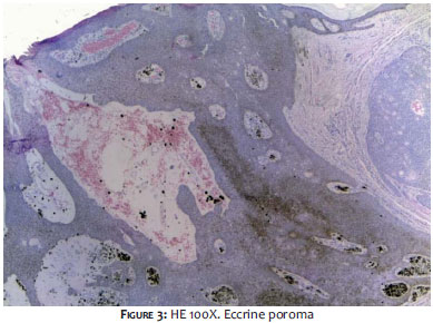
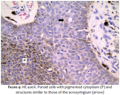
Under dermoscopic examination, the lesion showed features highly suggestive of malignant melanoma. A decision was made for diagnosis with surgical treatment. The patient underwent exeresis of the lesion, with the specimen sent for anatomical pathological examination. Histology showed poroid cells with pigmented cytoplasm (P) and structures similar to those of acrosyringium (arrow) (Figures 3 and 4). The histological examination was consistent with the diagnosis of pigmented eccrine poroma, with the clinical hypothesis of malignant melanoma being discarded. The patient is being followed-upwith at an ambulatory clinic, with good clinical outcome. There has been no recurrence of the lesion.
Eccrine poroma was first described by Pinkus et al. in 1956, whoderived the tumor's denomination from the sudoriparous duct. 2, 6 Ackerman histologically defines a group of four benign epithelial neoplasms composed of cells similar to those of the intradermal eccrine duct, the acrosyringium: hidroacanthoma simplex, eccrineporoma, dermal duct tumor and poroid hidradenoma-all being histopathologically classified based on their location relative to the epidermis. 1, 4, 7
The term poroma refers to a group of rare cutaneous adnexal tumors, composed by cells (cuticular and poroid) similar to those of the acrosyringium. 1, 2, 7 Eccrine poroma is a benign tumor of the eccrine or apocrine sweat gland. 1 It commonly occurs as nodules, or a sessile or pedunculate solitary papule the color of the skin. 1, 3, 4 There are clinical variants that include poromatosis, linear eccrine poroma and pigmented eccrine poroma. 4, 6, 7 It can sometimes be pigmented, with a bright red or violet color, being pruriginous or painful.
It affects individuals of different races, especially Caucasians between the ages of 40 and 60 years. 2 It affects both genders equally, with a slight predominance in men. The progression of the lesion may vary from weeks to years. 6 It rarely precedes the development of porocarcinoma. Usually located on the soles or palms, it can affect other body areas. 4, 5,8 It can also ulcerate on pressure points and areas of trauma, in general showing slow and asymptomatic growth. 2, 6
The pigmented variety of eccrine poroma occurs by persistence of melanocytes in the acrosyringium, with absence of a known cause. 8 Typically, in the acrosyringium, during the embryonic stage there is a presence of melanocytes, which recede at the end of thatphase. 6With the improvement in the diagnostic accuracy of dermatoscopy examinations for the various types of skin tumors, some lesions can be identified prior to histological examination. 7 Dermoscopy is a non-invasive examination, useful in the diagnosis of pigmented skin lesions. It helps in the early diagnosis of malignant melanoma lesions, allows for the differentiation of pigmented benign and malignant lesions from malignant melanoma, and is useful in the diagnosis of pigmented basal cell carcinoma. Although there are several dermoscopic studies of pigmented lesions, there are not many studies regarding eccrine poroma in its pigmented variety. 9
Pigmented eccrine poroma can clinically simulate various skin lesions, including pigmented basal cell carcinoma, seborrheic keratosis and malignant melanoma, due to its clinical, dermoscopic, and histologic variety. 7, 10Various types of dermoscopic structures associated with melanocytic and nonmelanocytic lesions are observed in pigmented poromas due to varying amounts of melanin in those lesions-which makes it clinically and dermoscopically indistinguishable from melanoma and, in some cases, from non-melanoma skin tumors. 7
Histologically, eccrine poroma reveals aggregates of uniform basaloid cells, which can irradiate from the basal layer of the epidermis to the dermis. 3, 5
Due to its being a benign lesion, eccrine poroma has a good prognosis. The treatment of choice is complete surgical excision. The recurrence of the lesion is uncommon. 2, 4, 6
Due to its pigmentation, the pigmented eccrine poroma may in some occasions clinically simulate a malignant melanoma. 6 It is important to highlight that the clinical features of eccrine poroma are not specific and may clinically resemble other skin tumors. Pyogenic granuloma, hemangioma, basal cell carcinoma, nodular melanoma and amelanotic melanoma can be cited among differential diagnoses, with histological examination being required to confirm the diagnosis. 1, 5-7
1. Allende I, Gardeazabal J, Acebo E, Díaz-Pérez JL. Poroma ecrino pigmentado. Actas Dermosifiliogr. 2008;99(6):493-501.
2. Pareyón LAR, Rojas PB, Ramos-Garibay A. Poroma ecrino pigmentado. Presentación poco habitual. Rev Cent Dermatol Pascua. 2001;10(2):70-2.
3. Taylor RS, Perone JB, Kaddu S, Kerl H. Tumores Anexiais e Hamartomas da Pele. In: Wolff K, Goldsmith LA, Katz SI, Gilchrest BA, Paller AS, Leffell DJ, editors. 7a ed. Fitzpatrick's Tratado de Dermatologia. p:1075-6.
4. Sano DT, Yang JJH, Fregonesi NCFP, Lima Jr CLH, Freua SRG, Pegas JRP. Poroma écrino simulando melanoma maligno: relato de caso. 67º Congresso da Sociedade Brasileira de Dermatologia. 2012. Pôster eletrônico mini-comunicação. PE-073.
5. Altamura D, Piccolo D, Lozzi GP, Peris K. Eccrine poroma in an unusual site: A clinical and dermoscopic simulator of amelanotic melanoma. J Am Acad Dermatol. 2005;53(3):539-41.
6. Rivera OL, Mora S, Gutiérrez, Novales J. Poroma ecrino simulando um melanoma maligno. Reporte de un caso y revisión de la literatura. Rev Cent Dermatol Pascua. 1999;8(1):35-8.
7. Minagawa A, Koga H. Dermoscopy of Pigmented Poromas. Dermatology. 2010;221(1):78-83.
8. Ohata U, Hara H, Suzuki H. Pigmented Eccrine Poroma Occurring on the Scalp. Derivation of Melanocytes in the Tumor. Am J Dermatopathol. 2006;28(2):138-41.
9. Kuo HW, Ohara K. Pigmented Eccrine Poroma: A Report of Two Cases and Study With Dermatoscopy. Dermatol Surg. 2003;29(10):1076-9.
10. Kakinuma H, Kobayashi M. Eccrine poroma: another cause of a pigmented scalp nodule. Br J Dermatol. 2002;146(3):523.
This study was performed at Complexo Hospitalar Padre Bento de Guarulhos (CHPBG) - Guarulhos (SP), Brazil.
 All content the journal, except where identified, under the Creative Commons Attribution 4.0 International licence - ISSN-e 1984-8773


 Read in Portuguese
 Portuguese PDF
 Print
Print
 Send this article by email
 How to cite this article
 Submit a comment
 Mendeley
 Pocket