


Maria Otília Teixeira Abalí1; Bruna Souza Félix Bravo2; Dina Zylbersztejn3
Keywords: LASERS; INTENSE PULSED LIGHT THERAPY; CICATRIX; BURNS.
Scars resulting from burns have the potential to cause significant disruption tobearers due to their often disfiguring clinical appearance, to the entailed functional impairment, and to the social embarrassment they produce. The approach to treatingscars includes several therapeutic options, such as pressure therapy, intralesional corticosteroid therapy, cryotherapy, silicones, topical treatments, and surgical corrections. These techniques-combined or not-neverthelesshave limited results, especially regarding the clinical appearance of scars.
Laser therapy has emerged as a therapeutic option for approaching scars. Published studies fromthe 1970s have highlighted that analysis of characteristics of the scar area, such as texture, thickness and color, constituted decisive parameters in pre-laser treatment evaluation. The improvement of this technique occurred pari passuto thedevelopmentof the treatment of atrophic scars usingablative (CO2 and Erbium:YAG) and non-ablative (1,320nm Nd:YAG)lasers and, more recently, fractional lasers. In the literature, the use of laser therapy for hypertrophic scars is conflicting and despite the gradual replacement of the Argon, 1,064nm Nd:YAG and 10,640nm CO2 lasersfor the 585nm and 595nm Pulsed Dye Laser (PDL) with promising results, further studies with a greater degree of evidence are still necessary. 1-10
In the present study, intense pulsed light (IPL) is used as a therapeutic option in the approachto scars caused by burns. Although there are publications suggesting the use of IPL as a therapeutic option in the approach of hypertrophic and keloid scars, its use for the treatment of scars after burns still remains unexplored and discussions about its indication for this purpose remain scarce. 1, 2, 4
To evaluate the response of IPL on scars after burns, based on the clinical parameters described in the international Vancouver scale used to assess scars. 11, 12
A prospective study was conducted from March 2012 to March 2013, at the Cosmetic Dermatology ambulatory of the Instituto de Dermatologia Prof. Rubem David Azulay, Santa Casa de Misericordia do Rio de Janeiro, with the approval of the Medical Ethics Committee of the institution. Six patients of both genders (4 women and 1 man), with ages between 21 and 48 years (mean = 33 years), with varied distribution of photo-types according to the Fitzpatrick classification (Table 1), who showed scarring from thermal burns which had occurred more than six months before and who had undergone prior conventional treatment in centers for treatment of burns, and who were not under ongoing topical treatment at the time of the study, were included in the present research.
The exclusion criteria in the selection of patients included: contraindications to the use of IPL, pregnancy or lactation, presence of symptoms of pain, burning and/or itching in the scar area, use of oral retinoids in the previous six months, and use of medication that induced photosensitivity in the previous three months.
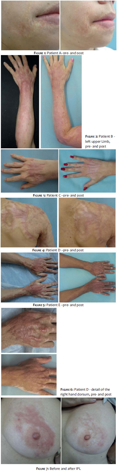
After the evaluation of the above criteria, all patients were informed of the study's objectives and were enrolled in the project according to their interest in participating. All participants read and signed a free and informed term of consent. Photographic records were always carried out in the same room and with the same photographic background, preferably by the same researcher physician, with a Nikon Cool Pix P100 (26x Zoom) camera, before and after the treatment. (Figures 1 to 7)
Patients underwent five IPL sessions at monthly intervals over the entire area of the scar using a Lip Sq tip (Square-wave Pulse system), which features an integrated cooling system through a sapphire tip, with 540nm cutoff filter from the Etherea® platform (Industra Technologies, São Carlos, SP, Brazil).
Before each session, the target area was cleansed with a lotion with no alcohol and without the prior use of a topical anesthetic. The parameters used in each session were defined according to the patient's tolerance regarding discomfort, with the data being recalculated according to the clinical results obtained in previous sessions. The fluence used was 12-18 J/cm2 (mean =14.6 J/cm2) and the pulse duration was 10 or 20 ms. (Table 1). The results were analyzed by three groups of evaluators: three researcher physicians, the patients included in the study, and three observer physicians. The first two groups carried out evaluations before and three months after the end of the study, while the third group carried out its assessment based on photographic material taken before and after treatment.
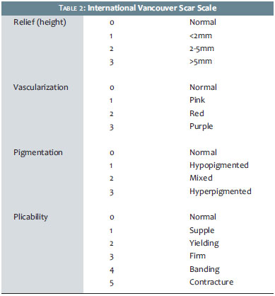
The clinical course of the scars was assessed by a group of evaluators through the international Vancouver scale for scars, which includes flexibility, vascularization (degree of erythema), relief and color (melanin pigmentation). (Table 2) In order to facilitate the patients' self-assessment, five questions were formulated with possible answers based on numerical scales derived from the clinical criteria or the Vancouver scale. Also, an overal lrating, ranging from 0 (excellent) to 10 (very bad), was used by the three evaluation groups to grade the overall assessment of the scar.
The descriptive analysis presented the observed data (expressed as median, minimum, and maximum) in the form of tables.
The before-and-after variation-assessed through a questionnaire, the Vancouver scale, and a numerical scale-was analyzed through of the Wilcoxon signed-rank test. The criterion determining the significance was set at 5%, i.e. when the p-value was less than or equal to 0.05, there would be no statistical significance.
The statistical analysis was performed with assistance of the SAS 6.11 software (SAS Institute, Inc., Cary, North Carolina, USA).
All patients selected completed the study, having answered the questionnaire before and after the treatment with an aim at verifying whether there was significant variation in the criteria assessed by the questionnaire (based on the Vancouver scale for scars). Similarly, the study aimed at validating the presence of a significant variation in the data obtained on that scale (according to the researcher physicians) and on the numerical scale (according to the patients, researcher physicians, and observer physicians).
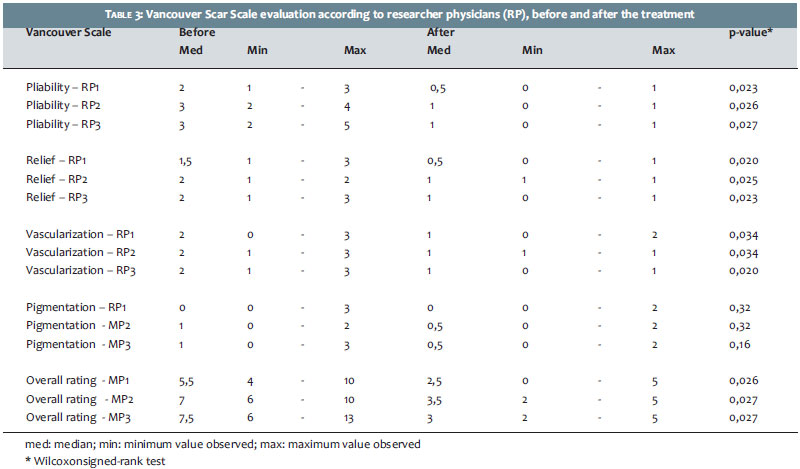
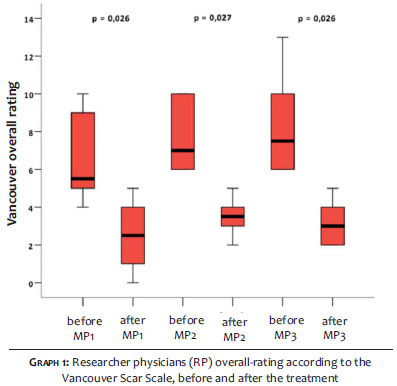
The variables assessed by the Vancouver scale were originally measured in an ordinal scale, i.e.a gradation with qualitative interpretation. However, the reduced sample size (n = 6), prevented the processing of appropriate statistical methods. Therefore, the present study proposedan exploratory analysis of the data froma numerical point of view, aiming mainly at the impact of the treatment after five monthly IPL sessions. Table 3 provides the median (minimum-maximum) rating of the Vancouver scale according to three researcher physicians (RP1, RP2, and RP3) at timesbefore and after the treatment and the corresponding descriptive level (p-value) of the statistical test.
Statistical analysis was performed through the Wilcoxon signed-rank test.
The patient self-evaluation before and after the treatment showed a significant decrease (at the 5% level) in the evaluation of all aspects of the questionnaire. That statistical validation translates the clinical improvement seen in all parameters observed by the patients after the treatment, such as dyschromias, hypertrophy, and flexibility of the scarred area, using criteria based on the Vancouver scale.
According to the researcher physicians, the ratings of the Vancouver scale for scarsshowed significant decrease (at the 5% level) before and after the treatment, except for the variable pigmentation, which had initially showed little expression, as shown in table 3 and graph 1.
The assessment done according to the numerical scale and corresponding to the overall rating attributed to the three evaluation groups before and after the treatment, presented a significant reduction (at the level of 5%) for all evaluators.
Regarding adverse effects, all patients had erythema and slight, tolerable discomfort during the sessions, with no need for any specific treatment. Burning sensation for a few hours after the session was reported by two patients, however without leading to changes in the schedule of the treatment. One patient had blisters after the 4th session, resolving without sequelae.
The introduction of laser therapy has emerged as a new tool in the therapeutic approach to scars. Based on the principle of selective photothermolysis, which acts on specific chromophores, it enabled a more specific approach to the assessment of parameters prevailing in each lesion, such asvariation in color, plicability and relief. 1-4, 6, 8, 13-6
The broad spectrum of the IPL's light beam (from 515nm to 1,200nm) allows exertion on the different chromophores present in scars-such as the hemoglobin present in the neovascularization of the intense cicatricial tissue and the melanin resulting from the stimulus of melanogenesis-enabling the-treatment of the erythema and the dyschromia, respectively. Another effect of IPL described in studies on its use in photo-rejuvenation is the possible induction of collagen remodeling through the photo-stimulation of the fibroblasts and metallo-proteinases of the dermal matrix. 17-24
In the literature, there are few studies aimed at evaluating the use of IPL on scars, more specifically after burns. Its use for hypertrophic or keloid scars, isolatedly or comparatively to laser therapy, was described by Bellew et al., who approached hypertrophic scars with PDL and IPL, finding improvements in the appearance of scars with both techniques, without demonstrating superiority of one over the other. In 2008, Erol et al. treated 109 patients with IPL-the scars had different etiologies, with 19 patients suffering from thermal injury. The results presented demonstrated improvement of those scars regarding dyschromia, relief, pliability and texture of the scar tissue, through clinical and photographic parameters. More recently, Isaac et al., aiming at determining safety standards and evaluating the degree of satisfaction and local complications after each session, demonstrated the use of IPL in hyperchromic scars after burns that had occurred more than two years before in 19 patients between 9 and 62 years of age, with IPL phototypes II-V. After 9 monthly sessions it was statistically demonstrated that there was an improvement in the level of patients' and observer physicians' satisfaction, in addition to the existence of a direct correlation between the degree of improvement and the number of sessions undergone.
Although recent studies have demonstrated benefits in the use of laser therapy in the early treatment of scars caused by elective procedures, the ideal time to start the therapeutic procedures remains unclear. Bellew et al. demonstrated clinical improvement of post-mammoplasty and abdominoplasty early hypertrophic scars using PDL and IPL in the proliferative phase of formation of the scar tissue (6-8 weeks after the injury was caused). 25, 26
The use of IPL during the study proved to provide clinical improvement in all parameters evaluated, such as dyschromias, pliability, and reduction of hypertrophic areas. The improvement of hypertrophic scarring in all cases treated is noteworthy. Regarding dyschromias, the response was more significant in erythemas as compared to the brown color of scars. Despite the fact that hypochromic areas were not included in the rating scales of scars, no improvement was observed in this parameter. It was possible to gradually increase the intensity of treatment parameters, such as fluence and pulse duration-and to beinitially more conservative when compared to those used for photorejuvenation-without adding significant side effects.
In the present study, the authors chose to focus on the approach to scarring caused by burns that had happened over six months before. Howeverit is also possible to compare the use of IPL in earlier stages of scar proliferation in further studies. Its use in the initial phase would be an attempt to reduce the formation of hypertrophic scars, which translates clinically into relief alterations (dystrophic) caused by the imbalance in the synthesis and degradation of collagen present in the wound healing process.
In the authors' opinion, IPL is able to combine important characteristics, which suggests that this technique can be made available for patients with scars caused by burns. IPL technology is a technology familiar todermatologists, it is cost-effective when compared to other laser sources, and has been demonstrated to provide satisfactory clinical improvement-evaluated both objectively and subjectively-for the treated scars that were caused by burns. In this context, the present study represents a pilot study carried out in the authors' dermatologic service aimed at demonstrating both the benefits of IPL in treating this type of scar and stimulating further studies with more accurate assessment methods in order to create a protocol for the approach of patients affected by burns or bearing scars.
1. Bellew SG, Weiss MA, Weis RA. Comparison of intense pulsed light to 595-nm long-pulsed pulsed dye laser for treatment of hypertrophic surgical scars: a pilot study. J Drugs Dermatol. 2005;4(4),448-52.
2. Bouzari N, Davis SC,Nouri K. Laser Treatment ok keloids and hypertrophic scars. Int J Dermatol. 2007;46(1):80-8.
3. Isaac C, Salles AG, Soares MFD, Camargo CP, Ferreira MC. Efeitos da Luz Intensa Pulsada em Sequelas Cicatriciais Hipercrômicas Pós Queimadura. Rev Bras Cir Plást. 2006;21(3):175-9.
4. Erol O, Gurlek A, Agaoglu G . Treatment of Hypertrophic Scars and Keloids Using Intense Pulsed Light. Aesth Plast .Surg 2008;32(6):902-9.
5. Campos V, Mattos R, Filippo A, Torezan LA. Laser no Rejuvenescimento Facial. Surg Cosmet Dermatol. 2009;1(1):29-36.
6. Nymann P, Hedelund L, Haedersdal M. Intense pulsed light vs. long-pulsed dye laser treatment of telangiectasia after radiotherapy for breast cancer: a randomized split-lesion trial of two different treatments. Br J Dermatol. 2009;160(6):1237-41.
7. Babilas P, Schremi S, Szeimies RM, Landhaler M. Intense pulsed light (IPL): a review. Lasers Surg Med. 2010;42(2):93-104.
8. Elsaie ML, Choudhary S. Lasers fors Scars: A Rewiew and Evidence-Based Appraisal. J Drugs Dermatol. 2010;8(11):1355-62.
9. Bravo BSF ; Vale EC; Serra MC. Queimaduras. In: Azulay DR, Azulay RD, Azulay-Abulafia L. Dermatologia. 6 ed. Rio de Janeiro: Guanabara Koogan; 2013.p.43-50.
10. Dadalti P; Pinto JM. Fisiologia da Reparação Tecidual e algumas implicações Terapêuticas. In: Dermatologia. Azulay DR, Azulay RD, Azulay-Abulafia L. 6ºed. Rio de Janeiro: Guanabara Koogan; 2013.p.34-41.
11. Baryza MJ, Baryza GA. The Vancouver Scar Scale: An Administration Tool and Its Interrater Reliability. J Burn Care Rehabil. 1995;16(5):535-8.
12. Nedelec B, Shankowsky HA, Tredget EE. Rating the Resolving Hypertrophic Scar: Comparison of the Vancouver Scar Scale and Scar Volume. J Burn Care Rehabil. 2000;21(3):205-12.
13. Chang H, Wong DS, Ho WS, Lam LK. The Use of Pulsed Dye Laser for the Prevention and Treatment of Hypertrophic Scars in Chineses Persons. Dermatol Surg. 2004;30(7):987-94.
14. Bowen R. A Noovel Approach to Ablative Fractional Treatment of Mature Thermal Burn Scars. J Drugs Dermatol. 2010;9(4):389-92.
15. Cho SB, Lee SJ, Chung WS, Kang JM, Kim YK. Treatment of Burn Scar Using a Carbon Dioxide Fractional Laser. J Drugs Dermatol. 2010;9(2):173-75.
16. Waibel J, Beer Kenneth. Fractional Laser Ressurfacing for Thermal Burns. J Drugs Dermatol. 2008;7(1):59-61.
17. W WR, Shyu WL, Tsai JW, Hsu KH, Pang JH. Intense pulsed light effects on the expression of extracellular matrix proteins and transforming growth factor beta-1 in skin dermal fibroblasts cultured within contracted collagen lattices. Dermatol Surg. 2009;35(5):816-25.
18. Ross EV, Sminov M, Pankratov M, Altshuler G. Intense pulsed light and laser treatment of facial telangiectasias and dyspigmentation: some theoretical and practical comparisons. Dermatol Surg. 2005;31(9):1188-98.
19. Adamic M, Troilius A, Adath M, Drosper M, Dahmane R. Vascular lasers and IPLS: guidelines for care from the European Society for Laser Dermatology. J Cosmet Laser Ther . 2007;9(2):113-24.
20. Cao Y, Huo R, Feng Y, Li Q, Wang F. Effects of intense pulsed light on the biological properties and ultrastructure of skin dermal fibroblasts: potential roles in photoaging. Photomed Laser Surg. 2011;29(5):327-32.
21. Mandelbaun SH, Santis EP, Mandelbaun MH. Cicatrização 1: conceitos atuais e recursos auxiliares - Parte 1. An Bras Dermatol. 2003;78(4):93-410.
22. Goldberg DJ, Sarradet D, Hussain M, Krishtul A, Phelps R. Clinical, Histologic, and Ultrastructural Changes after Nonablative Treatment with a 595-nm Flashlamp-Pumped Pulsed Dye Laser: Comparison of Varying Settings. Dermatol Surg. 2004;30(7):979-82.
23. Alster TS, Williams CM. Treatment of keloid sternotomy scars with 585nm flashlamp-pumped pulsed-dye laser. Lancet. 1995;345(8959):1198-2000.
24. Mendonça RJ, Coutinho-Netto J. Aspectos celulares da cicatrização. An Bras Dermatol. 2009;84(3):257-6.
25. Liew SH, Murison M, Dickson WA. Prophylactic treatment of deep dermal burn scar to prevent hypertrophic scarring using the pulsed dye laser: a preliminary study. Ann Plast Surg. 2002;49(5):472-5.
26. Kouri K, Jimenez GP, Harrison-Balestra C, Elgart GW. 585-nm pulsed dye laser in the treatment of surgical scars starting on the suture removal day. Dermatol Surg. 2003;29(1):65-73.
This study was performed at the Dermatologic Surgery and Cosmiatry Ambulatory, Instituto Professor Rubem David Azulay - Santa Casa da Misericordia do Rio de Janeiro (RJ) - Brazil.
 All content the journal, except where identified, under the Creative Commons Attribution 4.0 International licence - ISSN-e 1984-8773


 Read in Portuguese
 Portuguese PDF
 Print
Print
 Send this article by email
 How to cite this article
 Submit a comment
 Mendeley
 Pocket