


Carla Gregório Barbosa de Oliveira1; Simão Cohen2; Valter Alves3
Tattooing has existed in humankind's culture since the onset of civilization. Tattoo removal attempts are also very ancient. The following current methods are reported for the removal of tattoos: dermabrasion, surgical excision, and laser procedures. The most commonly used lasers for tattoo removal are: QS-Nd:YAG (1,064 and 532nm), QS-Ruby (694nm) and QS-Alexandrite (755nm). The present review is aimed at studying the action mechanism of lasers for tattoo removal and the correct indication for each type of pigment, in addition to describing complications and the best manner of preventing them.
Keywords: LASERS; LASER THERAPY; TATTOOING.
Tattoos have been present in the culture of mankind since the beginning of civilization. They are permanent signs on the body that have different meanings: amulets, symbols of status, declarations of love, statements of religious beliefs, adornments, and even sometimes a form of punishment. The first described reports of tattoos date from 2,000 BC, and were found in Egyptian mummies. In 1991 a mummy from the ice age (around 5,200 years old) was found, which had several tattoos on its body. One development during the history of tattoo artistry was the introduction of different colored pigments, allowing more complex tattoos. Attempts to remove tattoos are also very old.
Types of tattoos
Tattoos can be divided into five categories: professional, amateur, cosmetic, medical, and traumatic. The professional type is performed using professional devices that contain vibratory needles, and pigments of various colors. The granules of pigment are deposited superficially in the dermis. Amateur tattoos are performed with needles or improvised devices and pen ink, charcoal, and soot are usually used as pigments. The use of cosmetic tattoos has increased in recent times, especially in eyebrows, eyelids (eyeliner), lip (contour), reconstruction of breast areola tissue and of other scars. Mostly brown, black, pink, and red pigments are used in those cases.
Traumatic tattoos occur when pigment is deposited in the skin through abrasion or resulting from the force of an explosion. The materials (asphalt, gunpowder, etc.) remain housed in the dermis after the trauma, lending a black or bluish hue to the skin, depending on the depth at which they settle.
Medical tattoos are used, for example, in radiotherapy protocols.2
Tattoo removal techniques
The oldest tattoo removal techniques date back to 543 BC, having been developed by the Greeks, who performed abrasion followed by the application of salts and chemicals.3 Dermabrasion has also been widely used. The principle of this technique is based on the local destruction of the skin and the consequent removal of the tattoo's pigment. The use of trichloroacetic acid in high concentrations has also been described. These two techniques do not always lead to complete removal of the tattoo and have a high risk of skin depigmentation and unaesthetic scars.4
Surgical removal of tattoos can also be performed. Nevertheless, linear scars may result from the procedure, and often tattoos are too large or in difficult to access sites. 5 Surgical removals can be indicated for patients who have allergic reactions to the pigments of tattoos. In such cases, removal attempts using laser can cause hypersensitivity reactions and even anaphylactic shock. Another therapeutic option described in this situation is the CO2 laser.
The first report of laser based tattoo removal using QS Nd:YAG laser was published in 1965 by Goldman et al.5,6 However, the lack of a thorough understanding of the physics of this laser type, combined with unpredictable clinical results,led it to fall into disuse at that time.
At the end of the 1970's and in the beginning of the 1980's, the most widely used lasers for tattoo removal were the carbon dioxide (CO2) and argon types. 6 Given that these lasers have water as the chromophore and that they are not selective, the problem of inconsistent clinical outcomes, with the possibility of the formation of unaesthetic scars and hypopigmentation arose again.7,8 The argon laser emits blue or green light and has a wavelength of 488 or 514nm.
Early in the 1980's there was great progress following the publication of the theory of selective photothermolysis by Anderson and Parrish.9 In this manner, selective Q-switched lasers (QS) would only destroy specific targets, with minimal damage to the underlying tissue. The theory previously proposed by Goldman was put into practice, inaugurating the use of QS Ruby laser for removing tattoos.10
Devices with pulses in the magnitude of milliseconds, such as those employing intense pulsed light, should not be used for tattoo removal, for they heat the granules of the pigment, allowing that heat to spread to adjacent tissue, causing damage. Attempts at removing tattoos with these devices usually results in scarring and does not completely remove the pigment. For best results Q-switched lasers must be used.
Q-Switched Laser
The way that Q-switched lasers operate in tattoo removal is not completely understood. In a study of tattoos treated with the use of QS lasers (with evaluation through electron microscopy) the destruction of pigment contained in the cells, with the fragmentation of target-pigments, can be observed. That pigment is then phagocytised, and an inflammatory response is responsible for transporting those cells into the lymphatic tissue. QS Ruby laser was the first QS laser to become commercially available, followed next by the QS Nd:YAG and QS Alexandrite lasers. These three lasers are still currently used-and it is important to note that each has a different wavelength. In order to select the correct laser to be used, the following criteria must be considered: the patient's skin phototype, laser pulse duration, spot size, and fluence.2
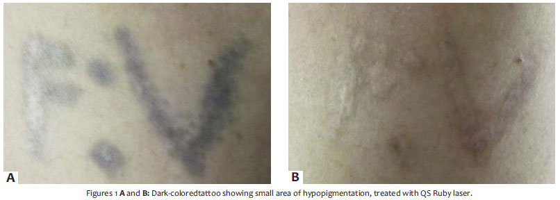
QS Ruby lasers have a wavelength of 694nm, emit red light, and are better absorbed by the black and dark blue colors. Very dark and amateur tattoos usually respond considerably well to this laser type. Medical tattoos can also have a good response. 2 After treatment with this laser type, transient hypopigmentation that resolves spontaneously in variable periods can occur (Figure 1).
Zelickson et al. carried out a study of 47 black or blue tattoos treated simultaneously with QS Ruby, QS Nd:YAG and QS Alexandrite lasers, with QS Ruby showing superior results. Kilmer and Anderson also demonstrated that QS Ruby laser is effective in treating green ink, although other studies have shown that QS Alexandrite treats this color more efficiently.11,12
QS Nd:YAG laser has a wavelength of 1,064 nm, emits green light and, through the KTP crystal (potassium titanyl phosphate), also doubles the 1,064 frequency, emitting a 532nm wavelength. This versatility allows for the treatment of dark pigments (such as black and dark blue) using 1,064nm; red, yellow, and orange pigments can also be treated with 532nm.
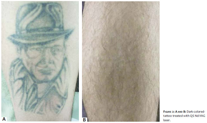
The longer wavelength lends greater penetrating power to this type of laser, thus better protecting the melanocytes of the epidermis, and as a result it is a laser type suitable for higher skin phototypes. Some studies comparing QS Ruby with QS Nd:YAG have demonstrated that the latter has less tendency to cause blisters and less chance of residual hypopigmentation 13 (Figure 2).
Kilmer et al. conducted a study of 39 tattoos treated with QS Nd:YAG laser, with fluences of 6 to 12j/cm2. A response of 75%was obtained for the black pigment in 77% of treated tattoos, in addition to a 90% clearance in 28% of the patients after four sessions, without secondary hypopigmentation.14
As already mentioned, QS Nd:YAG's wavelength can be doubled to 532nm with the use of KTP. This wavelength is well absorbed by red, orange, and yellow pigments. This finding was published by Anthony and Harland, who conducted a study in which seven patients with an allergy to red tattoo pigment were treated. 532nm QS Nd:YAG was used in conjunction with topical corticosteroids with good response.15 It is worth noting that this wavelength is absorbed by epidermal melanocytes, therefore, a chance of hypochromia exists with this laser type.
QS Alexandrite laser was launched in 1993 by Anderson et al. It has a 755nm wavelength. Fitzpatrick and Goldman published a series of 25 patients with professional and amateur tattoos with a 95% response when used for black and blue tattoos, with an average of 8.9 sessions.16 This laser was proven superior to QS Ruby and QS Nd:YAG for the removal of green pigment. Nevertheless, as it is well absorbed by epidermal melanocytes, it also brings with it the risk of residual hypochromia.12
Generally, darker and amateur tattoos respond well to all three types of lasers mentioned above, for, by definition, the black color absorbs the wavelengths of all visible light. Red and green pigments are well absorbed by 532nm QS Nd:YAG and 755nm QS Alexandrite, respectively. However, modern tattoos are often composed of a mixture of colors that can be complex and highly variable, with very similar colors even having completely different compositions, and thus very different absorption spectra. The variation in chemical composition and absorption spectrum can result in tattoos that are resistant and even unresponsive to laser treatment.
Colors such as yellow and orange are known to be very resistant, and colors such as red and green have a highly variable response. There is no theory that explains this incomplete response. It is believed that the wavelength used is not adequate for those colors.14
The paradoxical darkening after tattoo removal laser sessions is also described. Peach et al. completed a study involving 184 non-black tattoos and observed a change of color in 33 of them. That alteration ranged from light gray to a complete darkening of the tattoo. The tattoos treated contained white, yellow, and shades of red pigments, becoming gray or completely dark after the sessions.17 The exact mechanism that explains this change in color is not known. Cosmetic tattoos in shades of red and brown usually contain iron oxide in their composition and the oxidation of this component following QS Ruby laser sessions has already been demonstrated in vitro.18
The titanium dioxide found in white and shades of red tattoos is also responsible for the poor response to lasers. Titanium dioxide corresponds to 95% of the pigment in white tattoos, which in turn are used in conjunction with other tattoos to highlight color and brightness. 19 Some cases of resistance to green and blue are also attributed to the presence of titanium dioxide.19
In certain cases, treatment with ablative lasers can be indicated to remove pigments that have a risk of darkening or those resistant to treatment. 20
Complications after laser treatment for tattoos, such as dermatitis, granulomatous reactions, lichenoid reactions, and pseudolymphomatous reactions, including lymphadenomegalias, have been reported.21-23 Moreover, there is a concern about the pigments' degradation following laser. Vasold et al. recently showed the formation of products containing the "Azo" radical, which is known to be carcinogenic and cytotoxic.24
Considerations regarding the treatment
The patient must be informed about the number of sessions required to remove the tattoo (six to ten, or possibly more sessions) and about the possibility of their incomplete removal. The number of sessions depends on the tattoo's color, and the age and depth of the pigment.2
It is important to instruct patients about protection against the sun-since melanin absorbs the laser and therefore there is a greater chance of damage to adjacent skin, with blistering, hypopigmentation, and scarring. If the patient is tanned or has a higher phototype, 1,064nm QS Nd:YAG is recommended, for it has a greater protective effect of epidermal melanocytes, due to the longer wavelength.2 It is also important to pay close attention to the spot size of the device used in the treatment-the larger the spot size, the less energy will be deposited superficially in the skin and the lower the chance of causing damage to epidermal melanocytes. In patients with higher phototypes or who are tanned, skin-whitening treatment is recommended before the sessions. These treatments can be carried out with creams containing tretinoin, hydroquinone, and corticosteroids (triple combination).
Regarding any medication that may interfere with the effect of the laser, patients with rheumatoid arthritis who use a gold salts treatment may develop chrysiasis due to the exposure to laser. Patients taking isotretinoin should discontinue its use six months before the start of treatment in order to reduce the chance of formation of hypertrophic scars. Recent studies, nonetheless, have not suggested that isotretinoin may increase the chance of alteration in the healing process.
Anesthesia before the sessions is indicated, with the possible use of topical anesthetics in the form of creams with 5% lidocaine, with occlusion before the session or use of infiltrative and even regional block anesthesia. Cooled air can be used during the session to provide comfort. When the tattoo is considerably large, it is recommended that it be divided into parts, treating one part per session.
The color of the tatoo and the patient's phototype will be the main criteria in selecting the laser type to be used. As already indicated, QS Ruby, (1,064nm) QS Nd:YAG and QS Alexandrite are the most effective lasers in the treatment of dark blue and black tattoos. The carbon contained in the pigment of amateur tattoos also responds well, typically requiring fewer treatments than colored professional tattoos. However, in patients with higher phototypes,1,064nm QS Nd:YAG laser is indicated, as the longer wavelength interacts less with the epidermal melanin, resulting in a lower probability of hypopigmentation.
Colorful tattoos have unpredictable responses to treatment. In general, QS lasers will treat most of the colors, even though certain colors may be highly resistant to treatment (particularly yellow and orange).
Some lasers can treat certain colors more effectively-as 532nm QS Nd:YAG and QS Alexandrite lasers for red and green pigments, respectively-but as tattoo pigments are complex compounds with varying compositions, the successful treatment of colorful tattoos is often difficult. As the response of a tattoo to laser cannot be predicted, a test point can be indicated at the physician's discretion before the full treatment.
During the session, the laser will cause the "whitening out" of the color in the treated area. This phenomenon seems to be attributed to the vapor and gas bubbles (frost) resulting from the fast heating of the tissue, which usually resolves 20 minutes after the session.25,26 It indicates the end point of the session, and, if not observed, it is likely that the treatment has not been enough. It is also common to observe petechiae and even purpura following laser sessions.
The formation of crusting that lingers seven to ten days is common in the period immediately following the treatment. The patient should be instructed about the appropriate dressing of the wound in order to minimize the risk of infection, and about protection against exposure to the sun. In the case of formation of blisters, the patient must be instructed not to rupture them outside of a sterile environment. A new session can be carried out at an average interval of four weeks.
A very common mistake when performing tattoo removal with lasers is the reduction of the spot size and increase of the fluence when the tattoo becomes more resistant to the treatment. It is important to bear in mind that in those cases, decreasing the spot size will entail a more superficial, and consequently more aggressive laser, therefore increasing the risk of scarring. In such cases it is often even preferable to change the laser type, given that many Q-Switched lasers absorb black or any other type of pigment.
In most cases, the treatment of tattoos will occur in multiple sessions. More recently, studies were carried out that suggest multiple treatments in the same session, spaced long enough to resolve the frosting effect (usually 20 minutes). Kossida et al. conducted a study in which a black tattoo underwent four passes of QS Alexandrite laser, separated by 20-minute intervals. After three months, the tattoos that underwent that protocol showed better response than those that underwent a single application. This technique became known as R20.27
Other techniques described could serve as adjuvants to therapy with laser. Weiss et al., for instance, described good results using QS Ruby in conjunction with CO2 laser for removing tattoos. They believe that the CO2 laser would provide a type of abrasion of the tissue containing the tattoo, thus stimulating the inflammatory response of macrophages for removing the pigment. 28 Also, the use of imiquimod combined to QS laser has been reported in two studies with humans with better response than that of the isolated use of laser.29,30
Scheibner et al. carried out a study using QS Ruby for the treatment of 163 tattoos (101 amateur and 62 professional tattoos). A 5-8mm spot size and 2 to 4J/cm2 fluence was used. On average three sessions were carried out for each lesion. In this study it was possible to observe that amateur tattoos responded better than the professional tattoos. There was complete resolution of 4 amateur tattoos, while 84 responded almost completely, with significantly decreased pigment in 11, and only 2 with unsatisfactory response. In the group of professional tattoos, 2 achieved complete response, 5 responded almost completely, 18 had a significant decrease of pigment, 25 presented minimal response, and 12 had almost no response. Professional tattoos contained colored pigments (yellow, red, green) that responded less than black pigment. Those authors did not report any cases of scarring after the treatment.31
How to avoid complications
Tissue damage: The main parameters related to the damage of tissue are the use of appropriate wavelength and the fluence of the laser. The fluence is a measure of the energy density, measured in J/cm2. Ideally, the minimum fluence necessary to cause the whitening of the lesion should be used. With the use of very high fluences, the skin absorbs great amounts of energy and the formation of blisters and scars are possible.
Greater caution must be taken when using high fluences in skin with darker phototypes, for the laser is more intensely absorbed in such cases, increasing the risk of undesired side effects. In darker phototypes, the most suitable laser is the 1,064nm QS Nd:YAG, which has greater penetration and protects the epidermis.
Paradoxical darkening: Cosmetic tattoos are made from a mixture of red, white, brown, and black pigments. Many of these tattoos, when treated with Q-switched lasers, exhibit a paradoxical darkening immediately after the treatment. White tattoos also exhibit this behavior due to the presence of zinc and titanium dioxide. When the paradoxical darkening of a tattoo that is being treated occurs, a number of measures can be taken. The sessions can be carried on using QS laser, or ablative lasers can be used and even the surgical removal of the lesion can be used in more resistant cases. There are reports with good outcomes for cases with the use of QS Nd:YAG in red or brown tattoos, although with an unfavorable response for yellow and white tattoos. It is worth noting that white tattoos almost always darken when exposed to laser, and in such cases it is possible to consider the association with ablative lasers, such as 10,600nm CO2 or 2,940nm Erbium:YAG.
Traumatic tattoos: Tattoos resulting from trauma usually contain carbon and graphite, and usually respond very well to all types of QS lasers. If the particles are too large, a nanosecond laser may not be enough, with ablative lasers like Erbium:YAG being recommended in those cases. 30
A great deal of caution must be taken with tattoos resulting from explosives, for the laser's energy can be enough to trigger the explosion of these particles, which are flammable and will result in scarring (pock-like).2
Allergic Reactions: The color to which patients have the most allergic reaction is red. However, as a point of caution, red can often be concealed in a mixture with other colors, for instance, with white to form a pink pigment. An allergy in such a case can manifest as eczema, which can become an intensely pruriginous nodule. Yellow tattoos can also cause photoallergies for they contain cadmium, which is a highly photoallergenic compound. Q-switched laser treatment of tattoos producing an allergic response is not recommended, as there is the possibility of triggering a systemic allergy, including anaphylactic shock. In such cases intralesional infiltration of corticosteroid is recommended, with some cases described with the use of ablative laser (CO2).32-37
Picosecond lasers: This laser type has a shorter pulse duration than that of Q-switched lasers. This shorter duration allows the laser to reach the pigment more effectively, and with less interaction with surrounding tissues. An article by Ross et al. demonstrated that 12 of 16 black tattoos treated using 1,046nm Nd:YAG obtained a better response with one pulse with duration of 35 picoseconds than one pulse with duration of 10 nanosseconds. In that study, 16 tattoos underwent four treatments with four-week intervals. In 12 of the 16 tattoos, the picosecond laser showed better results.33
Also, substances that enhance the skin's optical properties are being developed, allowing lasers to more easily reach tattoos in the dermis. These topical or injectable substances are being developed with the aim of reducing the dispersion of light due to dermal collagen, allowing better removal of tattoos with fewer side effects.35
New Pigments: Since 1999 a new type of tattoo pigment has been marketed in the United States-the Infinitink® (Freedom Ink, USA), created specifically for obtaining better response to laser treatment. It is composed of bioabsorbable dyes encapsulated in polymethylmethacrylate (PMMA) spheres. These granules also contain additional pigments specially designed for absorption by certain wavelengths.2 Tattoos created with Infinitink® can be removed in a much shorter time than traditional tattoos.
The removal of tattoos was revolutionized with the invention of lasers, and the improvement of that technology has led to better and more predictable results. Nevertheless, more research regarding the safety of tattoo pigments is still needed. Currently, studies are more focused on developing faster lasers (picosecond) and on the more efficient targeting of tattoo pigment by lasers. In the future, these new technologies will generate safer and more effective procedures.
It is important to note that there is no legal requirement for manufacturers to disclose the ingredients of pigments or to use a pure formulation in the dyes used in tattoos. In addition to being a health risk, this makes the removal of a tattoo an even more challenging procedure. Knowledge of the formulation of these pigments could help guide treatments and predict the likelihood of a response or change in the color of tattoos.
1. Smithsoanianmag.com [página na internet]. Lineberry C. Tattoos. [acesso 04 abril 2013]. Disponível em: http://www.smithsonianmag.com/history-archaeology/tattoo.html.
2. Kent, KM, Graber, EM. Laser tattoo removal: A Review. Dermatol Surg. 2012;38(1):1-13
3. Manchester G. Tattoo removal. A new simple technique. Calif Med. 1973;118(3):10-2.
4. Bernstein E. Laser tattoo removal. Semin Plast Surg. 2007;21(3):175-92.
5. Goldman L, Rockwell RJ, Meyer R, Otten R, Wilson RG, Kitzmiller KW. Laser treatment of tattoos. A preliminary survey of three year's clinical experience. JAMA. 1967;201(11):841-4.
6. Yules RB, Laub DR, Honey R, Vassiliadis A, Crowley L. The effect of Q-switched ruby laser radiation on dermal tattoo pigment in man. Arch Surg. 1967;95(2):179-80.
7. Reid R, Muller S. Tattoo removal by CO laser dermabrasion. Plast Reconstr Surg. 1980;65(6):717-28.
8. Apfelberg D, Maser M, Lash H. Argon laser treatment of decorative tattoos. Br J Plast Surg. 1979;32(2):141-4
9. Anderson R, Parrish J. Microvasculature can be selectively damaged using dye lasers: a basic theory and experimental evidence in human skin. Lasers Surg Med. 1981;1(3):263-76.
10. Reid W, McLeod P, Ritchie A, Ferguson-Pell M. Q-switched Ruby laser treatment of black tattoos. Br J Plast Surg. 1983;36(4):455-9.
11. Zelickson BD, Mehregan DA, Zarrin AA, Coles C, Hartwig P, Olson S, et al. Clinical, histologic, and ultrastructural evaluation of tattoos treated with three laser systems. Lasers Surg Med. 1994;15(4):364-72.
12. Kilmer S, Anderson R. Clinical use of the Q-switched ruby and the Q-switched Nd:YAG (1064 nm and 532 nm) lasers for treatment of tattoos. J Dermatol Surg Oncol. 1993;19(4):330-8.
13. DeCoste S. Comparison of Q-switched ruby and Q-switched Nd:YAG laser treatment of tattoos. Lasers Surg Med. 1991;11(Suppl 3):11.
14. Kilmer SL, Lee MS, Grevelink JM, Flotte TJ, Anderson RR. The Qswitched Nd:YAG laser effectively treats tattoos. A controlled, dose-response study. Arch Dermatol 1993;129(8):971-8.
15. Antony FC, Harland CC. Red ink tattoo reactions: successful treatment with the Q-switched 532 nm Nd:YAG laser. Br J Dermatol. 2003;149(1):94-8.
16. Fitzpatrick RE, Goldman MP. Tattoo removal using the alexandrite laser. Arch Dermatol. 1994;130(12):1508-14.
17. Peach A, Thomas K, Kenealy J. Colour shift following tattoo removal with Q-switched Nd-YAG laser (1064/532). Br J Plast Surg. 1999;52(6):482-7.
18. Anderson RR, Geronemus R, Kilmer SL, Farinelli W, Fitzpatrick RE. Cosmetic tattoo ink darkening. A complication of Qswitched and pulsed-laser treatment. Arch Dermatol. 1993;129(8):1010-4.
19. Ross EV, Yashar S, Michaud N, Fitzpatrick R, Geronemus R, Tope WD, et al. Tattoo darkening and nonresponse after laser treatment: a possible role for titanium dioxide. Arch Dermatol. 2001;137(1):33-7.
20. Wang C, Huang C, Yang A, Chen CK, Lee SC, Leu FJ. Comparison of two Q-switched lasers and a short-pulse erbiumdoped yttrium aluminum garnet laser for treatment of cosmetic tattoos containing titanium and iron in an animal model. Dermatol Surg. 2010;36(11):1656-63.
21. Ferguson J, Andrew S, Jones C, August P. The Q-switched neodymium:YAG laser and tattoos: a microscopic analysis of laser-tattoo interactions. Br J Dermatol. 1997;137(3):405-10.
22. Engel E, Vasold R, Santarelli F, Maisch T, Gopee NV, Howard PC, et al. Tattooing of skin results in transportation and light-induced decomposition of tattoo pigments-a first quantification in vivo using a mouse model. Exp Dermatol. 2010;19(1):54-60
23. Kazandjieva J, Tsankov N. Tattoos: dermatological complications. Clin Dermatol. 2007;25(4):375-82.
24. Vasold R, Naarmann N, Ulrich H, Fischer D, König B, Landthaler M, et al. Tattoo pigments are cleaved by laser light-the chemical analysis in vitro provide evidence for hazardous compounds. Photochem Photobiol. 2004;80(2):185-90.
25. Alissa A. Concomitant use of laser and isotretinoin, how safe. Grapevine, TX: American Society for Laser Medicine and Surgery; 2011.
26. Taylor CR, Gange RW, Dover JS, Flotte TJ, Gonzalez E, Michaud N, et al. Treatment of tattoos by Q-switched ruby laser. A dose-response study. Arch Dermatol. 1990;126(7):893-9.
27. Kossida T, Rigopoules D, Katsambas A, Anderson R. Optimal tattoo removal in one treatment session with nanoseconddomain laser pulses. Grapevine, TX: American Society for Laser Medicine and Surgery; 2011.
28. Weiss E, Geronemus R. Combining fractional resurfacing and Q-switched ruby laser for tattoo removal. Dermatol Surg. 2011;37(1):97-9.
29. Ricotti CA, Colaco SM, Shamma HN, Trevino J, Palmer G, Heaphy MR Jr. Laser-assisted tattoo removal with topical 5% imiquimod cream. Dermatol Surg. 2007;33(9):1082-91.
30. Elsaie ML, Nouri K, Vejjabhinanta V, Rivas MP, Villafradez-Diaz LM, Martins A, et al. Topical imiquimod in conjunction with Nd:YAG laser for tattoo removal. Lasers Med Sci. 2009;24(6):871-5.
31. Scheibner A, Kenny G, WhiteW, Wheeland RG. A superior method of tattoo removal using the Q-switched ruby laser. J Dermatol Surg Oncol. 1990;16(12):1091-8.
32. Cambier B, Rogge F. Traumatic tattoo: use of the variable pulsed erbium:YAG laser. Photomed Laser Surg. 2006;24(5):605-9.
33. Ross V, Naseef G, Lin G, Kelly M, Michaud N, Flotte TJ, et al. Comparison of responses of tattoos to picosecond and nanosecond Q-switched neodymium: YAG lasers. Arch Dermatol. 1998;134(2):167-71.
34. Bernstein EF. Laser Tattoo Removal. Semin Plast Surg. 2007;21(3):175-192.
35. Bernstein EF, Kornbluth S, Brown DB, Black J. Treatment of spider veins using a 10 millisecond pulse-duration frequency-doubled neodymium YAG laser. J Dermatol Surg. 1999;25(4):316-320.
36. Anderson RR, Geronemus R, Kilmer SL, Farinelli W, Fitzpatrick RE. Cosmetic tattoo ink darkening. A complication of Q-switched and pulsed-laser treatment. Arch Dermatol. 1993;129(8):1010-4.
37. Jimenez G, Weiss E, Spencer JM. Multiple color changes following laser therapy of cosmetic tattoos. Dermatol Surg. 2002;28(2):177-9.
Study conducted at the Faculdade de Medicina do ABC (FMABC)-Santo André (SP), Brazil.
 All content the journal, except where identified, under the Creative Commons Attribution 4.0 International licence - ISSN-e 1984-8773


 Read in Portuguese
 Portuguese PDF
 Print
Print
 Send this article by email
 How to cite this article
 Submit a comment
 Mendeley
 Pocket