


Marcia Regina Monteiro1; Ivarne Luis dos Santos Tersario2; Sheyla Varela Lucena3; Gioconda Emanuella Diniz de Dantas Moura3; Denise Steiner4
Introduction: Cutaneous fillings are a common procedure in today's dermatology, with the majority being carried out with hyaluronic acid isolated or combined with other substances. Objective: To study the effects of adding hyaluronic acid and polyethylene glycol to cultures of human dermal fibroblasts. Methods: The study evaluated: cell proliferation and production of type 1 collagen (COL1A1) in the presence or absence of anti-CD44 antibodies (membrane receptor for hyaluronic acid); the synthesis of metalloproteinase-1 (mmp-1), of tissue factor inhibitor of metalloproteinase-1 (TIMP - 1) and of transforming growth factor-α (TGF-α) through the analysis of gene expression via PCR (polymerase chain reaction); cell proliferation through the detection of the incorporation of a thymidine analogue in the cellular DNA. Results: Increased proliferation of fibroblasts and collagen synthesis were observed in the cultures exposed to hyaluronic acid, partially inhibited by the presence of anti-CD44 antibodies in the cultures. The exposure of cultures to hyaluronic acid enhances the production of TIMP-1 and TGF - α, and reduces the expression of MMP-1. These effects were not noticed in the cultures exposed to polyethyene glycol. Conclusion: In an in vitro setting, the addition of hyaluronic acid to cultures of human dermal fibroblasts increases proliferation and synthesis of COL1A1, TIMP-1 and TGF-α, decreasing that of MMP-1. The addition of anti-CD44 to the cultures reduces cell proliferation and collagen synthesis, which may indicate the role of that receptor in mediating those events.
Keywords: FIBROBLASTS, HYALURONIC ACID, EXTRACELULAR MATRIX.
Dermal fillings are one of the most commonly performed procedures in modern cosmetic dermatology. Most fillings are performed with various modalities of hyaluronic acid. More recently, a new filling material, polyethylene glycol (PEG) has been introduced. As a result, it has became necessary to evaluate the effect of exposing cultures of human dermal fibroblasts to two types of hyaluronic acid (HA)-one associated with a sugar and PEG. Furthermore, similar experiments were performed with the addition of an Anti-CD44 antibody to the cultures. HA is a glycosaminoglycan, which naturally forms the extracellular matrix of connective tissue and is present in the vitreous humor, synovial fluid, cartilage and fascia. It is constituted by the polyanionic repetition of disaccharide units of glucuronic acid and N-acetyl glucosamine, connected by alternate links.1
In order to increase the cutaneous filling capacity and longevity of results, manufacturers use substances to bind HA polymers-called cross-link agents-that prolong the degradation time of HA in the skin.2 The most commonly used agent is BDDE (1-4 butanediol diglycidyl ether).
PEG is a synthetic material that has had several applications in medicine, including as the basis for the release of biomolecules and as a skeleton for the integration of cells in regenerative medicine.3,4
More recently, a new product for cutaneous filling that consists of a PEG hydrogel (whose cross-link agent is the diacrylate) has been introduced.
Finally, it is important to highlight that antibodies Anti-CD44, added to the cultures, were used in the present study in order to investigate whether the interaction of HA with fibroblasts via its receptor (CD44) would have any interference in the evaluated parameters.
CD44 is part of a family of transmembrane glycoproteins that mediate cellular responses to the micro-environment-more specifically, the interaction between cells and the extracellular matrix. CD44 family proteins are involved in cellular growth, differentiation, survival, and motility processes. One of the cellular interactions mediated by CD44 protein occurs with AH present in the extracellular matrix.5
The present in vitro study was aimed at evaluating the effects of the addition of compounds used in cutaneous fillings in cultured human dermal fibroblasts. To assess such effects, the following parameters were chosen: cell proliferation, and the expression of collagen type 1 (COL1A1), tissue inhibitor of metalloproteinases (TIMP1), metalloproteinase 1 (MMP1) and transforming growth transformation (TGF-β), by those cells. Cell proliferation and collagen production were also assessed in the presence of AntiCD44 antibody, which acts as a receptor of HA in thecell membrane of the fibroblasts.
References6-8 indicating that the use of HA fillers may induce local fibroblasts to increase their collagen synthesis through mechanisms not yet fully known, can be found in the literature. In the present study, the addition of Anti-CD44 to the cultures reduced cell proliferation and collagen production. This result suggests that the CD44 receptor may be involved in the stimulation of collagen production by fibroblasts when in the presence of HA.
Human dermal fibroblasts purchased from ATCC were incubated for one hour in a 5% CO2 atmosphere at 37ºC in DMEM supplemented with 10% fetal bovine serum (FBS), 100U/ml penicillin and 100µg/ml streptomycin.
The fibroblasts (105 cells/compartment) were incubated in the presence of a product based on polyethylene glycol diacrylate (PEG)-(Scientech. Corp., Italy)-at 1/10 (v/v); isolated HA-(Allergan, Santa Barbara - CA, USA)-0.1 mg/ml or HAstabilized with DEAE Sephadex A25-Prollenium (HA+D)-(MEDICAL Technologies Inc, Ontario, Canada) - at 0.1 mg/ml for 24 hours at 37ºC, with the parameters being compared with the control cultures.
After 24 hours of incubation, the following gene expression parameters were studied:
COL1A1 (collagen type 1)
TIMP-1 (tissue inhibitor of metalloproteinase 1)
MMP-1 (metaloproteinase1)
TGF-β (transforming growth factor β)
After 96 hours in culture, the cell proliferation of fibroblasts was evaluated in the presence of the described substances and when incubated with antibodies Anti-CD44 (Clone DF1485)(Dako comp.). The expression of COL1A1 gene was analyzed through PCR with the aid of the primers 5'-GGGATTCCCTGGACCTAAAG-3' (forward primer) and GGAACACCTCGCT-CTCCAG (reverse primer). The expression of MMP1 gene was analyzed through PCR with the aid of the primers 5'-GCTAACCTTTGATGCTATAACTAC-GA- 3' (forward primer) and TTTGTGCGCATGTAGAATCTG (reverse primer). The expression of TIMP1 gene was analyzed with assistance/the aid of the primers 5'-GAAGAGCCTGAACCACAGGT-3' (forward primer) and CGGGGAGGAGATGTAGCAC (reverse primer), and, finally, the expression of TGF-β3 was analyzed with the aid of the primers 5'-GAATTCTATGCACTTGCAAAGGGCTCTGG-3' (forward primer) and GTCGACTTATTATCAGCTGCACTTACAC (reverse primer). To evaluate the production of collagen, MMP, TIMP, and TGF-β, total RNA was isolated (Trizol, Invitrogen) from cells at 70% confluence, transcribed and reversely amplified using Taqman Assay-on-Demand primers (Applied Byosystems) and One-Step Master Mix (Applied Byosystems). The amplified sequences were detected using the ABI Prism 7900HT detector (Perkin-Elmer-Cetus), according to the product instructions.
The gene expression of collagen type 1, MMP1, TIMP1, TGF-β3 were evaluated through the Real Time PCR technique, and cell proliferation was measured through BrdU incorporation into DNA and subsequent evaluation by detection with chemiluminescence using the Cell Proliferation ELISA BrdU kit (Roche). Cells were incubated with the compounds for 48 hours at 96-Black-F-Bottom well plates (Nunc, Roskilde, Denmark).
Quantification experiments through real time PCR
These experiments were used to measure the expression rate of genes COL1, TIMP1, MMP-1, and TGF-β3 of fibroblast cultures exposed to the products (HA and PEG), compared to the cultures that were not exposed. Results were presented in terms of rates of variation in the gene expression or cell proliferation (increase or decrease) of the exposed cultures compared to those that had not been exposed. The gene expression of β-actin and GAPDH was used as a control of the experimental quantification variability.
Statistical analysis
The comparison of gene expression and cell proliferation between treated and untreated samples was carried out in terms of the rate of increase (mean ± SD) of independent experiments, performed three times, through the Tukey's post hoc test (ANOVA) with P < 0.05. The statistical analysis was performed with the Graph Pad Prism 5® software.
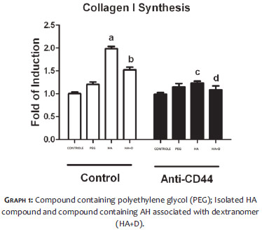
Evaluation of type 1 collagen synthesis through the expression of COL1A1 gene
After 24 hours, a statistically significant increase in the gene expression of COL1A1 in cultures exposed to all compounds studied was observed when compared to the controls. The cultures exposed to the HA compound (a) were those which presented greater increase in the gene expression of collagen when compared to the control cultures, followed by the cultures exposed to the HA compound associated with the dextranomer (b).
When the Anti-CD44 antibody was added to the cultures, a significant reduction of COL1A1 gene expression was observed only in cultures exposed to the HA compounds (c, d). (Graph 1)
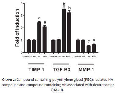
Production of MMP -1, TIMP -1, and TGF-β
After 24 hours, a statistically significant increase in the gene expression of TIMP-1 (a) and TGF-β (b) was observed only in cultures where HA had been added.
Likewise, after 24 hours, there was a statistically significant reduction in the gene expression of MMP1 only in the cultures exposed to the AHs (c).
These parameters showed no significant alteration in the cultures exposed to the PEG compound when compared with controls. (Graph 2)
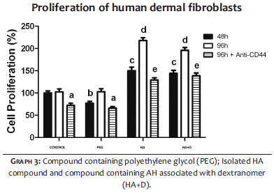
Cell proliferation
After 48 hours, a statistically significant increase of fibroblasts was observed when compared to controls, only in cultures with AH (c). This result became more pronounced after 96 hours (d).
After 96 hours, the proliferation of fibroblasts exposed to compounds containing AH was significantly reduced when cultures were incubated with Anti-CD44 antibodies (e). However, despite this effect, the cell proliferation of these cultures was still greater than that observed in control cultures and in cultures exposed to PEG (a).
The cultures exposed to PEG (b) showed an initial reduction in fibroblast proliferation (48h) when compared to controls. After 96 hours, proliferation was comparable to that found in the controls (a).
The proliferation of fibroblasts after 96 hours (with and without the addition of Anti-CD44) was similar in the controls and in cultures exposed to PEG. (Graph 3)
This in vitro study demonstrated that 24 hours of exposure of cultured human dermal fibroblasts to the various compounds used in cutaneous fillers led to an increase in collagen production, evidenced by increased expression of COL1A1 gene, as compared to the controls. It was also observed that the increased expression of collagen was more pronounced in cultures exposed to compounds of AH.
Concomitantly, gene expression was inhibited when cultures exposed to AHs were treated with the Anti-CD44 antibody, which is a cell membrane receptor of AH. This result suggests that the receptor CD44 may be involved in the mechanism of stimulation of collagen production by fibroblasts in the presence of HA.
An interesting observation is that the exposure of the cultures to HA increases production of TIMP-1 and TGF-β, and reduces the expression of MMP-1.This data may indicate that the increased expression of collagen in the cultures exposed to AH is due to a greater stimulation from growth factors, coupled with the reduction of degradation (decrease in MMP-1 and increase in TIMP-1).
The results of this in vitro study are in agreement with the findings in the literature.6,7 Other studies show that the use of HA for dermal filling seems to stimulate the production of collagen at the site of its application.6 There has been speculation about the possible mechanisms involved in this process. There is evidence that the mechanical distension of fibroblasts at the site of application of HA could be one of the stimuli. 8,9
The present study was an in vitro analysis in which human dermal fibroblast cultures were exposed to different compounds available for cutaneous filling. The authors studied the gene expression of collagen type I, metalloproteinase I, tissue inhibitor of metalloproteinase, and TGF-β, as well as the proliferation of fibroblasts in these conditions.
It could be observed that the addition of HA to the cultures resulted in increased fibroblast proliferation and greater gene expression of collagen, TGF-β, and TIMP-1, and decreased expression of MMP-1.
An important finding of this study links the reduction of fibroblast proliferation and collagen production to the addition of CD44 antibodies to cultures exposed to HA, indicating that this receptor may be involved in the mechanism leading to those effects.
The addition of PEG to the cultures did not lead to an increased proliferation of fibroblasts, nor did it alter the gene expression of studied proteins.
Further studies are needed to deepen the understanding of the role of the macromolecules used in cutaneous fillings in the biology of fibroblasts.
1. 1 Hascell V, Laurent T. Hyaluronic: structure and physical properties. [Acessed 28 March 2008]. Available from: http://glycoforum.gr.jp/science/hyaluronan/HA01/HA01E.html.
2. Beasley KL, Weiss MA, Weiss RA. Hyaluronic Acid Fillers: A Comprehensive Review. Facial Plast Surg. 2009;25(2):86-94.
3. Mole BJ. Remake, une nouvelle famille de gels injectables à dureé prolongeé. Med Esth Chir. 2008;35(140):211-5.
4. Peppas NA, Hilt JZ, Khademhosseini A, Langer R. Hydrogels in biology and medicine: From molecular principles to bionanotechnology. Adv Mater. 2006;18(11):1345-60.
5. Ponta H, Sherman L, Herrlich PA. CD44: from adhesion molecules to signalling regulators. Nat Rev Mol Cell Biol. 2003;4(1):33-45.
6. Wang F, Garza LA, Kang S, Varani J, Orringer JS, Fisher GJ, et al. In vivo stimulation of de novo collagen production caused by cross-linked hyaluronic acid dermal filler injections in photodamaged human skin. Arch Dermatol. 2007;143(2):155-63.
7. Rock K, Fischer K, Fischer J W. Hyaluronan Used for Intradermal Injections Is Incorporated into the Pericellular Matrix and Promotes Proliferation in Human Skin Fibroblasts in vitro. Dermatology. 2010;221(3):219-28.
8. Kessler D, Dethlefsen S, Haase I, Plomann M, Hirche F, Krieg T, et al. Fibroblasts in mechanically stressed collagen lattices assume a 'synthetic' phenotype. J Biol Chem. 2001;276(39):36575-85.
9. Silver FH, Siperko LM, Seehra GP. Mechanobiology of force transduction in dermal tissue. Skin Res Technol 2003;9(1):3-23.
 All content the journal, except where identified, under the Creative Commons Attribution 4.0 International licence - ISSN-e 1984-8773


 Read in Portuguese
 Portuguese PDF
 Print
Print
 Send this article by email
 How to cite this article
 Submit a comment
 Mendeley
 Pocket