


Doris Hexsel1,2; Clarissa Reinehr1,2; Patricia Caspary1,2; Gabriela Mosena1,2; Vitor Costa Fabris1,3
Funding: None
Conflict of interest: Doris Hexsel, MD: The author has acted as a speaker and consultant for Galderma. The author is also a consultant for Allergan. The other authors have no other conflict of interest to disclose.
Submitted on: 05/06/2026
Final decision: 07/09/2026
How to cite this article: Hexsel D, Reinehr C, Caspary P, Mosena G, Fabris V. Botulinum toxin for submandibular gland treatment: A scoping review of aesthetic and functional outcomes. Surg Cosmet Dermatol. 2026;18(3):e20260577.
Enlarged or ptotic submandibular glands (SmG) can negatively impact the mandibular angle and facial contour. Although botulinum toxin type A (BoNT-A) is well established as a treatment for parotid hypertrophy, its use for the treatment of SmG enlargement remains underexplored. A literature search was conducted in the PubMed, Cochrane, and LILACS databases, covering studies from November 1977 to November 2024. Seven studies met inclusion criteria, encompassing animal models, cadaveric studies, clinical trials, and case reports. Studies reporting SmG size found volumetric reduction following BoNT-A administration. Current evidence suggests BoNT-A injections into the SmG effectively reduce gland volume and may enhance facial contour.
Keywords: Submandibular Gland; Botulinum Toxins, Type A; Neck; Aging; Esthetics
The neck plays a crucial role in overall facial aesthetics, particularly among patients seeking rejuvenation procedures. Structural deformities in the cervical region can compromise the visual harmony of the neck by disrupting its natural contour. The submandibular glands (SmG), as prominent anatomical components of this area, significantly influence the cervicomental angle. When these glands are enlarged or ptotic, they may detract from cervical aesthetics by distorting this angle and contributing to a poorly defined neck contour.1,3
Since its initial aesthetic application for the treatment of facial rhytides, botulinum toxin type A (BoNT-A) has demonstrated therapeutic efficacy in a variety of conditions, including rosacea and chronic parotid sialadenitis.4,5 Given the existing evidence supporting its use in the management of parotid gland hypertrophy,1 it is reasonable to hypothesize that botulinum toxin may also be effective in addressing enlargement of other salivary glands, such as the SmG.
This scoping review aims to explore the potential role of BoNT-A in the treatment of SmG hypertrophy. Outcomes of interest included gland function, gland size, improvement in cervical aesthetics, and impact on salivary production.
A literature search was conducted on November 11, 2024, using the PubMed, Cochrane Library, and LILACS databases to identify studies evaluating botulinum toxin treatment for the submandibular glands. The search encompassed articles published from November 1977 to November 2024. The search strategy employed the terms: "botulinum toxin" AND "submandibular gland."
Inclusion criteria comprised original studies in English that addressed clinical, anatomical, histological, or ultrasonographic aspects of the salivary glands before and after BoNT-A injections, conducted in human or animal models and focused on the treatment of hypertrophic or ptotic salivary glands. Exclusion criteria included studies involving salivary gland tumors and those exclusively addressing sialorrhea without reference to glandular hypertrophy. Extracted variables included the study model (human or animal), study design, intervention details, treatment and control groups, study protocol, type of BoNT-A used, reported outcomes, and adverse events.
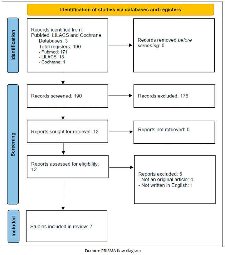
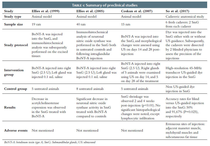
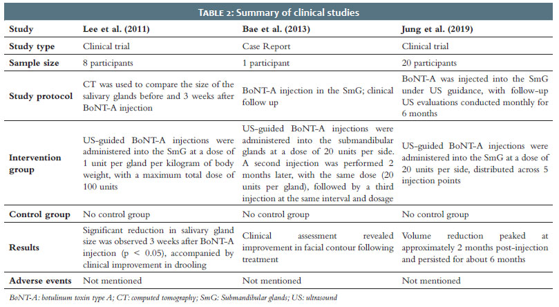
Figure 1 presents the search results following the PRISMA (Preferred Reporting Items for Systematic Reviews and Meta-Analyses) flow diagram. Tables 1 and 2 summarize the 7 studies included in this review, consisting of 4 preclinical studies conducted in cadavers and animal models, 1 human case report, and 2 clinical trials. The clinical studies did not include a control group. All studies that evaluated SmG size before and after the treatment, both preclinical and clinical, reported a reduction in SmG size following treatment with BoNT-A.
The treatment was generally considered to be safe, and the adverse events described in the studies were attributed to errors in the injection technique (erroneous sites of injection adjacent to the masseter muscle, mylohyoid muscles, and subcutaneous fat tissue).
In the case report by G.Y. Bae et al., the authors conducted a preliminary evaluation prior to BoNT-A injections into the SmG to exclude alternative causes of gland enlargement, such as alcohol use, eating disorders, and tumoral lesions.6
In the studies included in the review, different imaging modalities were employed to localize and measure the SmG: Bae et al. and Lee et al. used computed tomography (CT), while Jung et al. used ultrasonography (US).6,7 In the study by Jung et al., US was used not only to measure the gland but also to guide BoNT-A injection placement.7
In the clinical studies included in this review (n = 3), the indications for SmG treatment with BoNT-A were as follows: (1) aesthetic concerns in patients with visibly prominent SmG, and (2) evaluation of the effect of BoNT-A injections on SmG volume reduction in patients with cerebral palsy.6-8
In the study by So J et al. (2017), the SmG were localized using anatomical landmarks without US guidance. The authors established a reference line from the mandibular angle to the gnathion and administered injections at a point located 20%–35% along this line from the mandibular angle, 1.5 cm below the inferior mandibular border, at a depth of 2 cm.9
The clinical study by Jung et al. (2019) combined anatomical landmarks with US guidance. The investigators divided the distance from the gnathion to the mandibular angle into thirds and performed US assessment in the lateral two-thirds of this line to evaluate the mediolateral diameter of the gland and guide the injections.7
All 3 human studies included in this review used US guidance for BoNT-A injections into the SmG.6-8 Jung et al. performed US-guided injections in the SmG of 20 patients. Each gland received 20 units of onabotulinum toxin type A (Botox® - BTX), diluted in 0.2 mL of saline, distributed across 5 injection points spaced 5 mm apart, using a 30-gauge, 0.5-inch needle, resulting in a total dose of 40 units per patient.7
Lee et al. treated 8 patients with cerebral palsy, also under US guidance. BTX was injected at a dose of 1 unit/kg per gland, with a maximum of 100 units per patient. Five patients required sedation, while the remaining 3 underwent the procedure without anesthesia. The injection technique, whether via single or multiple injection points, was not specified in the study protocol.8
In the case report by Bae et al., US-guided injections of 20 units of BoNT-A were administered into each SmG. The toxin was diluted at 4 units per 0.1 mL saline (100 units in 2.5 mL saline). The brand of BoNT-A and the injection technique (eg, number and distribution of injection points) were not reported. Treatment was repeated at 2 and 4 months following the initial injection.6
The main potential adverse effects of BoNT-A injection into the SmG include weakness of adjacent muscles, which may result in dysphagia and dysarthria, as well as intravascular or neurovascular injury due to improper injection technique.7 The most commonly reported erroneous injection sites are the mylohyoid muscles and subcutaneous fat tissue. To minimize the risk of BoNT-A diffusion into surrounding structures, Jung et al. recommend using high-concentration, low-volume injections, which may reduce the likelihood of adverse events such as dysphagia and dysarthria.7
BoNT-A injections into the SmG can be performed with or without US guidance. In a cadaveric study by Song JI et al., the accuracy of US-guided injections was compared to injections based solely on anatomical landmarks. The results demonstrated that US guidance significantly improved injection accuracy (p = 0.0025).9 The study involved fresh cadavers and compared 2 techniques: non-guided injections, based on a reference line from the mandibular angle to the gnathion, with injection points placed 20%–35% along this line from the mandibular angle, 1.5 cm below the inferior mandibular border, at a depth of 2 cm; and US-guided injections, using a high-resolution 45-MHz transducer. Accuracy improved from 50% with the landmark-based method to 91.67% with US guidance.9 The authors concluded that the use of US enhances injection precision and may also reduce the risk of adverse events, such as weakness of adjacent muscles and intravascular or neurovascular puncture during the procedure. The most common sites of erroneous injection identified were the mylohyoid muscles and subcutaneous fat tissue.9
BoNT-A injection into the SmG can result in several clinical effects. From an aesthetic standpoint, the most relevant outcome is reduction in gland size, which contributes to a smoother and more defined jawline. Studies employing CT imaging before and after BoNT-A injection have demonstrated a significant decrease in SmG volume.6-8
In the study by Lee Z et al., the SmG of 8 patients with cerebral palsy were injected with BoNT-A at a dose of 1 unit/kg per gland, followed by CT imaging to assess morphological changes 3 weeks post-injection. The results revealed a statistically significant reduction in SmG size (p < 0.05).8 Specifically, the mean area of the right SmG decreased from 3.19 cm2 to 2.76 cm2, and that of the left SmG from 2.98 cm2 to 2.76 cm2 after 3 weeks.8 This finding supports the authors' clinical observation of gland size asymmetry in nearly all cases.
In the study by Jung et al., 20 patients received 20 units of BoNT-A per SmG, administered at 5 injection points under ultrasound guidance. A reduction in the mediolateral diameter of the SmG was observed, with the maximum effect occurring 2 months post-injection and persisting for approximately 6 months.7 The authors hypothesized that the observed glandular shrinkage may result from functional denervation, although the precise underlying mechanism remains unclear.7
Most studies investigating histological changes in the SmG following BoNT injection have been conducted in rat models. Coskun et al. evaluated morphological alterations in the rat SmG at 14 and 28 days after BoNT-A injection and reported a significant reduction in anteroposterior diameter and glandular circumference at both time points (p < 0.05), with maximal shrinkage observed on day 28. The only notable histopathological finding was lymphocytic infiltration.10
In another study, Ellies et al. demonstrated a significant reduction in neuronal nitric oxide synthase levels in SmG treated with BoNT-A (2.5 units of Botox® diluted in 0.1 mL saline) compared with untreated and sham-injected glands.11 This suggests that nitric oxide pathways play a critical role in the neural regulation of salivary gland function. These findings are consistent with earlier work by Ellies et al. (1999), who reported a decrease in acetylcholinesterase concentration in the SmG of BoNT-treated rats, further supporting the hypothesis of functional denervation as a mechanism for glandular atrophy.11,12
Studies suggest that SmG size does not significantly increase with age; however, age-related ptosis occurs gradually, making the glands more prominent over time.13 Additional, yet unidentified, factors may contribute to SmG descent or hypertrophy with aging. Submandibular gland ptosis correlates positively with age, is common in older adults, and accurate clinical assessment requires anatomical or imaging guidance due to overlying tissue laxity.13 Diagnosing SmG ptosis can be challenging because the laxity of overlying soft tissues may obscure clinical visualization.3
Therefore, the use of anatomical landmarks or imaging modalities (eg, US or CT) is recommended to ensure accurate gland localization and safe BoNT-A injection.
To aid in clinical evaluation, Lee et al. proposed a grading system for SmG ptosis based on the distance from the inferior border of the mandible to the lowest point of the gland: Grade I, < 20 mm; Grade II, 20–25 mm; Grade III, 25–30 mm; Grade IV: > 30 mm.14 This grading system may be useful for pretreatment assessment before administering BoNT-A injections.
In a cross-sectional study, Lee et al. analyzed 100 CT scans (50 men and 50 women), from patients aged 18 to 91 years, to assess SmG ptosis. A linear correlation was found between age and gland descent (p < 0.001), with an average descent rate of 0.17 mm per year. Notably, 92.6% of patients aged 65 years or older exhibited Grade III or IV ptosis, with no significant difference between men and women.14
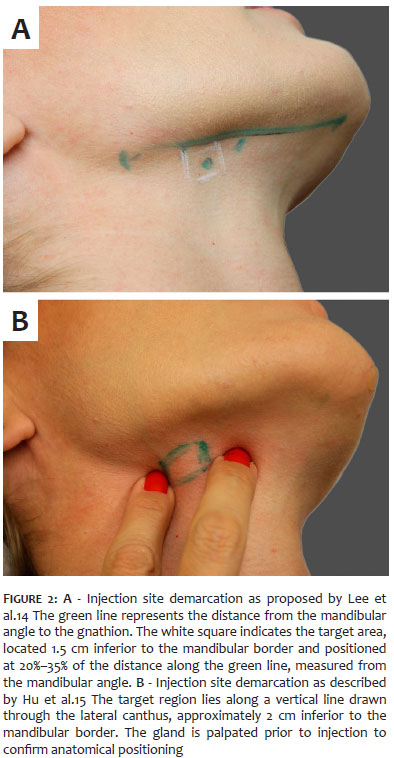
Lee et al. also defined a "safe zone" for SmG injection (Figure 2a), recommending BoNT-A administration between 20%–35% of the distance from the mandibular angle to the gnathion, 1.5 cm below the mandibular border, using a 2 cm deep injection (2-cm needle).14 Hu et al. described a technique involving palpation of the gland followed by injection using a 0.5-inch needle (Figure 2b). The gland is targeted along a vertical line intersecting the lateral canthus, while maintaining a 2-cm distance from the mandibular border to minimize the risk of intravascular injection. BoNT-A is delivered via a single injection point.15 Hu et al. recommended a dose ranging from 15 to 30 units of BoNT-A per gland.15 Reported injection depths vary from 0.5 to 2.0 cm across different studies.14-16
The grading system proposed by Lee et al. provides a practical framework for assessing submandibular gland ptosis and may guide pretreatment planning for BoNT-A injections.14 Defined injection zones, depths, and doses for BoNT-A, as described by Lee et al. and Hu et al., allow for safe and effective treatment of the submandibular gland while minimizing the risk of complications.14,15
Glandular atrophy, particularly after repeated botulinum toxin injections, is a potential concern associated with this treatment. None of the studies included in this review evaluated the effects of repeated treatment sessions. Nevertheless, a reduction in gland size was observed after a single injection, suggesting a decrease in gland volume, but true glandular atrophy cannot be confirmed based on the available evidence.17,18 Although no cases of xerostomia were reported in the included studies,6-8 reduced salivary function remains a plausible concern, particularly with repeated treatments.17 Experimental and clinical evidence from studies involving other major salivary glands, particularly the parotid gland, suggests that botulinum toxin-induced reductions in gland volume and secretory function are generally transient and reversible after the pharmacological effect subsides.17,18 Therefore, if clinically significant glandular hypofunction or xerostomia occurs, discontinuation of treatment would likely allow recovery of gland size and salivary function over the following months.17,18
The primary limitation of this scoping review is the limited number of clinical and preclinical studies currently available on this topic. Nonetheless, this review represents an important step toward guiding the standardization of the technique and fostering greater consistency and dialogue among future studies in this field.
The studies included in this scoping review suggest that BoNT-A injections can effectively reduce the size of the SmG, resulting in improved visibility of the jawline and enhanced cervicofacial contour. However, a thorough diagnostic evaluation is essential to identify the underlying cause of cervical contour irregularities. Excluding alternative etiologies of submandibular fullness, such as tumoral, infectious, or systemic conditions, is critical to ensure that BoNT-A treatment is both appropriate and safe.
Doris Hexsel
ORCID: 0000-0002-0615-9026
Approval of the final version of the manuscript, Conception and design of the study, Preparation and writing of the manuscript, Effective participation in the conduct of the study, Critical revision of the manuscript
Clarissa Reinehr
ORCID: 0000-0003-1811-4519
Preparation and writing of the manuscript, Acquisition, analysis and interpretation of data, Critical review of the literature, Critical revision of the manuscript
Patricia Caspary
ORCID: 0009-0000-9205-3051
Preparation and writing of the manuscript, Acquisition, analysis and interpretation of data, Critical review of the literature, Critical revision of the manuscript
Gabriela Mosena
ORCID: 0000-0002-4907-2713
Preparation and writing of the manuscript, Acquisition, analysis and interpretation of data, Critical review of the literature, Critical revision of the manuscript
Vitor Costa Fabris
ORCID: 0000-0002-3540-2769
Approval of the final version of the manuscript, Preparation and writing of the manuscript, Effective participation in the conduct of the study, Critical review of the literature, Critical revision of the manuscript
1. Minelli L, Yang HM, van der Lei B, Mendelson B. The surgical anatomy of the jowl and the mandibular ligament reassessed. Aesthetic Plast Surg. 2023;47:170-80.
2. Auersvald A, Auersvald LA. Management of the submandibular gland in neck lifts: indications, techniques, pearls, and pitfalls. Clin Plast Surg. 2018;45:507-25.
3. Ramirez OM. Multidimensional evaluation and surgical approaches to neck rejuvenation. Clin Plast Surg. 2014; 41:99-107.
4. Alsaati AA, Alsaadoun D, Kinkar LI. The efficacy and safety of Botulinum Toxin A for the treatment of rosacea: a systematic review. Cureus. 2023;15:e51304.
5. Strohl MP, Chang CF, Ryan WR, Chang JL. Botulinum toxin for chronic parotid sialadenitis: a case series and systematic review. Laryngoscope Investig Otolaryngol. 2021;6:404-13.
6. Bae GY, Yune YM, Seo K, Hwang SI. Botulinum toxin injection for salivary gland enlargement evaluated using computed tomographic volumetry [published correction appears in Dermatol Surg]. Dermatol Surg. 2013;39:1404-7.
7. Jung GS, Cho IK, Sung HM. Submandibular gland reduction using Botulinum Toxin Type A for a smooth jawline. Plast Reconstr Surg Glob Open. 2019;7:e2192.
8. Lee ZI, Cho DH, Choi WD. Effect of botulinum toxin type a on morphology of salivary glands in patients with cerebral palsy. Ann Rehabil Med 2011;35:636-40.
9. So JI, Song DH, Park JH. Accuracy of ultrasound-guided and non-ultrasound-guided botulinum toxin injection into cadaver salivary glands. Ann Rehabil Med. 2017;41:51-7.
10. Coskun BU, Savk H, Cicek ED. Histopathological and radiological investigations of the influence of botulinum toxin on the submandibular gland of the rat. Eur Arch Otorhinolaryngol 2007;264:783-7.
11. Ellies M, Schütz S, Quondamatteo F, Laskawi R. The effect of local injection of botulinum toxin A on the immunoreactivity of nNOS in the rat submandibular gland: an immunohistochemical study. Int J Pediatr Otorhinolaryngol 2006;70:59-63.
12. Ellies M, Laskawi R, Getz W. Immunohistochemical and morphometric investigations of the influence of botulinum toxin on the submandibular gland of the rat. Eur Arch Otorhinolaryngol. 1999; 256:148-52.
13. Rohrich RJ, Rios JL, Smith PD, Gutowski KA. Neck rejuvenation revisited. Plast Reconstr Surg. 2006;118:1251–63.
14. Lee JH, Lee BN, Kwon SO. Anatomical localization of submandibular gland for botulinum toxin injection. Surg Radiol Anat 2010;32:945-9.
15. Hu H, Kim SB, Wan J. Anatomical guidelines and technical tips for neck aesthetics with botulinum toxin. Arch Plast Surg. 2024;51:447-58.
16. Lee MK, Sepahdari A, Cohen M. Radiologic measurement of submandibular gland ptosis. Facial Plast Surg 2013; 29:316-20.
17. Lakraj AA, Moghimi N, Jabbari B. Sialorrhea: anatomy, pathophysiology and treatment with emphasis on the role of botulinum toxins. Toxins (Basel). 2013;5(5):1010-31.
18. Jost WH, Friedman A, Michel O, Oehlwein C, Slawek J, Bogucki A, et al. SIAXI: Placebo-controlled, randomized, double-blind study of incobotulinumtoxinA for sialorrhea. Neurology. 2019;92(17):e1982-91.
 All content the journal, except where identified, under the Creative Commons Attribution 4.0 International licence - ISSN-e 1984-8773




 Read in Portuguese
 Portuguese PDF
 Print
 Send this article by email
 How to cite this article
 Submit a comment
 Mendeley
 Pocket
 Share on Facebook
 Share on Twitter