


Vivian Zague1; Ana Lúcia Tabarini Alves Pinheiro2; Juliana Rodrigues Pinto2; Viviane Cristina Albarici2; Gustavo Facchini2; Samara Eberlin2
Funding: None
Conflict of interest: None
Research Ethics Committee approval CAAE Number: 70137923.4.0000.5514
Submitted on: April 29, 2026
Final decision: July 9, 2026
How to cite this article: Zague V, Pinheiro ALTA, Pinto JR, Albarici VC, Facchini G, Eberlin S. Clinical aesthetic effects of oral collagen oligopeptides on wrinkle appearance in women aged 45-60 years: a randomized, double-blind, placebo-controlled trial. Surg. Cosmet Dermatol. 2026;18(3):e20260573.
INTRODUCTION: Oral collagen supplementation has been investigated as a strategy to support skin appearance, although clinical effects are generally modest. In aesthetic dermatology, both the magnitude and temporal pattern of changes are relevant.
OBJECTIVE: To assess the effects of oral supplementation with collagen oligopeptides on facial wrinkle appearance in women aged 45-60 years.
METHODS: A randomized, double-blind, placebo-controlled clinical trial was conducted in 102 women. Participants received collagen oligopeptides (2.5 g/day) or a placebo for 12 weeks. Wrinkle intensity was the primary endpoint, assessed using standardized digital imaging. Dermal density and collagen-related optical signals were analyzed as exploratory outcomes.
RESULTS: A significant reduction in wrinkle intensity was observed in the active group at week 8. A reduction was already apparent at week 4, although not statistically significant, and values remained numerically lower at week 12. The placebo group showed minor variations. Exploratory parameters showed numerical increases but were not statistically significant.
CONCLUSION: Collagen oligopeptide supplementation was associated with a reduction in wrinkle intensity, with an early effect that was partially maintained over time, indicating a measurable aesthetic benefit.
Keywords: Skin Aging; Collagen; Dietary Supplements; Peptides; Dermis
Skin aging arises from a complex interplay between intrinsic biological mechanisms, such as genetic programming, hormonal fluctuations, and chronological decline, and extrinsic stressors including ultraviolet exposure, environmental pollution, and behavioral factors. Together, these factors progressively impair dermal architecture, leading to reduced elasticity, thinning of the dermis, and the formation of clinically visible wrinkles.1 Among the various manifestations of skin aging, wrinkle appearance remains one of the most relevant and perceptible endpoints in aesthetic dermatology, serving as a key parameter for assessing the efficacy of interventions. Given the multifactorial nature of skin aging, nutritional interventions and functional food ingredients capable of modulating extracellular matrix (ECM) homeostasis have gained increasing attention as complementary strategies for maintaining skin health. In this context, the ability of such interventions to produce measurable changes in skin appearance, particularly under controlled clinical conditions, represents an important translational challenge in the field.1–3
Collagen, the predominant fibrillar protein of the dermal ECM, plays a central structural role in maintaining the mechanical integrity and firmness of the skin. With advancing age, collagen synthesis decreases while enzymatic degradation accelerates, resulting in fragmentation of collagen fibers and progressive loss of tensile strength. These age-related alterations in collagen dynamics are closely associated with clinically observable deterioration in skin appearance, contributing to skin laxity, reduced tone, and deeper wrinkle formation.2
Among nutritional interventions targeting ECM integrity, hydrolyzed collagen peptides (CPs) have gained increasing attention. Derived from enzymatic cleavage of native type I collagen, these peptides possess low molecular weight and are efficiently absorbed following oral ingestion. After ingestion, specific collagen-derived peptides can be detected in plasma and have been reported to accumulate within the dermis, where they are thought to function as biochemical messengers, potentially interacting with fibroblast activity and supporting ECM-related processes, as suggested by experimental and translational studies.3–6
Experimental data provide biological plausibility for the proposed mechanisms. In vitro studies have shown that collagen peptides can enhance fibroblast activity, increasing type I procollagen synthesis while reducing the expression or activity of matrix metalloproteinases (MMP-1 and MMP-2). Zague et al.7 showed that collagen hydrolysates promoted ECM protein deposition in fibroblasts from both sun-protected and photoaged skin, with greater effects under UV-induced stress, supporting a potential modulatory role of collagen-derived peptides under conditions of dermal challenge. Low-molecular-weight peptides containing hydroxyproline are particularly relevant because they have been shown to interact with fibroblast-associated pathways and modulate ECM-associated processes, providing a structure-function rationale based primarily on experimental models, which is relevant to the interpretation of functional food applications.7,8
More recently, Zague et al.9 expanded these findings by integrating molecular and clinical evidence. In a randomized controlled trial involving 85 women, daily ingestion of collagen oligopeptides (Col-OP, 2.5 g/day) was associated with improvements in skin firmness and elasticity compared with placebo, while hydration showed a consistent, though not statistically significant, improvement. Complementary gene expression analyses indicated upregulation of extracellular matrix components (procollagen I, decorin, biglycan) and TIMP-1, alongside downregulation of MMP-1 and versican, providing molecular-level support for a coordinated modulation of dermal matrix-related pathways, which may contribute to the clinical effects observed.9
Taken together, these results strengthen the biological plausibility of collagen peptides as potential modulators of ECM homeostasis and overall skin quality. Nevertheless, inconsistencies among studies, stemming from differences in peptide profiles, dosage regimens, and methodological designs, have led to substantial variability in reported clinical outcomes. Recent meta-analyses emphasize the need for standardized formulations and rigorously controlled trials to confirm efficacy and to further characterize, rather than definitively establish, the molecular basis of observed effects.9–11
Given the need for objective imaging-based assessments and dermal biomarkers capable of sensitively capturing ECM-related changes in vivo, this study tested the hypothesis that daily oral supplementation with collagen oligopeptides (2.5 g/day for 12 weeks) would promote a measurable reduction in facial wrinkle intensity, with particular emphasis on the temporal pattern of response, compared with placebo. Changes in dermal density and collagen-related optical markers were evaluated as complementary, exploratory endpoints, to support biological plausibility and provide an integrated assessment of structural and optical skin-related changes, rather than to establish definitive mechanistic effects.
The investigational product consisted of food-grade collagen oligopeptides (Col-OP; manufacturer details withheld for blinded review), supplied as a powder and derived from bovine type I collagen by controlled enzymatic hydrolysis. The investigational product was supplied in single-dose sachets, each containing 2.5 g of Col-OP, to be dissolved in water before oral intake. The placebo was also supplied as a powder in identical sachets and was matched in appearance, taste, and texture to maintain blinding. The resulting peptide mixture had an average molecular weight of approximately 4 kDa, with about 40% of fragments below 1 kDa. The peptide profile of Col-OP was analytically characterized, confirming a reproducible distribution of low-molecular-weight collagen-derived oligopeptides consistent with the specifications of a standardized bovine type I collagen hydrolysate. This level of physicochemical characterization was considered sufficient for the purposes of the present clinical investigation. Although individual peptide sequences are proprietary, previous studies have shown that low-molecular-weight collagen-derived peptides can reach the bloodstream after oral ingestion12 and have been associated with modulation of ECM-related pathways, including fibroblast activity and MMP/TIMP balance.7,9 These prior observations provide biological plausibility but should not be interpreted as direct mechanistic evidence within the present clinical trial.
The placebo was an isocaloric, organoleptically identical powder composed of maltodextrin and containing no collagen peptides. The placebo was matched in appearance, taste, and texture to ensure adequate blinding of participants and investigators. No bioactive compounds, flavors, or other functional excipients with potential biological activity were included.
The protocol was reviewed and approved by the Research Ethics Committee of Universidade São Francisco, located in Bragança Paulista, São Paulo, Brazil (CAAE number 70137923.4.0000.5514; approval no. 6.123.303, issued on June 16). All volunteers provided written informed consent prior to enrollment. The study was conducted in accordance with the principles of the Declaration of Helsinki and Good Clinical Practice (GCP) guidelines, and in compliance with all applicable national regulatory requirements. This study was a single-center, randomized, double-blind, placebo-controlled clinical trial. One hundred and two women aged 45–60 years, representing Fitzpatrick skin phototypes II–V, were screened and randomized (49 to the Col-OP group and 53 to the placebo group). The computer-generated sequence used an equal allocation probability (1:1), without blocking or forced balancing of final group sizes; therefore, a small numerical imbalance resulted by chance, with 49 participants allocated to the Col-OP group and 53 to the placebo group. Baseline clinical assessments were performed prior to randomization to confirm comparability between treatment arms.
Exclusion criteria included pregnancy or lactation, any active dermatological condition, or ongoing medical or cosmetic procedures that could affect the outcomes. The use of topical retinoids, chemical peels, or similar interventions within 4 weeks prior to baseline was prohibited. Throughout the trial, participants were instructed to avoid excessive UV exposure, artificial tanning, and dietary or topical products aimed at anti-aging effects. The use of basic skincare products habitually used by the participants, limited to cleansers, moisturizers, and sunscreens, was permitted, provided that these products had been part of their routine before enrollment and remained unchanged throughout the study. Participants were further instructed to maintain consistent dietary habits, contraceptive methods, and skincare routines throughout the study period. Adherence to these restrictions was monitored at each study visit, and noncompliance was prospectively defined as a criterion for exclusion from the study.
Participants were randomly assigned with an intended 1:1 allocation probability to receive either Col-OP or placebo for 12 weeks using a computer-generated randomization sequence. The unequal 49:53 distribution reflected the use of simple randomization and did not result from exclusions according to predefined eligibility criteria. Both test materials were provided in identical sachets, each containing a single daily dose (2.5 g) to be dissolved in water and consumed in the morning. Randomization was computer-generated using a predefined random sequence, and both participants and investigators remained blinded to group allocation throughout the study. Allocation concealment was ensured using sequentially numbered, identical sachets prepared by an independent pharmacist, and compliance was monitored at each visit through systematic sachet counts and standardized adherence questionnaires. Study visits were conducted at baseline, week 4, week 8, and week 12 under controlled environmental conditions.
Study visits were conducted at baseline and at weeks 4, 8, and 12. All instrumental assessments were performed under standardized environmental conditions following predefined operating procedures.
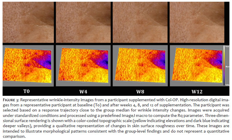
Wrinkle intensity was quantified using standardized high-resolution digital imaging coupled with dedicated analytical software (ImageJ 1.47v, National Institutes of Health, Bethesda, MD, USA). All photographs were acquired under controlled illumination, at a fixed camera distance, and with standardized facial positioning using a mechanical head stabilizer. Image processing was performed using a pre-specified macro that quantified the Rq parameter (root-mean-square roughness). Rq corresponds to the root-mean-square of the profile-height deviations within the analysis area and was used as the primary quantitative descriptor of wrinkle intensity and the primary clinical endpoint of the study. Dermal density was assessed using a 22-MHz high-frequency ultrasound device, the Ultrascan UC22 (Courage + Khazaka Electronic GmbH, Köln, Germany). Three consecutive measurements were obtained at each anatomical site and averaged to minimize technical and operator-dependent variability.
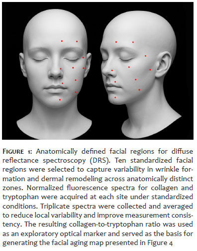
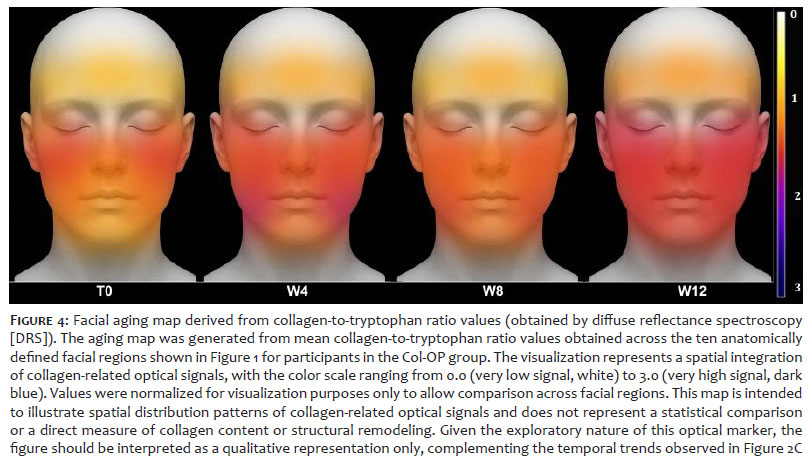
In parallel, collagen-related optical signals were indirectly estimated by diffuse reflectance spectroscopy (DRS) using the FluoroMax-4 spectrofluorometer (HORIBA Scientific, Edison, NJ, USA). The excitation wavelength was set to 290 nm, emission spectra were recorded from 300–500 nm, and all spectra were normalized to the maximal tryptophan fluorescence peak prior to ratio calculation. Spectra were collected in triplicate at each anatomical site and averaged to minimize instrument-related variability. The collagen-to-tryptophan ratio was calculated as a noninvasive, exploratory optical marker associated with dermal collagen-related signals and should not be interpreted as a direct measure of collagen biosynthesis or structural remodeling. Based on collagen-to-tryptophan ratio values obtained from 10 anatomically defined facial regions (Figure 1), an exploratory aging map was generated. The resulting map (Figure 4) was rendered as a color-coded image, with a scale ranging from 0.0 (indicating very low collagen-related signal, represented in white) to 3.0 (indicating very high collagen-related signal, represented in dark blue).
Wrinkle intensity was designated as the primary clinical endpoint of the study, with particular emphasis on its temporal evolution throughout the intervention period. Dermal density and the DRS collagen-to-tryptophan ratio were defined as secondary, exploratory endpoints intended to provide supportive biological context and an integrated interpretation of instrumental findings, rather than definitive mechanistic evidence.
All evaluators were trained before study initiation and followed standardized operating procedures throughout the study. Instrument calibration and quality control procedures were performed according to the manufacturers' recommendations.
Data were analyzed using analysis of covariance (ANCOVA) and linear mixed-effects models, incorporating group, time, and their interaction (group × time) as fixed factors and participant as a random effect. The mixed-effects framework was used to evaluate the overall temporal trajectory of responses across study visits, allowing for the assessment of time effects and potential group × time interactions, in addition to between-group comparisons at individual time points. Between-group contrasts were expressed as adjusted means and change-from-baseline estimates, accompanied by 95% confidence intervals and standardized effect sizes (Hedges' g), to facilitate interpretation of effect magnitude alongside statistical significance, with particular emphasis on the primary endpoint. P-values were corrected for multiple comparisons within each endpoint across time points using the Holm-Bonferroni method. All models included baseline values as covariates where appropriate.
Model assumptions, including normality, homoscedasticity, and independence of residuals, were evaluated using standard diagnostic procedures, such as inspection of residual distributions, assessment of variance homogeneity, and screening for influential observations. For percentage-based outcomes (collagen-to-tryptophan ratio), residual distributions were additionally inspected for skewness; no substantial deviations from model assumptions were identified. Missing data were handled under the assumption of missing at random (MAR). The linear mixed-effects models were estimated using restricted maximum likelihood, allowing the inclusion of all available observations without the need for data imputation. All statistical tests were two-sided, with a predefined significance level of α = 0.05. Primary analyses followed the intention-to-treat principle using available-case analysis under the mixed-effects framework. Sensitivity analyses using mixed-effects models were performed to confirm the robustness of the primary ANCOVA findings. All statistical procedures were defined a priori before data unblinding and applied consistently across endpoints. All analyses were performed using R software version 4.3.1 (R Foundation for Statistical Computing, Vienna, Austria).
A post-hoc power analysis was conducted using the observed effect size for wrinkle intensity at week 8 (Hedges' g ≈ 0.28). Based on the final sample size (n = 49 in the active group and n = 53 in the placebo group), α = 0.05, and a two-tailed ANCOVA design, the statistical power achieved was estimated at approximately 65% for the observed effect size. This level of statistical power reflects the ability to detect of modest treatment effects as limited by the sample size and is consistent with effect magnitudes typically reported in nutritional and aesthetic intervention trials. However, it remains below the conventional 80% threshold, indicating limited sensitivity for detecting small between-group differences, particularly for secondary exploratory endpoints and later time-point comparisons, and should be interpreted accordingly.12
Expected effect sizes for planning were derived from previous randomized controlled trials evaluating collagen peptides, including Proksch et al.,13 Asserin et al.,14 and Zague et al.,9 which reported effect sizes between 0.25 and 0.45 for wrinkle- and elasticity-related endpoints using daily doses of 2.5–5 g. Assuming a conservative anticipated effect size of 0.30, α = 0.05, and 80% statistical power, a minimum of 45–50 participants per group was estimated to be required. The final sample size of the present study fell within this recommended range for detecting modest primary endpoint effects, while acknowledging reduced sensitivity for secondary exploratory outcomes and small between-group differences at later time points.
Out of 102 randomly assigned women, 49 were allocated to Col-OP and 53 to placebo. This 49:53 distribution reflected the use of simple computer-generated randomization with equal allocation probability and no forced block balancing, rather than post-randomization exclusion according to eligibility criteria. Four participants (2 from each group) discontinued before the 12-week visit, all for personal reasons unrelated to treatment or adverse events. No treatment-related adverse events leading to discontinuation were reported, and no serious adverse events were observed during the study period. No protocol deviations related to product intake or prohibited concomitant treatments were reported. Baseline demographic and clinical parameters, including age, Fitzpatrick phototype, and wrinkle severity, were generally comparable across groups, indicating adequate balance between groups (Table 1). No statistically significant baseline differences were observed between groups for key demographic or clinical variables. The per-protocol sample included 98 women, and all analyses followed the intention-to-treat principle with available-case analysis under the mixed-effects framework for repeated measures. Overall study retention was high, with balanced discontinuation across treatment arms, supporting the internal validity of the trial.
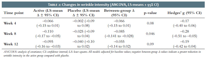
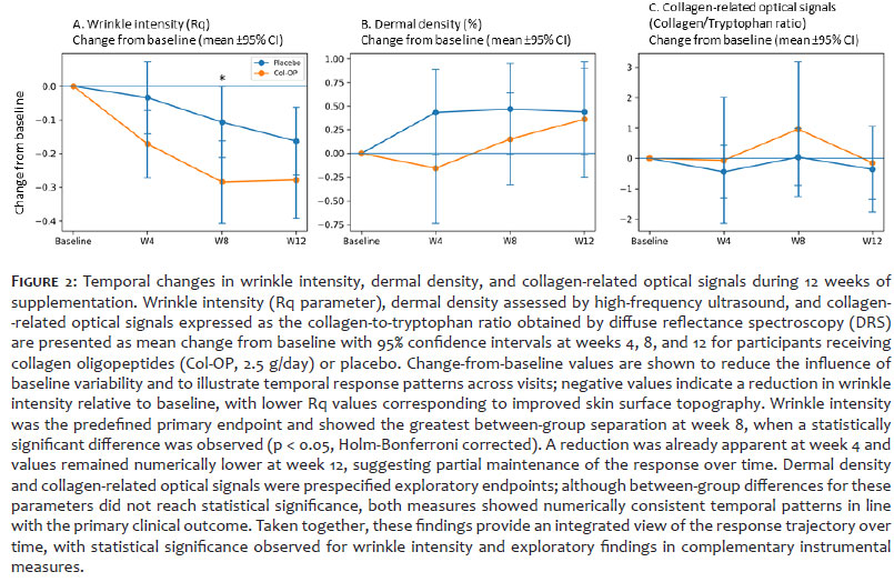
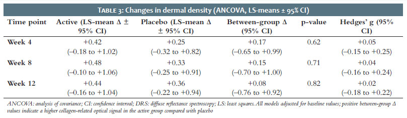
A statistically significant between-group difference in change-from-baseline wrinkle intensity was observed at week 8, when Col-OP supplementation was associated with a reduction in wrinkle intensity compared with placebo. This time point represented the greatest separation between groups during the intervention period. Detailed adjusted mean changes and effect size estimates are presented in Table 2. Within the active group, a directional reduction was apparent by week 4 and values remained numerically lower at week 12, whereas participants receiving placebo showed a reduction primarily at the final assessment. This temporal pattern is consistent with an early onset of response followed by a peak separation at the intermediate time point and partial maintenance over time. Figure 2A depicts the mean temporal trend, and Figure 3 presents representative images from one participant, illustrating the qualitative pattern of reduction in wrinkle depth and surface roughness, consistent with the group-level findings.
Although the standardized effect size was small (Hedges' g), negative between-group Δ values indicate a greater reduction in wrinkle intensity in the active group compared with placebo. The consistency of directional effects across multiple analytical approaches supports the robustness of the observed temporal pattern, despite the modest magnitude of change. These findings were consistent in direction across sensitivity analyses using linear mixed-effects models, which reproduced the overall temporal pattern observed in the ANCOVA results. The modest effect magnitude likely reflects inter-individual variability in baseline wrinkle characteristics and response kinetics. Such variability is commonly observed in aesthetic clinical trials involving heterogeneous skin-aging profiles. No adverse events related to imaging procedures or participant discomfort were reported.
Ultrasound assessments revealed small increases in dermal density following Col-OP intake (Figure 2B). Although the active group exhibited numerically higher values across all post-baseline visits, these changes did not reach statistical significance, as the 95% confidence intervals included zero at every time point (Table 3). Both groups demonstrated minor numerical fluctuations over time, without a consistent directional pattern clearly favoring the active treatment. The corresponding effect size was negligible, indicating a limited detectable effect on dermal density within the 12-week intervention period. ANCOVA and mixed-effects sensitivity analyses produced consistent, non-significant results. Within-group variability was observed across visits, remaining within the expected range for ultrasound-based dermal density measurements. High-frequency ultrasound is known for operator- and site-dependent variability, which may contribute to measurement variability in dermal assessments. Overall, the ultrasound data indicate no statistically significant changes in dermal density over the study period.
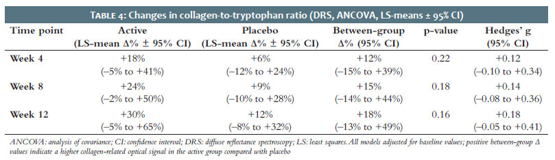
Measurements obtained using DRS, used here as a noninvasive optical surrogate for collagen-related signal rather than as a direct measure of collagen biosynthesis or structural remodeling, indicated a numerical increase in collagen-to-tryptophan ratios in the Col-OP group compared with placebo (Figure 2C). The collagen-to-tryptophan ratio increased progressively throughout the intervention, reaching approximately 30% above baseline at week 12 in the Col-OP group, whereas the placebo group showed an increase of approximately 12%. Although between-group differences did not reach statistical significance, the temporal pattern remained consistently favorable across all post-baseline visits (Table 4). Both groups exhibited minor variability across visits, although the active group demonstrated a numerically more consistent pattern over time. The standardized effect size was small, indicating a limited detectable signal under the conditions of this study. Sensitivity analyses using mixed-effects modeling produced similar non-significant patterns, confirming the consistency of the observed temporal profiles across analytical approaches.
Figure 4 illustrates the aging map generated from mean collagen-to-tryptophan ratio values in the Col-OP group, providing a complementary visual representation of spatial variation in collagen-related optical signals; the color scale was normalized for visualization purposes. This visualization was included to facilitate qualitative interpretation of spatial patterns rather than quantitative comparison. The map reflects mean values within the active group and does not represent a formal statistical comparison. Given the exploratory nature of this optical biomarker, these findings should be interpreted as exploratory and supportive rather than as conclusive evidence of ECM-associated changes.
In this randomized, double-blind, placebo-controlled clinical trial, daily supplementation with standardized collagen oligopeptides (2.5 g/day) met the predefined primary clinical endpoint by significantly reducing wrinkle intensity compared with placebo at week 8. Improvements were already evident at week 4 and remained numerically favorable at week 12, suggesting an early onset followed by partial maintenance of the response. Dermal density showed no detectable changes, whereas collagen-related optical signals demonstrated consistent numerical increases throughout supplementation, providing complementary support for the overall pattern of clinical findings.
Although the observed effect size for wrinkle reduction was modest, the corresponding change in Rq roughness reflects a measurable modification in skin surface topography. In the context of nutritional and cosmetic dermatology, such variations are frequently reported and may be associated with perceptible improvements in fine wrinkle appearance under controlled conditions. The temporal profile observed here, with early changes followed by a clearer separation at week 8, is consistent with a gradual response pattern typically associated with oral supplementation. These findings suggest that relatively small quantitative shifts may reflect dynamic changes in skin surface characteristics that are not fully captured by secondary structural endpoints within a 12-week period.
The present findings are broadly consistent with previous experimental and clinical studies investigating collagen-derived peptides. Evidence from in vitro models indicates that these peptides can modulate ECM-related pathways, including collagen synthesis and matrix metalloproteinase activity.7,8 Clinical studies have also reported improvements in parameters such as elasticity, hydration, and wrinkle appearance after 8 to 12 weeks of supplementation.13,14 However, the magnitude of these effects is generally modest, reflecting the progressive and cumulative nature of nutritional interventions compared with procedural or topical approaches.
For example, Proksch et al. reported improvements in skin elasticity at doses ranging from 2.5 to 5 g/day, along with residual effects observed after discontinuation.13 In a subsequent analysis, reductions in periorbital wrinkle volume were observed at weeks 4 and 8, accompanied by increased levels of procollagen I and elastin. Similarly, Asserin et al. described improvements in skin hydration, dermal density, and collagen network organization, supported by ex vivo findings.14 Taken together, these studies suggest a temporal pattern in which early changes may emerge within the first month and become more evident between weeks 8 and 12, consistent with the temporal profile observed in the present study.
Additional evidence from other clinical contexts supports the broader relevance of collagen supplementation. A longer-term randomized trial in cellulite found improvements in skin waviness and dermal density, particularly in individuals with lower BMI.15 More recently, supplementation with hydrolyzed fish cartilage has been associated with improvements in wrinkle-related parameters and dermal echogenicity.16 These findings indicate that the effects of collagen-derived peptides may extend across different anatomical sites and structural outcomes, although inter-study variability remains substantial.
In this context, the results of the present study are generally in line with the literature, although the magnitude of change was comparatively smaller. Several factors may contribute to this difference, including variations in peptide composition, dosage, baseline characteristics of participants, and analytical methodologies. In particular, the lack of statistically significant effects on dermal density contrasts with some previous reports.14,15
It should be considered, however, that the present study was not specifically powered to detect differences in secondary endpoints, particularly those related to structural outcomes. In addition, the 12-week duration may not have been enough to capture slower ECM remodeling dynamics.
The spectroscopic findings should be interpreted as exploratory and as still providing complementary biological context for the clinical outcomes. Collagen-related optical signals showed a progressive numerical increase throughout supplementation, reaching approximately 30% above baseline after 12 weeks in the active group, whereas in the placebo group it increased by approximately 12%. Although between-group differences did not reach statistical significance, the consistent temporal pattern observed across all post-baseline visits suggests that subtle ECM-related changes may accompany the observed reduction in wrinkle intensity. Given the inherent variability associated with optical measurements, larger studies incorporating complementary molecular biomarkers, such as procollagen I, TIMP-1, and MMPs, are warranted to further characterize the biological relevance of these findings.
The strengths of this study include its randomized, double-blind design, the use of objective imaging-based endpoints, and the application of a predefined statistical framework combining ANCOVA and mixed-effects modeling. The integration of multiple assessment modalities, including imaging, ultrasound, and spectroscopy, also provides a broader perspective on skin-related changes. However, several limitations should be acknowledged. These include the moderate sample size for secondary endpoints, the single-center design, and the use of proprietary image-processing methods, which may limit comparability across studies. In addition, the absence of direct biomarker validation restricts mechanistic interpretation, and the study population, limited to women aged 45–60 years, may limit the generalizability of the findings.
From a translational standpoint, the convergence of modest clinical improvements with existing biological plausibility supports further investigation in larger and more diverse populations. Future trials would benefit from longer follow-up periods, standardized imaging methodologies, and the inclusion of validated structural and molecular endpoints. Such approaches may help to better define the relationship between measurable biological changes and clinically perceptible outcomes.
In summary, daily supplementation with collagen oligopeptides for 12 weeks was associated with a small but measurable reduction in wrinkle intensity, reaching statistical significance at week 8. No significant changes were observed in dermal density, and collagen-related optical signals showed a consistent numerical increase without statistical significance.
These findings are consistent with a modest, time-dependent response profile and should be interpreted in light of the limited effect size and the exploratory nature of the secondary endpoints. Importantly, these findings complement our previous randomized clinical study using the same collagen oligopeptide ingredient, which found improvements in skin firmness, elasticity, and ECM-related gene expression, thereby expanding the clinical evidence across complementary skin-aging endpoints. Further studies are warranted to better define the magnitude, durability, and clinical relevance of these effects.
Daily supplementation with a standardized collagen oligopeptide (Col-OP; 2.5 g/day for 12 weeks) was associated with a statistically significant reduction in wrinkle intensity at week 8 compared with placebo. Although the magnitude of change was limited, the temporal pattern is consistent with an early response followed by partial maintenance over time. No significant effects were observed for dermal density, while collagen-related optical signals showed a consistent numerical increase that did not reach statistical significance. These findings should be interpreted as exploratory and supportive.
Overall, the results indicate that oral collagen supplementation may contribute to measurable, albeit modest, improvements in skin appearance under the conditions of this study. Further research is needed to better define the magnitude, durability, and biological relevance of these effects.
During the preparation of this manuscript, the authors used ChatGPT (OpenAI, GPT-4 or a later version) as an AI-assisted tool to support language refinement and improve clarity of expression. The tool was used exclusively for editing purposes, including grammar correction, sentence restructuring, and improvements in readability in English. No AI tools were used for data analysis, data generation, interpretation of results, or generation of scientific content. All outputs were critically reviewed and edited by the authors, who take full responsibility for the accuracy, integrity, and originality of the final manuscript.
Os autores agradecem a todas as participantes do estudo e à equipe clínica por suas contribuições.
Vivian Zague
ORCID: 0009-0001-1958-0207
Statistical analysis, Approval of the final version of the manuscript, Conception and design of the study, Preparation and writing of the manuscript, Acquisition, analysis and interpretation of data, Effective participation in the conduct of the study, Critical review of the literature, Critical revision of the manuscript.
Ana Lúcia Tabarini Alves Pinheiro
ORCID: 0000-0002-0226-2544
Statistical analysis, Conception and design of the study, Acquisition, analysis and interpretation of data, Intellectual participation in the propaedeutic and/or therapeutic approach to the cases studied, Critical review of the literature.
Juliana Rodrigues Pinto
ORCID: 0000-0002-6387-7243
Statistical analysis, Conception and design of the study, Preparation and writing of the manuscript, Acquisition, analysis and interpretation of data, Critical review of the literature, Critical revision of the manuscript.
Viviane Cristina Albarici
ORCID: 0000-0001-6181-8859
Statistical analysis, Conception and design of the study, Preparation and writing of the manuscript, Acquisition, analysis and interpretation of data, Critical review of the literature, Critical revision of the manuscript.
Gustavo Facchini
ORCID: 0000-0003-0111-7596
Statistical analysis, Approval of the final version of the manuscript, Conception and design of the study, Preparation and writing of the manuscript, Acquisition, analysis and interpretation of data, Critical review of the literature, Critical revision of the manuscript.
Samara Eberlin
ORCID: 0000-0001-7001-801X
Statistical analysis, Approval of the final version of the manuscript, Conception and design of the study, Preparation and writing of the manuscript, Acquisition, analysis and interpretation of data, Effective participation in the conduct of the study, Critical review of the literature, Critical revision of the manuscript.
1. Fisher GJ, Kang S, Varani J, Bata-Csorgo Z, Wan Y, Datta S, et al. Mechanisms of photoaging and chronological skin aging. Arch Dermatol. 2002;138:1462–70.
2. Varani J, Dame MK, Rittie L, Fligiel SEGG, Kang S, Fisher GJ, et al. Decreased collagen production in chronologically aged skin: roles of age-dependent alteration in fibroblast function and defective mechanical stimulation. Am J Pathol. 2006;168:1861–8.
3. Ohara H, Matsumoto H, Ito K, Iwai K, Sato K. Comparison of quantity and structures of hydroxyproline-containing peptides in human blood after oral ingestion of gelatin hydrolysates from different sources. J Agric Food Chem. 2007;55:1532–5.
4. Mari WK, Muneshige S, Shin K, Yasuki T, Hideyuki S, Fumiki M, et al. Absorption and effectiveness of orally administered low molecular weight collagen hydrolysate in rats. J Agric Food Chem. 2010;58:835–41.
5. Taga Y, Iwasaki Y, Shigemura Y, Mizuno K. Improved in vivo tracking of orally administered collagen hydrolysate using stable isotope labeling and lc-ms techniques. J Agric Food Chem. 2019;67:4671–8.
6. Virgilio N, Schoen C, Steen B van der, Kleinnijenhuis AJ, Holthoon FL van, Vleminckx S, et al. Unravelling the bioavailability of amino acids and bioactive peptides from collagen hydrolysate in coffee in healthy volunteers. Food Res Int. 2025;211:116478.
7. Zague V, Amaral JB, Rezende Teixeira P, Oliveira Niero EL, Lauand C, Machado-Santelli GM. Collagen peptides modulate the metabolism of extracellular matrix by human dermal fibroblasts derived from sun-protected and sun-exposed body sites. Cell Biol Int. 2018;42:95–104.
8. Fisher GJ, Quan T, Purohit T, Varani J, Kang S, Voorhees JJ. Collagen fragmentation promotes oxidative stress and elevates matrix metalloproteinase-1 in fibroblasts in aged human skin. Am J Pathol. 2009;174:101–14.
9. Zague V, Pinheiro ALTA, Pinto JR, Facchini G, Eberlin S. Oral Collagen oligopeptides as a modulator of skin health: a Comprehensive Evaluation of Clinical and Molecular Effects. J Med Food. 2025;28:869–76.
10. Brueckheimer PJ, Silva TC, Rodrigues L, Zague V, Filho CI, Silva C. The effects of type I collagen hydrolysate supplementation on bones, muscles, and joints: a systematic review. Orthop Rev (Pavia). 2025;17:129086.
11. Miranda RB, Weimer P, Rossi RC. Effects of hydrolyzed collagen supplementation on skin aging: a systematic review and meta-analysis. Int J Dermatol. 2021;60:1449–61.
12. Iwai K, Hasegawa T, Taguchi Y, Morimatsu F, Sato K, Nakamura Y, et al. Identification of food-derived collagen peptides in human blood after oral ingestion of gelatin hydrolysates. J Agric Food Chem. 2005;53:6531–6.
13. Proksch E, Schunck M, Zague V, Segger D, Degwert J, Oesser S. Oral intake of specific bioactive collagen peptides reduces skin wrinkles and increases dermal matrix synthesis. Skin Pharmacol Physiol. 2014;27:113–9.
14. Asserin J, Lati E, Shioya T, Prawitt J. The effect of oral collagen peptide supplementation on skin moisture and the dermal collagen network: evidence from an ex vivo model and randomized, placebo-controlled clinical trials. J Cosmet Dermatol. 2015;14:291–301.
15. Schunck M, Zague V, Oesser S, Proksch E. Dietary supplementation with specific collagen peptides has a body mass index-dependent beneficial effect on cellulite morphology. J Med Food. 2015;18:1340–8.
16. Franco PMBGM, Barboza CRS, Kakuda L, Cadioli GF, Costa GMD, Bouvret E. Oral supplementation with hydrolyzed fish cartilage improves the morphological and structural characteristics of the skin: a double-blind, placebo-controlled clinical study. Molecules. 2021;26:1134.
 All content the journal, except where identified, under the Creative Commons Attribution 4.0 International licence - ISSN-e 1984-8773




 Read in Portuguese
 Portuguese PDF
 Print
 Send this article by email
 How to cite this article
 Submit a comment
 Mendeley
 Pocket
 Share on Facebook
 Share on Twitter