


Rafael Reinert1,2; André Felipe Kroenke1,2; Emanoelle Machado1; Marcos Felippe Takano de Saidneuy1,4; Lauren Menna Marcondes1,4,5;Murilo Gamba Beduschi1,3
Funding source: None
Conflict of interest: None
Submitted on: 11/11/2025
Final decision: 03/10/2026
How to cite this article: Reinert R, Kroenke AF, Machado E, Saidneuy MFT, Marcondes LM, Beduschi MG. Frontoplasty and hair transplantation with FUE technique in the same surgical time. Surg Cosmet Dermatol. 2026;18(1):e20260539.
Frontal fibrosing alopecia (FFA) is a condition characterized by cicatricial alopecia affecting the frontal hairline, causing aesthetic and psychosocial repercussions. This case report describes a 47-year-old patient with no comorbidities and stable FFA. After two years of clinical treatment, she underwent frontoplasty combined with Follicular Unit Extraction (FUE) hair transplantation, with implantation of 2,100 follicular units. Combining the two surgical techniques in a single procedure proved safe and effective, with good aesthetic results after 15 months. Disease stabilization and a multidisciplinary intervention were key. A combined approach of medical and surgical treatment is feasible and enhances results in selected cases.
Keywords: Alopecia; Hair; Atrophy; Lichen planus
Frontoplasty and hair transplantation techniques emerged in the 1990s.1,2 Both procedures aim to improve quality of life and restore self-esteem through the treatment of dermatologic and morphologic conditions.3
Frontal fibrosing alopecia (FFA) is one of the classic forms of cicatricial alopecia. This pathology requires prolonged pharmacological treatment because it causes progressive scarring damage to the skin, compromising the viability of local skin appendages.1,3 In conjunction with pharmacological treatment, an invasive approach may be chosen to restore hair growth in areas affected by the disease.1,3
To demonstrate the feasibility of performing frontoplasty and hair transplantation using the Follicular Unit Extraction (FUE) technique at the same surgical time.
This is an observational, descriptive case report addressing the treatment of FFA.
A 47-year-old woman with no comorbidities and an 8-year history of FFA underwent medical treatment for 2 years with hydroxychloroquine sulfate 400 mg/day, intralesional injections of triamcinolone, and clobetasol shampoo twice weekly, achieving remission of disease activity.
After 12 months of disease stabilization under continuous treatment, hair transplantation using the FUE technique was planned in association with frontoplasty to lower the anterior hairline. The shave FUE technique was selected based on the technological context available at the time, when it represented the most predictable, safe, and reproducible method for follicular unit extraction, with a low transection rate, making it the best technical option available.
The procedure was performed in an operating room under local anesthesia with sedation by a multidisciplinary team consisting of a dermatologist, a plastic surgeon, and an anesthesiologist. To maximize patient safety and comfort during the procedure, perioperative corticosteroid therapy, analgesia, and antibiotic therapy were administered.
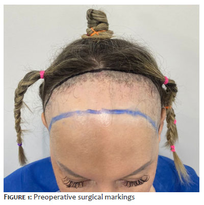
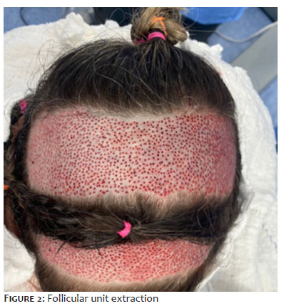
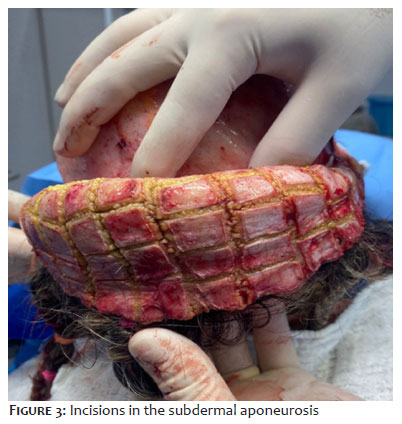
Initially, surgical planning and marking of the donor area were performed to determine the number of follicular units (FUs) to be harvested (Figure 1). A total of 2,188 FUs were harvested from the occipital region (Figure 2) using a motorized Trivellini® system with a 0.9-mm punch, a diameter reported in the literature to minimize follicular trauma and preserve graft integrity. During graft preparation, a scalp flap was elevated in the subgaleal plane, with relaxing incisions performed to increase tissue mobility and elasticity (Figure 3).
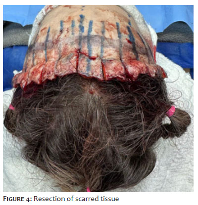
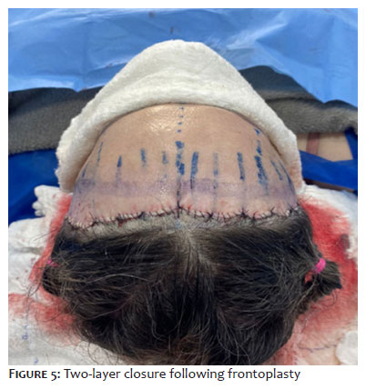
After measurement of the advancement achieved through detachment, an elliptical segment of skin was excised from the frontal region, measuring approximately 4 cm in width, corresponding to the advancement obtained, and 20 cm in length (Figure 4). Simultaneously, a scalp biopsy was performed to monitor the underlying disease. Histopathological examination found mild perivascular and perifollicular inflammatory infiltrates, followed by closure in two layers (Figure 5).
Positioning of the new hairline was based on facial proportions, the anatomy and dynamics of the frontalis muscle, and individualized aesthetic parameters, taking into account the anticipated advancement obtained through frontoplasty and resection of scarred tissue. The recipient area was prepared using 0.8-mm premade incisions, respecting the natural angulation and direction of hair growth. Implantation density was adjusted according to the recipient site, using 50 follicular units/cm² in well-vascularized scarred areas and 40 follicular units/cm² in regions with fibrosis or atrophy, seeking to optimize graft survival.
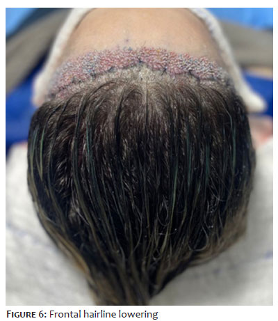
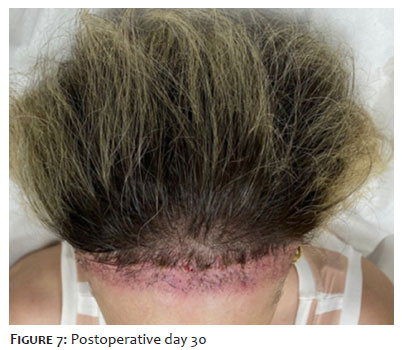
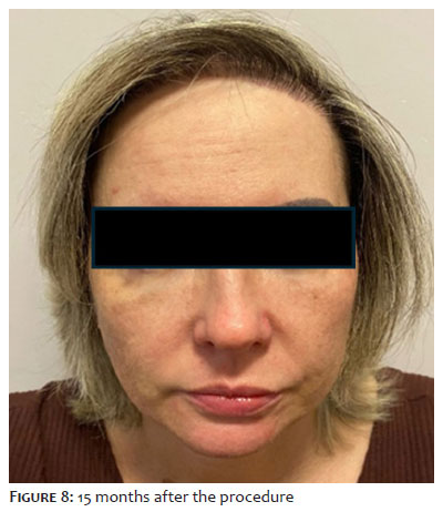
The follicular units were implanted using blunt implanters, a technique that provides greater control of implantation depth, reduces graft trauma, and minimizes risks (Figure 6). The design and orientation of the grafts followed anatomical and aesthetic principles, respecting the natural pattern of female frontal hair growth, with special attention given to the transition between the scarred area and non-scarred scalp. Figure 7 documents the outcome 30 days after the procedure. Following surgery, the patient continued using clobetasol shampoo twice weekly until the 15-month postoperative follow-up visit (Figure 8), reducing use to once weekly afterward. During longitudinal follow-up, a multidisciplinary approach was adopted, including psychological follow-up
FFA was first identified and described by Kossarden in 1994 as a primary lymphocytic cicatricial alopecia characterized by irreversible destruction of hair follicles. Clinically, the disorder affects the frontotemporal hairline and is characterized by perifollicular erythema, follicular hyperkeratosis, and scarring with hairline recession, features that aid in the clinical diagnosis.4,5
The absence of a standardized treatment protocol for alopecia limits the treatment; however, studies have shown that the combination of antimalarial agents and intralesional corticosteroid therapy yields a positive response in up to 60% of cases, whereas oral corticosteroid therapy is ineffective. The use of 5-alpha-reductase inhibitors, within the recommended maximum daily dosage, leads to disease stabilization.6,7
After disease stabilization has been achieved through medical treatment for a minimum period of 12 months, aesthetic reconstructive surgery may be performed to improve the affected area. Accordingly, hair transplantation can be performed as an isolated procedure in areas affected by cicatricial alopecia.8,9
A multicenter study involving 52 patients reported satisfaction rates exceeding 80% when hair transplantation was performed in areas with stabilized disease following pharmacological treatment.9 A different study found improvements in both patient satisfaction and self-esteem following frontal hairline-lowering surgery among women.10 Therefore, in collaboration with the multidisciplinary team and to achieve a superior aesthetic outcome, excision of scarred tissue by means of frontoplasty combined with advancement of the hairline through individual follicular unit transplantation was selected.11
FU extraction was performed using the shave FUE technique, chosen to minimize the transection rate, as the literature reports damage rates below 4% with this approach.12 Hair density planning was designed to preserve a natural appearance while respecting aesthetic standards and the average follicular unit density recommended for the patient’s ethnicity, resulting in a final density of approximately 40 to 45 FUs/cm².13
The ultimate aesthetic outcome and the true survival of the transplanted follicular units can be adequately observed 12 months postoperatively. However, the possibility of loss of transplanted FUs and recurrence of inflammatory activity in the treated area cannot be ruled out.7,9,10 For this reason, continued pharmacological treatment and regular follow-up with a dermatologist are necessary for adjustment and maintenance of therapy with clobetasol shampoo.14
This study shows that pharmacological treatment is crucial for stabilizing and promoting regression of FFA, thereby allowing appropriate planning for aesthetic correction of the affected area. Hair transplantation can be performed over scarred tissue, although complete graft survival cannot be guaranteed. Combining this procedure with surgical excision of the affected region may result in superior aesthetic and functional outcomes. This case report contributes to the literature by highlighting the feasibility of performing both procedures in the same surgical time, promoting improved aesthetic results and enhancing patient self-esteem.
This treatment strategy emphasizes the importance of initial pharmacological treatment followed by the potential benefits of combining hair transplantation with surgical resection of the fibrotic area, providing both aesthetic and psychological benefits for patients.
Rafael Reinert
ORCID: 0009-0002-2357-435X
Approval of the final version of the manuscript.
André Felipe Kroenke
ORCID: 0009-0004-7581-9368
Effective participation in the conduct of the study.
Emanoelle Machado
ORCID: 0009-0008-7433-3517
Critical review of the literature.
Marcos Felippe Takano de Saidneuy
ORCID: 0009-0009-3815-4439
Approval of the final version of the manuscript, Preparation and writing of the manuscript.
Lauren Menna Marcondes
ORCID: 0009-0009-5124-0470
Acquisition, analysis and interpretation of data.
Murilo Gamba Beduschi
ORCID: 0000-0002-5218-9509
Critical revision of the manuscript.
1. Graça Neto L, Araujo LRR, Graça ACM. Frontoplastia endoscópica subperiostal com fixação temporal por fios de sutura. Rev Bras Cirurgia Plástica, v. 37, p. 3-8, 2022.
2. Cintra GS, Cristovão AX, Silva MVA, Corazza AV. Técnicas de Transplante Capilar e suas Principais Complicações: uma Revisão Integrativa. Arch Health Invest. 2022;11(5):827–31.
3. Schmidt M, Ramelli E, Atlan M, Cristofari S. Étude anatomique scanographique du sinus frontal des patients transgenres en vue d’une chirurgie de frontoorbitoplastie de féminisation [Frontal sinus anatomical scanographic study of transgender patients for feminization frontoorbitoplasty surgery]. Ann Chir Plast Esthet. 2023;68(2):93-8.
4. Kossard S. Postmenopausal frontal fibrosing alopecia: scarring alopecia in a pattern distribution. Arch Dermatol. 1994;130(6):770–4.
5. Jouanique C, Reygagne P. L'alopécie frontale fibrosante [Frontal fibrosing alopecia]. Ann Dermatol Venereol. 2014;141(4):272-8.
6. Marmor MF, Kellner U, Lai TY, Melles RB, Mieler WF, American Academy of Ophthalmology. Recommendations on screening for chloroquine and hydroxychloroquine retinopathy. Ophthalmology. 2016;123(6):1386-94.
7. Vañó-Galván S, Molina-Ruiz AM, Serrano-Falcón C, Arias-Santiago S, Rodrigues-Barata AR, Garnacho-Saucedo G. Frontal fibrosing alopecia: a multicenter review of 355 patients. J Am Acad Dermatol. 2014;70(4):670-8.
8. Unger W, Unger R, Wesley C. The surgical treatment of cicatricial alopecia. Dermatol Ther. 2008;21(4):295-311.
9. Vañó-Galván S, Villodres E, Pigem R, Navarro-Belmonte MR, Asín-Llorca M, Meyer-González T. Hair t.ransplant in frontal fibrosing alopecia: A multicenter review of 51 patients. J Am Acad Dermatol. 2019;81(3):865-6.
10. Park JH, You SH, Kim N. Frontal hairline lowering with hair transplantation in Asian women with high foreheads. Int J Dermatol. 2019;58(3):360-4.
11. Jimenez F, Alam M, Vogel JE, Avram M. Hair transplantation: Basic overview. J Am Acad Dermatol. 2021;85(4):803-14.
12. Avram MR, Watkins S. Robotic Hair Transplantation. Facial Plast Surg Clin North Am. 2020;28(2):189-96.
13. Epstein GK, Epstein J, Nikolic J. Follicular Unit Excision: Current Practice and Future Developments. Facial Plast Surg Clin North Am. 2020;28(2):169-76.
14. Mendes-Bastos P, Camps-Fresneda A. Hair Transplantation for Frontal Fibrosing Alopecia: Part of the Solution? Actas Dermosifiliogr. 2016;107(1):3-4.
 All content the journal, except where identified, under the Creative Commons Attribution 4.0 International licence - ISSN-e 1984-8773




 Read in Portuguese
 Portuguese PDF
 Print
Print
 Send this article by email
 How to cite this article
 Submit a comment
 Mendeley
 Pocket
 Share on Facebook
 Share on Twitter