


Luíza Maria Grangeiro de Sousa1; João Alfredo Monte Melo de Barros2; Rafael Freire de Lira Sobral3; Ana Beatriz Albuquerque Nunes4 Jader Freire Sobral Filho5; Maria Tereza de Medeiros Leite Espínola5
Funding source: None
Conflict of interest: None
Submitted on: 08/02/2025
Final decision: 02/09/2026
How to cite this article: How to cite this article: Sousa LMG, Barros JAMM, Sobral RFL, Nunes ABA, Espínola MTML, Sobral Filho JF. Amelanotic melanoma in a tattoo: A great masquerader. Surg Cosmet Dermatol. 2026;18(2):e20260504.
Amelanotic malignant melanoma (AMM) is a rare and diagnostically challenging variant, especially when located on tattooed skin, where visual clues may be obscured. We report the case of a young male patient with AMM over a deltoid tattoo, initially misdiagnosed as a benign lesion.
Histopathology revealed melanoma in situ with a Breslow thickness of 0.7 mm. This case deviates from the typical AMM profile and raises questions about the potential role of tattoos in skin carcinogenesis. Although a causal link is unproven, the rising prevalence of tattoos highlights the need for clinical vigilance and further research into this possible association.
Keywords: Melanoma; Melanoma, Amelanotic; Tattooing
Amelanotic malignant melanoma (AMM) presents a particular challenge in the diagnosis of cutaneous melanoma due to its lack of pigmentation,1 accounting for 2% to 20% of all melanomas.2 The growing popularity of tattoos adds complexity to dermatologic diagnosis because of their association with various skin lesions, both benign and malignant. Identification of AMM can be challenging given that it does not exhibit the typical features of melanoma, such as asymmetry, irregular borders, and color variation. This challenge is even greater when AMM occurs in tattooed areas, where the visual clues used to identify skin abnormalities may be obscured.3 In this report, we present a case that illustrates AMM arising in the context of a tattoo, emphasizing the importance of clinicians remaining vigilant for melanomas associated with tattoos. Early recognition and diagnosis are essential, since diagnostic delays have been associated with adverse outcomes.
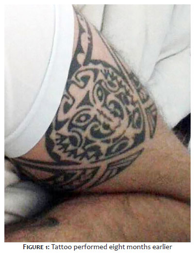
A 27-year-old male patient with Fitzpatrick skin phototype II sought care at our service for evaluation of a red patch with occasional pruritus on a tattoo located over the left deltoid region, which had appeared approximately four months earlier. The tattoo had been performed eight months prior, without immediate complications or reactions, and the lesion was not present at that time (Figura 1). The patient reported that previous dermatologists had suspected either an allergic reaction or a bacterial infection and had prescribed treatment with betamethasone and amoxicillin for 7 days. Despite treatment, the lesion persisted.
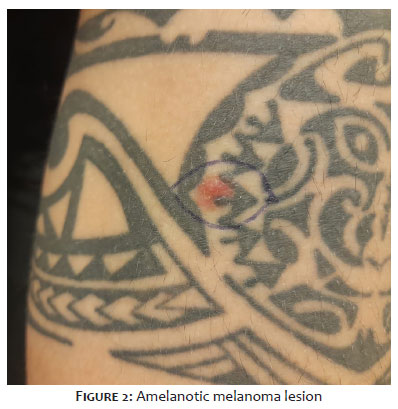
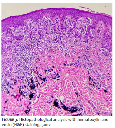
Clinical examination revealed an erythematous lesion 1.5 cm in diameter on the left deltoid region, overlying a black-pigmented tattoo and exhibiting regular oval borders. The appearance of the lesion was consistent with amelanotic melanoma (Figura 2). Histopathological analysis revealed lentigo maligna in situ with an epithelioid cell pattern. In addition, a mild inflammatory process was observed surrounding the tumor, as well as absence of mitoses (Figura 3). Breslow thickness was 0.7 mm, classifying the lesion as Clark level III. No lymphatic invasion, perineural invasion, or regression was detected. A second excision was performed to widen the safety margins. No evidence of residual melanoma was identified in the scar tissue.
The patient remains under clinical follow-up, with quarterly visits scheduled for five years to ensure adequate monitoring. As part of follow-up care, the patient was also instructed to perform regular self-examinations of all skin lesions, assessing them for asymmetry, borders, color, diameter, and progression
AMM is a subtype of cutaneous melanoma characterized by little to no clinically perceptible pigmentation.1 Its diagnosis is complicated by the absence of the cardinal warning signs of melanoma, which may cause it to resemble less severe skin lesions, including inflammatory disorders and benign tumors (benign melanocytic lesions, pyogenic granulomas, nevi, and seborrheic keratoses), as well as malignant neoplasms (basal cell carcinoma, Bowen’s disease, and squamous cell carcinoma).1,6 This complexity may result in inappropriate treatment. In the present case, the patient had previously been treated for other conditions, resulting in delays in biopsy and subsequent treatment. The impact of this diagnostic challenge on melanoma mortality is a key factor to consider. AMMs are frequently diagnosed at advanced stages, resulting in lower overall survival rates than those of pigmented malignant melanomas (PMMs).4 However, when Breslow thickness is taken into account, AMM becomes comparable to PMM.5 This suggests that, once Breslow thickness is considered, the mortality risk becomes more closely aligned between the two subtypes, highlighting the importance of this specific parameter in assessing the prognosis of melanomas with varying degrees of pigmentation.
It remains unclear whether the advanced stage is primarily due to delayed diagnosis or whether amelanotic melanoma inherently exhibits features associated with greater Breslow thickness, mitotic activity, ulceration, and the nodular subtype.2,4,5 This case adds an intriguing dimension, as the lesion presented with a Breslow stage I, without mitoses or ulceration, challenging conventional expectations. Typically, AMM is associated with older age and is most often found in chronically sun-exposed areas.2,5,7 The present case diverges from this pattern, as the patient was only 27 years old and the lesion was located in a sun-protected site. This departure from the typical AMM characteristics raises the possibility that tattooing may play a role in carcinogenesis. However, it is important to note that this relationship has not been conclusively demonstrated.
Despite the increasing prevalence of tattoos, only a limited number of reported cases have involved melanoma in tattoos. The mechanisms underlying malignant transformation in tattoos remain unclear, giving rise to several hypotheses. Pigment toxicity, chronic inflammatory responses, and trauma may trigger malignant transformations, whereas exposure to ultraviolet (UV) radiation may induce oxidative stress. In addition, melanoma cells may be disseminated during the tattooing process.8 Dark tattoos may not exert a direct carcinogenic effect but still interfere with the early detection of malignant transformations, making it more difficult to identify the “ugly duckling” sign. Although increased UV light absorption by dark tattoo pigments has been proposed as a potential factor in carcinogenesis, the present case introduces uncertainty, given that the tumor site was not exposed to sunlight.10
The findings of this case are consistent with the clinical patterns reported in the literature, showing that malignancies in tattoos tend to occur at a younger age compared to typical epidemiological data for melanoma diagnosis in the general population.9,10
In conclusion, this case of AMM deviates from the conventional pattern and underscores the importance of early and accurate diagnosis given its deceptive presentation. As tattoos continue to increase in popularity, investigating possible associations with melanoma becomes critical, reinforcing the existing clinical uncertainty and the need for ongoing vigilance.
Luíza Maria Grangeiro de Sousa
ORCID: 0000-0002-0067-959X
Approval of the final version of the manuscript, Conception and design of the study, Preparation and writing of the manuscript, Acquisition, analysis and interpretation of data, Effective participation in the conduct of the study, Intellectual participation in the propaedeutic and/or therapeutic approach to the cases studied, Critical review of the literature, Critical revision of the manuscript.
João Alfredo Monte Melo de Barros
ORCID: 0000-0002-4681-0311
Approval of the final version of the manuscript, Conception and design of the study, Preparation and writing of the manuscript, Critical revision of the manuscript.
Rafael Freire de Lira Sobral
ORCID: 0009-0006-5732-9968
Author’s contribution: Critical revision of the manuscript.
Ana Beatriz Albuquerque Nunes
ORCID: 0009-0005-3551-0670
Critical revision of the manuscript.
Maria Tereza de Medeiros Leite Espínola
ORCID: 0009-0004-2990-1125
Author’s contribution: Critical revision of the manuscript.
Jader Freire Sobral Filho
ORCID:0000-0001-7822-8994
Approval of the final version of the manuscript, Acquisition, analysis and interpretation of data, Effective participation in the conduct of the study, Intellectual participation in the propaedeutic and/or therapeutic approach to the cases studied, Critical revision of the manuscript.
1. Pizzichetta MA, Talamini R, Stanganelli I, Puddu P, Bono R, Argenziano G, et al. Amelanotic/hypomelanotic melanoma: clinical and dermoscopic features. Br J Dermatol. 2004;150(6):1117–24.
2. Thomas NE, Kricker A, Waxweiler WT, Dillon PM, Busam KJ, From L. Comparison of Clinicopathologic Features and Survival of Histopathologically Amelanotic and Pigmented Melanomas: A Population-Based Study. JAMA Dermatol. 2014;150(12):1306–14.
3. Jaimes N, Braun RP, Thomas L; Marghoob AA. Clinical and dermoscopic characteristics of amelanotic melanomas that are not of the nodular subtype. J Eur Acad Dermatol Venereol. 2011;26(5):591–6.
4. Strazzulla LC, Li X, Zhu K, Okhovat JP, Lee SJ; Kim CC. Clinicopathologic, misdiagnosis, and survival differences between clinically amelanotic melanomas and pigmented melanomas. J Am Acad Dermatol. 2019;80(5):1292–8.
5. Wee E, Wolfe R, Mclean C, Kelly JW, Pan Y. Clinically amelanotic or hypomelanotic melanoma: Anatomic distribution, risk factors, and survival. J Am Acad Dermatol. 2018;79(4):645-51.
6. Jaimes N, Braun RP, Thomas L, Marghoob AA. Clinical and dermoscopic characteristics of amelanotic melanomas that are not of the nodular subtype. J Eur Acad Dermatol Venereol. 2011;26(5):591–6.
7. Gualandri L, Betti R, Crosti C. Clinical features of 36 cases of amelanotic melanomas and considerations about the relationship between histologic subtypes and diagnostic delay. J Eur Acad Dermatol Venereol. 2009;23(3):283–7.
8. Tchernev G, Chokoeva AA. Melanoma in a Chinese dragon tattoo. The Lancet. 2015.
9. Leijs M, Schaefer H, Rübben A, Cacchi C, Rustemeyer T. Cutaneous Malignancies in Tattoos, a Case Series of Six Patients. Curr Oncol. 2021;28(6):4721–37.
10. Melanoma Arising in a Tattoo: Case Report and Review of the Literature. [online] Available at: https://www.mdedge.com/dermatology/article/78662/melanoma/melanoma-arising-tattoo-case-report-and-review-literature.
 All content the journal, except where identified, under the Creative Commons Attribution 4.0 International licence - ISSN-e 1984-8773




 Read in Portuguese
 Portuguese PDF
 Print
Print
 Send this article by email
 How to cite this article
 Submit a comment
 Mendeley
 Pocket
 Share on Facebook
 Share on Twitter