


Elisandra Barbara Pontes Carlos; Paulo Roberto Gorgatti Filho; Ruan Giorgenon; Hélio Amante Miot; Anna Carolina Miola
Funding: None
Conflict of interest: None
Submitted on: 01/09/2026
Final decision: 02/12/2026
How to cite this article: Carlos EBP, Gorgatti Filho PR, Giorgenon R, Miot HA, Miola AC. The role of high-frequency ultrasound in the evaluation of an acral subcutaneous nodule: a case report and literature review. Surg Cosmet Dermatol. 2026;18(1):e20260545.
Acral subcutaneous nodules pose a diagnostic challenge due to their broad differential diagnoses. We report a case of a foreign body granuloma presenting as a long-standing acral subcutaneous nodule on the thumb of a 64-year-old woman. High-frequency ultrasound revealed a homogeneous hypoechoic nodule with posterior acoustic enhancement, surrounding a central linear hyperechoic structure suggestive of a foreign body reaction. Excisional biopsy confirmed a granulomatous inflammatory process surrounding a vegetal fiber. This case highlights the value of high-frequency ultrasound as a noninvasive tool to support etiological diagnosis and guide surgical planning in chronic acral subcutaneous nodules.
Keywords: Ultrasonography; Granuloma, Foreign-Body; Epidermal Cyst
Acral subcutaneous nodules (ASNs) pose a diagnostic challenge given the broad range of conditions that may present with this clinical appearance. In this context, complementary imaging techniques, such as high-frequency ultrasound (HFUS), can aid in establishing an etiological diagnosis,1 since ASNs, although common in dermatological practice, may represent inflammatory, infectious, or neoplastic conditions that require distinct management strategies.
We report a case of a foreign body granuloma (FBG) presenting as an ASN on the thumb and discuss the clinical features and HFUS findings that helped elucidate the etiological diagnosis.
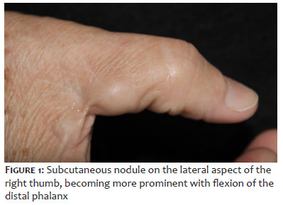
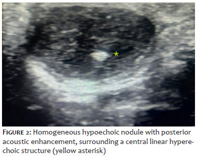
A 64-year-old White woman presented with a painless, slow-growing nodule that had progressively increased in size over 15 years. Physical examination revealed a 1.5 × 1.0 cm no¬dular lesion located on the lateral aspect of the proximal phalanx of the thumb, which was fibroelastic in consistency, mobile, and non-tender (Figure 1). The initial clinical differential diagnoses included giant cell tumor of the tendon sheath, epidermal inclusion cyst, lipoma, FBG, and low-grade fibromyxoid sarcoma. HFUS was performed using a 22 MHz transducer (LogicE GE®, GE Electronics®, Boston, MA, USA) and demonstrated a ho¬mogeneous hypoechoic nodule with posterior acoustic enhan¬cement, surrounding a central linear hyperechoic structure (Figure 2). No central vascular flow was detected on Doppler imaging. These findings were suggestive of an FBG.
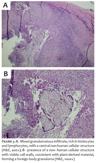
An excisional biopsy was subsequently performed. Histopathological examination revealed granulation tissue composed of aggregates of epithelioid histiocytes surrounded by a lymphocytic rim, with a central suppurative area and no cellular atypia. A central foreign body compatible with a vegetal fiber was identified (Figure 3), confirming the diagnosis of FBG. Upon further questioning, the patient recalled a prior injury involving "broom fibers."
Foreign body granulomatous reaction is defined as a chronic inflammatory response to exogenous materials introduced into the skin, such as sand, plant fibers, glass, or injectable substances (e.g., polymethyl methacrylate [PMMA]).2 In the present case, HFUS suggested the diagnosis of FBG through the visualization of a central hyperechoic structure corresponding to the retained foreign material.
The use of HFUS has expanded across all areas of dermatology. Owing to its high spatial resolution (0.1–0.2 mm), HFUS enables the assessment of small structures, improving diagnostic accuracy and facilitating the evaluation of the activity of several inflammatory, infectious, neoplastic, and degenerative skin disorders.1
A previous study compared the ultrasound findings of patients with FBG secondary to injectable fillers (e.g., PMMA) with those of patients with hyaluronic acid deposits.3 In FBG, lesions are characteristically oval with irregular, poorly defined margins and contain hyperechoic foci representing accumulated material surrounded by granulomatous inflammation — features consistent with those observed in the present case.4
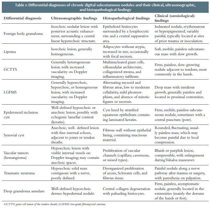
HFUS may also aid in differentiating ASNs of various etiologies. Lesions with heterogeneous central areas and increased Doppler flow may suggest a neoplastic etiology, whereas inflammatory lesions or benign tumors tend to be more homogeneous and avascular on HFUS. Cystic lesions, in turn, typically demonstrate posterior acoustic enhancement.5 Table 1 summa¬rizes the main HFUS and histopathological features of chronic ASN differential diagnoses.
In patients with chronic ASNs, obtaining an accurate clinical history may be challenging due to the often prolonged period between exposure and lesion development, hindering clinical characterization and diagnosis. In this setting, HFUS — a noninvasive, painless, rapid, and increasingly common procedure in dermatological practice — emerges as an important tool in the management of ASNs.
In conclusion, we report a case of ASN whose etiology was FBG, which could be anticipated by HFUS findings. Dermatologists should consider HFUS as part of the diagnostic workup of ASNs, thereby aiding surgical planning.
Elisandra Barbara Pontes Carlos
ORCID: 0009-0001-4150-2598
Approval of the final version of the manuscript, Preparation and writing of the manuscript, Acquisition, analysis and interpretation of data, Critical review of the literature.
Paulo Roberto Gorgatti Filho
ORCID: 0000-0003-4127-7172
Approval of the final version of the manuscript, Preparation and writing of the manuscript, Acquisition, analysis and interpretation of data, Critical review of the literature.
Ruan Giorgenon
ORCID: 0000-0001-7131-3280
Author's contribution: Approval of the final version of the manuscript, Preparation and writing of the manuscript, Acquisition, analysis and interpretation of data, Critical review of the literature.
Hélio Amante Miot
ORCID: 0000-0002-2596-9294
Approval of the final version of the manuscript, Preparation and writing of the manuscript, Intellectual participation in the propaedeutic and/or therapeutic approach to the cases studied, Critical revision of the manuscript.
Anna Carolina Miola
ORCID: 0000-0001-8926-734X
Approval of the final version of the manuscript, Conception and design of the study, Effective participation in the conduct of the study, Intellectual participation in the propaedeutic and/or therapeutic approach to the cases studied, Critical review of the literature, Critical revision of the manuscript.
1. Argalia G, Reginelli A, Molinelli E, Russo A, Michelucci A, Sechi A, et al. High- Frequency and Ultra-High-Frequency Ultrasound in Dermatologic Diseases and Aesthetic Medicine. Medicina (Kaunas) 2025;61(2):220.
2. Molina-Ruiz AM, Requena L. Foreign body granulomas. Dermatol Clin 2015;33:497-523.
3. Mlosek RK, Skrzypek E, Skrzypek DM, Malinowska S. High-frequency ultrasound-based differentiation between nodular dermal filler deposits and foreign body granulomas. Skin Res Technol 2018;24(3):417-22.
4. Valentim F de O, Miola AC, Miot HA. Low-grade fibromyxoid sarcoma: important differential diagnosis in acral tumor lesions. Surg Cosmet Dermatol 2022;14.
5. Kuwano Y, Ishizaki K, Watanabe R, Nanko H. Efficacy of diagnostic ultrasonography of lipomas, epidermal cysts, and ganglions. Arch Dermatol 2009;145(7):761-4.
 All content the journal, except where identified, is under a Creative Commons Attribution-NonCommercial 4.0 International license - ISSN-e 1984-8773


 Read in Portuguese
 Portuguese PDF
 Print
Print
 Send this article by email
 How to cite this article
 Submit a comment
 Mendeley
 Pocket