


Bahareh Nowruzi; Mahshid Alibabaei
Funding support: None.
Conflicts of interest: None.
Submitted on: 19/08/2024
Approved on: 16/01/2025
How to cite this article: Nowruuzi B, Alibabaei M. Study of the antimicrobial and antioxidant activity of anti-acne face wash, hand sanitizer gel, and soap containing phycocyanin and phycoerythrin. Surg Cosmet Dermatol. 2025;17:e20250396.
INTRODUCTION: The use of natural ingredients in skincare products has gained increasing attention due to the harmful effects and environmental risks associated with synthetic compounds.
OBJECTIVE: This growing concern has driven research into photosynthetic organisms as sustainable and eco-friendly sources of effective ingredients. Natural extracts — particularly those derived from plants, algae, and cyanobacteria — have drawn significant interest in the cosmetics industry. Cyanobacteria stand out for their low cultivation requirements, rapid growth, and ability to produce a wide range of bioactive metabolites, making them a sustainable and cost-effective resource.
METHODS: This study evaluated the effects of natural pigments — phycocyanin and phycoerythrin — extracted from Spirulina platensis and Nostoc sp. on three cosmetic products: soap, anti-acne face wash, and hand sanitizer gel. After culturing the cyanobacteria, the pigments were extracted, purified, and coated with chitosan as a stabilizer. These coated pigments were then used to formulate the cosmetic products, which were assessed for viscosity, pH, stability, antioxidant activity, and antibacterial properties.
RESULTS: The results showed that phycocyanin and the combined pigment content were higher in the anti-acne face wash and soap, whereas phycoerythrin and the total pigment concentration were greater in the hand sanitizer gel.
CONCLUSION: This study demonstrates the potential of cyanobacterial pigments to produce stable, enriched cosmetic products, offering sustainable and effective alternatives to synthetic ingredients.
Keywords: Cyanobacteria; Microwaves; Microbiology.
Cyanobacteria have been used for over 2,000 years, dating back to their application by the Chinese during periods of malnutrition. Since the 1950s, they have been increasingly utilized in biotechnology.1,2 Their chemical composition enhances the nutritional value of food and animal feed, plays a crucial role in aquaculture, and supports the development of cosmetic products.2 Microalgal biomass, including that of cyanobacteria, is used to produce a variety of valuable products, particularly high-protein dietary supplements for human nutrition, aquaculture, and nutraceutical applications.3
Cosmetics have become an essential part of daily routines, with growing consumer demand driven by concerns for skin health and aesthetics. Ethical issues surrounding the use of animal-derived ingredients, as well as the potential adverse effects of synthetic substances — such as allergies and environmental risks — have led to increased interest in cosmetic products formulated from photosynthetic organisms.4 Among these, microalgae stand out for their ability to promote skin healing and repair, along with anti-blemish and anti-inflammatory properties.5-7
The cyanobacterial genera Nostoc, Spirulina (also known as Arthrospira), and Aphanizomenon are particularly well-studied due to their high content of calcium, beta-carotene, phosphorus, iron, biotin, folic acid, pantothenic acid, and vitamin B12.8 Extracts and bioactive compounds derived from these species are under investigation for their protective effects on skin and hair.9 For example, beta-carotene from Desmonostoc muscorum, Leptolyngbya foveolarum, and Arthrospira platensis has demonstrated the ability to regulate UV-A-induced gene expression in human keratinocytes.10 It also modulates biological targets such as NF-κB, COX-2, and matrix metalloproteinase-9, owing to its anti-inflammatory activity.5
Cyanobacterial phycobiliproteins (PBPs), such as phycocyanin (PC), exhibit antioxidant, anti-inflammatory, and anti-aging properties and are already being utilized in cosmetics.7-11 These hydrophilic proteins, found in cyanobacteria and certain red algae, offer promising commercial applications as natural cosmetic ingredients.12 Structurally similar to bilirubin, PBPs are efficient scavengers of reactive oxygen species, making them potential antioxidant agents. They are composed of three main components: phycoerythrin (PE; a red pigment), allophycocyanin (a bluish-green pigment), and PC (a blue pigment). These fluorescent protein-pigment complexes have diverse commercial potential.13
Spirulina contains several key phytopigments — including PC, gamma-linolenic acid, phycocyanobilin, and phycoerythrobilin — which contribute to its antioxidant, anti-melanogenic, anti-wrinkle, and anti-aging properties.11 These compounds are already incorporated into cosmetic products such as lipsticks, eyeliners, and eyeshadows as natural colorants. In addition to its dermatological benefits, Spirulina is also recognized for its anti-inflammatory, neuroprotective, and hepatoprotective effects. It has been shown to reduce serum levels of alanine aminotransferase, aspartate aminotransferase, and malondialdehyde.4,14
Cyanobacterial extracts also contain peptides and proteins that are suitable for use in hair care products, including lotions, shampoos, permanent wave solutions, and hair coloring agents.15,16 Studies on Chlorogloeopsis sp. and Spirulina extracts have reported benefits such as increased shine, easier combing, improved hair restoration, and enhanced moisturization. For example, the Blue Green Algae Hair Rescue Conditioning Mask by Aubrey Organics has shown effectiveness in strengthening hair and preventing breakage and split ends.6
Active cyanobacterial extracts, such as Spirulina-derived Spiralin®, are currently in use in the cosmetics industry for skin protection. Products like Skinicer® Repair Cream and Spirularin® contain these extracts, which have demonstrated regenerative effects on damaged skin cells and collagen, as well as protection against UV radiation. Additionally, the PBP C-phycocyanin (C-PC), derived from Aphanizomenon flos-aquae, is used in NaturCyanin Bioactive® as a natural alternative to synthetic colorants, due to the attractive pink-purple hue it imparts.17
There remains limited research on cosmetic applications of other cyanobacterial genera, thus presenting a valuable opportunity for further exploration. This study evaluates the antioxidant and antimicrobial effects of soap, anti-acne face wash, and hand sanitizer gel formulated with chitosan-coated PC and PE.
Cyanobacterial strains Spirulina platensis and Nostoc sp., isolated from the cyanobacteria culture collection (CCC) of the ALBORZ Herbarium at the Science and Research Branch, Islamic Azad University, Tehran, were cultivated in modified Zarrouk and BG110 media, respectively. Cultures were maintained in illuminated growth chambers (300 µmol m-2 s-1) at 28 ± 2 ºC for 30 days.18,19
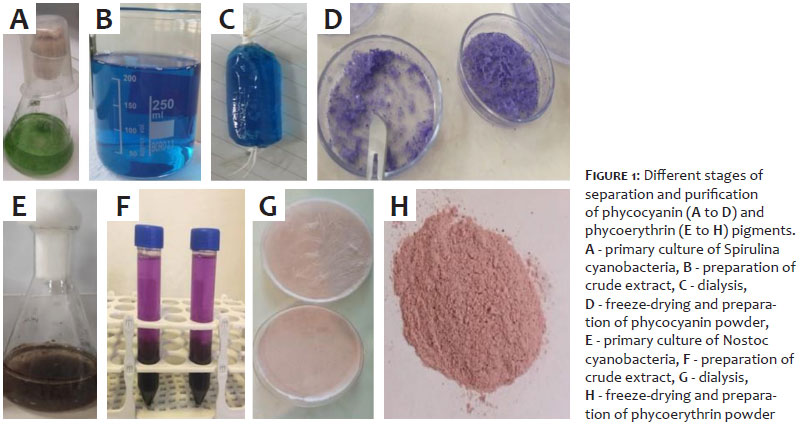
PC and PE were extracted and purified from 14-day-old log-phase cultures.20 The cyanobacterial cultures were centrifuged at 4,000 rpm to collect the biomass pellet. The pellet was resuspended in a specific buffer for PC and in potassium phosphate buffer for PE. The extraction procedure was repeated daily for four days until the cell biomass turned dark purple.
Crude pigment extracts were obtained by centrifugation at 5,000 rpm for 10 minutes. Purification followed the method of Afreen and Fatma, which involves the addition of solid ammonium sulfate to the crude extract to achieve 65% saturation.21 The mixture was centrifuged at 4,500 × g for 10 minutes, and the resulting pellet was resuspended in 50 mM acetic acid–sodium acetate buffer, followed by overnight dialysis. The dialyzed extracts were then filtered through a 0.45 μm membrane filter.
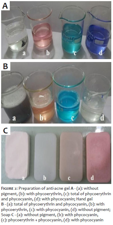
The absorption spectra of the purified pigments were measured using a Specord 200 spectrophotometer (Analytik Jena, Germany), scanning the samples across wavelengths ranging from 300 to 750 nm. Concentrations of PC and PE were determined based on absorbance measurements at 620 and 650 nm (for PC) and at 565 nm (for PE), using standard equations. Pigment purity was assessed at each step by calculating the purity ratios: A620/A652 for PC and A555/A280 for PE (Figure 1).22
(1) PC (µg·mL-1) = (OD620 - 0.70 × OD650) / 7.38
(2) APC (µg·mL-1) = (OD650 - 0.19 × OD620) / 5.65
(3) PE (µg·mL-1) = OD565 - 2.8 × [PC] - 1.34 × [APC]
PC and PE were encapsulated by combining them with a water-soluble chitosan (WSC) solution, using sodium tripolyphosphate as a cross-linking agent. The WSC solution was prepared by dissolving oligochitosan in distilled water at a concentration of 1 mg/mL. This mixture was maintained at 4 ºC for 24 hours to ensure complete hydration. Subsequently, 1 mL of PC + PE solution (in deionized water) was slowly added to 1 mL of WSC solution (1 mg/mL) under constant stirring at 25 ºC. Sodium tripolyphosphate (2 mg/mL) was then added in 0.5 mL aliquots, followed by pH adjustment to 7.0 using 1% HCl. Finally, 0.5 mL of polyethylene glycol (PEG) was added to complete the stabilization process.23
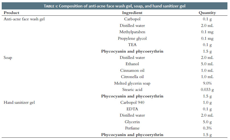
A series of experimental trials were conducted to optimize the laboratory formulations of soap, anti-acne face wash, and hand sanitizer gel. The final compositions were developed based on the best-performing ingredient combinations (Table 1). Four control treatments were included in the formulation process for all three products: (1) a formulation without any pigment, (2) a formulation containing only PC (1.5 g), (3) a formulation containing only PE (1.5 g), and (4) a formulation containing a combination of both pigments (1.5 g total) (Figure 2).24-26
The viscosity of the anti-acne face wash was evaluated using a digital Brookfield viscometer (spindle No. 64), operating at 10 rpm and a controlled temperature of 25 ºC. A predetermined amount of the product was transferred to a beaker, and the viscometer spindle was immersed in the sample to perform the measurement. All tests were conducted in triplicate to ensure accuracy and reproducibility.27
The pH of a 1% aqueous solution of the formulation was measured at a constant temperature of 25 ºC using a calibrated digital pH meter.28
A visual inspection was conducted to assess the physical characteristics of the formulation, including color, appearance, and consistency.29
The stability of the gel formulations was evaluated using the freeze–thaw cycle method. Samples were stored at 4 ºC, 25 ºC, 37 ºC, and 40 ºC for a period of 7 days, and any changes in physical appearance or texture were recorded.30
Homogeneity was assessed visually after transferring the gel into a designated container. Each sample was examined for uniformity in appearance, presence of any lumps or phase separation, and density distribution.31
The antimicrobial activity of the anti-acne face wash was evaluated using the turbidimetric method. A sterile nutrient agar medium was prepared aseptically and spread evenly onto Petri dishes. The acne-affected area on a volunteer's face was first cleansed with distilled water and allowed to air dry. By using a sterile cotton swab, distilled water was applied to a ruptured pimple to collect microbial content, which was then transferred onto the agar medium to inoculate it. Following incubation at 37 ºC for 24 hours, six sterile cotton balls (1 cm in diameter) were immersed for 5 minutes in various formulations — including a standard drug, the test formulations, and distilled water (control). A 50 mL volume of nutrient broth was prepared and sterilized; 5 mL of this broth was used as a blank reference in a UV spectrophotometer. The remaining broth was inoculated with the microbial culture from the Petri plate and divided into six sterile test tubes, each containing one of the cotton ball samples. Test tubes were incubated at 37 ºC for 24 hours. After incubrpmation, the absorbance of each sample was measured at 600 nm to assess microbial growth inhibition.32
A 10 g sample of soap was weighed immediately and recorded as the wet weight. The sample was then dried at a temperature below 115 ºC by using appropriate drying equipment until a constant weight was achieved. After cooling, the sample was reweighed and recorded as the dry weight. The moisture content was calculated using the following equation:
%W = 100 A – B/B × 100.
Where:
%W = Percentage of moisture in the sample
A = Weight of wet sample (g)
B = Weight of dry sample (g)33
A 40 mL portion of the soap solution was transferred into a 100 mL beaker. The spindle of a Brookfield digital viscometer was immersed into the solution, and viscosity was measured under standard conditions.34
An accurately weighed 5 g sample of soap was transferred to a 250 mL beaker. A volume of 100 mL of hot distilled water was added to completely dissolve the soap. Then, 40 mL of 0.5 N nitric acid was added until the solution reached a mildly acidic pH. The mixture was heated in a water bath to promote the separation of fatty acids, which formed a distinct upper layer. After cooling, the fatty acids were isolated using ice.
To extract residual fatty acids, 50 mL of chloroform was added to the remaining solution and transferred to a separating funnel. The mixture was shaken and allowed to separate into two phases. The lower aqueous layer was discarded. Another 50 mL of chloroform was added to the remaining solution to repeat the extraction process. The chloroform layers containing the fatty acids were combined.
The lipid content was transferred to a pre-weighed porcelain dish, evaporated, and the remaining residue was weighed. The percentage of total fatty matter was calculated by determining the mass difference.35
A 10% soap solution was prepared by dissolving 1 g of soap in 10 mL of distilled water in a volumetric flask. The pH was measured using a pre-calibrated digital pH meter.33
A 0.5 g soap sample was dispersed in 25 mL of distilled water and transferred to a measuring cylinder. The solution was diluted to a total volume of 50 mL. The mixture was then shaken manually with 25 strokes. The height of the foam formed above the aqueous solution was measured.33
A 25 mL sample of 1% soap solution was placed in a 100 mL graduated cylinder. The cylinder was manually sealed and shaken 10 times. Foam volume was recorded at 1-minute intervals over a 4-minute period.33
A 5 g sample of soap was dissolved in 50 mL of hot ethanol. The solution was filtered through a filter paper previously coated with tar and rinsed with an additional 20 mL of warm ethanol. The filter paper and retained residue were dried at 105 ºC for 1 hour. After drying, the weight of the residue was measured, and the percentage of alcohol-insoluble matter was calculated using the following formula:
% Alcohol-insoluble matter = (Weight of residue × 100) / Weight of sample36
Liquid soap samples were stored at temperatures of 25 ºC, 37 ºC, 40 ºC, and 50 ºC for a period of 1 month. Stability was monitored over this time frame. Samples that remained homogeneous and free of precipitation or crystallization were classified as stable. In contrast, samples that developed roughened crystals or visible precipitation were deemed unstable.37
Approximately 5 g of the soap sample were placed into a conical flask and mixed with 50 mL of neutralized alcohol. The mixture was subjected to reflux in a water bath for 30 minutes, then cooled to room temperature. Afterward, 1 mL of phenolphthalein indicator solution was added, and the mixture was titrated immediately with 0.1 N hydrochloric acid (HCl) until the pink color disappeared.37
The saponification value represents the number of mg of potassium hydroxide (KOH) required to saponify 1 g of fat or oil. Approximately 2 g of the soap sample were placed into a conical flask, followed by the addition of 0.5 M KOH solution. The mixture was stirred continuously and heated in a water bath at approximately 55 ºC. The temperature was then increased by an additional 100 ºC, and the mixture was boiled for about 1 hour.
Titration was performed using phenolphthalein as an indicator and 0.5 M HCl. The endpoint of the titration was determined by the disappearance of the pink color.38
The saponification value was calculated using the following formula:
Saponification value = (Average volume of KOH × 28.056) / Weight of oil (g)
Antimicrobial tests were conducted to evaluate the biological activity of the optimized soap formulations. The agar well diffusion method was used to assess their efficacy against Escherichia coli, Staphylococcus aureus, and Pseudomonas aeruginosa. Sterile nutrient agar plates were inoculated with the respective test organisms, and wells were created in the agar. Each formulation was placed into individual wells and allowed to diffuse for 2 hours. The plates were then incubated at 37 ºC for 24 hours. After incubation, the zones of inhibition (ZOI) around each well were measured and compared to assess antimicrobial effectiveness.39
Physical characteristics of the hand sanitizer gel were assessed through pH and viscosity measurements. pH was determined by using a pre-calibrated digital pH meter (Mettler Toledo, Philippines). Viscosity was measured with a Brookfield digital viscometer by immersing the spindle in a measured volume of gel placed in a glass container.26
The antibacterial efficacy of the hand sanitizer gel was evaluated against five bacterial species: two gram-positive bacteria (S. aureus and Enterococcus faecalis) and three gram-negative bacteria (E. coli, P. aeruginosa, and Salmonella typhi). Bacterial cultures were maintained on tryptone soya agar at 40 ºC.
Inoculum preparation followed the Clinical and Laboratory Standards Institute (CLSI) guidelines (M02-A12).39 Isolated colonies of each bacterial strain were selected from freshly prepared agar plates and incubated for 18–24 hours. These colonies were then transferred into tryptone soy broth to prepare bacterial suspensions. Turbidity of each suspension was adjusted to achieve a concentration of 1.0 to 2.0 × 10⁸ CFU/mL, verified using a UV-Visible spectrophotometer at 600 nm.
For the assay, 0.1 mL of each bacterial suspension was spread evenly onto Mueller-Hinton agar plates using a sterile spreader. Wells with a diameter of 6 mm were created using sterile borers. Into each well, 50 µL of the formulated gel and commercial hand sanitizer gels were introduced. Positive and negative controls were included using 70% ethanol and dimethyl sulfoxide (DMSO), respectively.
After a 5-minute diffusion period at room temperature, plates were incubated at 37 ºC for 18 to 24 hours. Following incubation, the ZOI surrounding each well were measured using an automatic colony counter set to inhibition zone mode.40
MIC of the hand sanitizer formulations was determined using the macrodilution method in sterile test tubes following the guidelines of the CLSI (M07-A08).41
The laboratory-prepared gel was serially diluted in Mueller-Hinton broth using a 1:2 ratio, resulting in a concentration range from 100% to 0.195%. The test inoculum was prepared in three steps: i) a cell suspension of each bacterial strain was prepared as described in the agar well diffusion assay, adjusted to a concentration of 1 to 2 × 10⁸ CFU/mL; ii) the suspension was diluted at a ratio of 1:150 to reach an intermediate density of 1 × 10⁶ CFU/mL; and iii) this suspension was further diluted 1:2 to obtain the final inoculum concentration of 5 × 10⁵ CFU/mL.
Each test tube in the dilution series received 1 mL of the hand sanitizer formulation and 1 mL of the bacterial inoculum, with the procedure performed within 15 min of inoculum preparation. The contents were mixed thoroughly. Serial dilution was conducted by transferring 1 mL from one tube to the next, maintaining a 1:2 dilution factor across the series. The final tube in the series contained the lowest concentration (0.195%).
The same procedure was followed to determine MIC for different pigment concentrations within the hand sanitizer. All tubes were incubated at 37 ºC for 16 to 20 hours. MIC was defined as the lowest concentration of the formulation at which no visible microbial growth was observed, as determined without the aid of instrumentation.40
MBC was determined by inoculating 0.1 mL of the bacterial suspension from each tube used in the MIC assay onto Mueller-Hinton agar plates using the spread plate technique. Following inoculation, plates were incubated at 37 ºC for 18 to 24 hours.
After incubation, the MBC was identified as the lowest concentration of the hand sanitizer gel at which no bacterial growth was observed. The absence of colony formation on the agar surface indicated complete bactericidal activity, confirming that the formulation effectively eliminated viable bacterial cells in the absence of antibiotics. MBC values were recorded for all pigment-containing hand sanitizer formulations against the five tested bacterial strains.40
Stability testing was performed by storing the hand sanitizer gel samples at three different temperatures (25 ºC, 37 ºC, 40 ºC) for a period of 1 month. Samples were routinely monitored for changes in physical characteristics such as color, odor, and phase separation.26
The antioxidant activity of extracts and products derived from cyanobacterial strains was evaluated using the 2,2-diphenyl-1-picrylhydrazyl (DPPH) radical scavenging assay. This method assesses the ability of antioxidant compounds to neutralize DPPH free radicals.
For the assay, 1 mL of Andrographis paniculata extract was mixed with 2 mL of a 0.1 mM DPPH solution prepared in methanol. The reaction mixture was incubated in the dark for 30 minutes to allow free radical scavenging activity to occur. Following incubation, an aliquot of the mixture was transferred into a cuvette, and absorbance was measured at 517 nm using a UV-Visible spectrophotometer.
A control sample was prepared by mixing 2 mL of the DPPH methanolic solution with 1 mL of methanol. All absorbance measurements were performed in triplicate for each sample.
The percentage of free radical scavenging activity (% inhibition) was calculated using the following formula42:
% DPPH = [(A_control - A_sample) / A_control] × 100
Where:
A_control = absorbance of the control
A_sample = absorbance of the test sample
Experimental data were analyzed using analysis of variance (ANOVA) in the IBM SPSS Statistics for Windows, version 24 (IBM Corp., Armonk, N.Y., USA). A 95% confidence level was applied to determine statistical significance. When significant differences were identified by ANOVA (p ≤ 0.05), the Tukey post-hoc test was used to compare the means.
Each treatment was tested in triplicate, and the results were expressed as mean values ± standard error of the mean (SEM).43
Spectrophotometric analysis revealed that PE exhibited maximum absorbance at 562 nm, with a secondary peak at 617 nm. PC showed its maximum absorbance at 621.9 nm. The calculated purity values were 0.845 for PE and 0.401 for PC (Figure 3).
One-way ANOVA followed by Tukey's post-hoc test indicated no statistically significant differences in viscosity among the different treatments over a 30-day period. However, formulations containing PC alone and PC + PE pigments exhibited the highest viscosity compared to the control.
Analysis of pH values using one-way ANOVA and Tukey's test showed no significant difference between treatments over the 30-day observation period. Notably, the control formulation recorded the highest pH value.
Gels formulated with PE, PC, and PC + PE displayed distinct coloration: pale pink, blue, and green, respectively, while the control gel remained colorless. All formulations were shiny, transparent, and exhibited a light, cooling sensation upon application to the skin (Figure 2).
One-way ANOVA followed by Tukey's test revealed no statistically significant differences in foamability among the various formulations over the 30-day evaluation period.
Assessment of physical stability — including color, odor, and consistency — at 4 ºC over 30 days showed that all formulations received the maximum score (5 out of 5) for all parameters.
At 25 ºC and 37 ºC, no significant differences in consistency were observed between the pigment-containing formulations and the control. However, on day 30 at 40 ºC, the control formulation showed a significant reduction in consistency compared to the pigment-containing treatments.
Color retention analysis at 25 ºC demonstrated that pigment-containing formulations maintained a significantly higher color intensity than the control throughout the 30-day period. At 37 ºC and 40 ºC, a significant difference in color intensity between the control and the pigment-treated samples was observed starting from day 20.
In contrast, odor assessments at 25 ºC, 37 ºC, and 40 ºC showed no significant differences between treatments and the control group over the entire 30-day period.
Visual inspection of the anti-acne gel formulations confirmed that all samples remained homogeneous throughout the entire evaluation period. No signs of phase separation, sedimentation, or texture inconsistencies were observed on any of the measurement days. According to one-way ANOVA followed by Tukey's post-hoc test, no significant difference in antimicrobial activity was observed between the control and the PE-containing gel until day 30. However, formulations containing PC and PC + PE showed significantly enhanced antimicrobial activity on days 15 and 5, respectively. Among all treatments, gels formulated with the combined pigments and those containing PC alone exhibited the highest bacterial inhibition rates.
Analysis using one-way ANOVA and Tukey's test showed that the highest antioxidant activity was observed on day 10 in gels formulated with PC + PE.
Results of one-way ANOVA and Tukey's test showed a significant decrease in moisture content over the 30-day period. Notably, on days 25 and 30, soaps enriched with PE alone and with the combination of both pigments retained significantly higher moisture content compared to the other formulations.
Both total fat content and pH levels showed a significant decrease over the 30-day period, according to one-way ANOVA and Tukey's test. However, no statistically significant differences in fat content were observed among the soaps prepared with different types of pigment.
One-way ANOVA and Tukey's test revealed a significant decline in foam height over the 30 days. However, no significant differences were observed between days 25 and 30. Additionally, there was no statistically significant difference in foam height among soaps formulated with different pigments on days 25 and 30.
One-way ANOVA and Tukey's test showed that foam height decreased significantly over the 30-day observation period. However, no statistically significant differences were found in foam retention between the various pigment-containing treatments and the control across different time points.
The analysis revealed a significant increase in alcohol-insoluble content compared to the control. However, no significant differences were observed between the different pigment-treated formulations.
One-way ANOVA followed by Tukey's test showed no significant differences in color or odor stability among the soap formulations over the 30-day period. Additionally, there were no statistically significant differences between pigment-containing treatments and the control in terms of color stability. At 25 ºC over 30 days, stable soap formulations showed no visible color changes or phase separation. Homogeneous samples were classified as stable, while those exhibiting sediment-like separation were considered unstable. Throughout the 30-day evaluation period, all soap formulations maintained structural integrity and received the maximum score of 5 for form stability.
No statistically significant differences were found in the percentage of alkali among the treatments when compared to the control, as determined by one-way ANOVA and Tukey's test.
Saponification value showed a statistically significant decrease compared to the control group, which indicates a reduction in the degree of saponification in pigment-treated soap formulations.
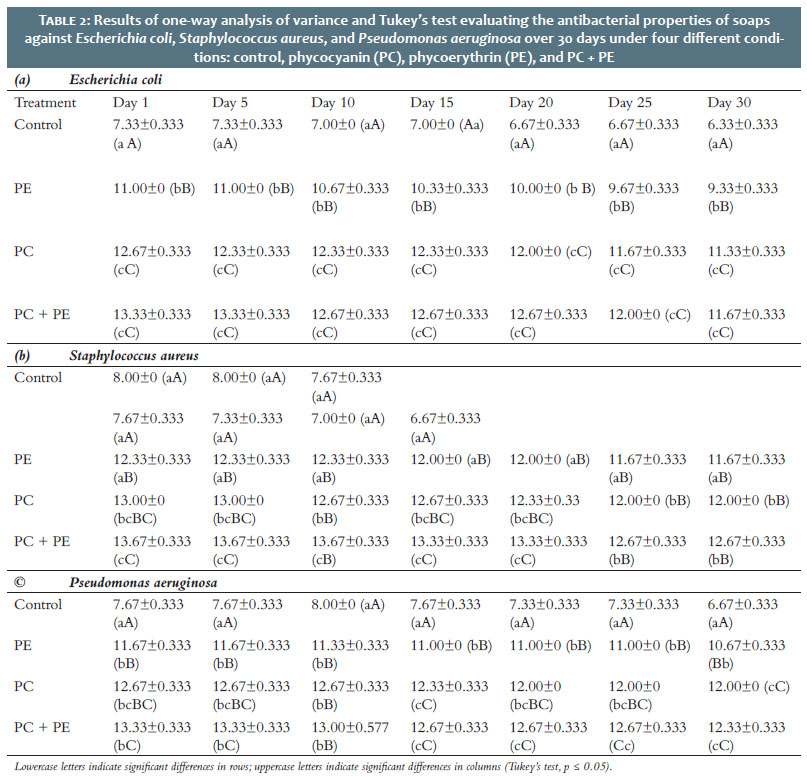
The antibacterial activities of soap formulations against E. coli, S. aureus, and P. aeruginosa were evaluated over a 30-day period using one-way ANOVA followed by Tukey's test. The soaps were prepared under four conditions: control (no pigment), PC, PE, and PC + PE (Table 2).
For E. coli, no significant differences in antibacterial activity were observed over the 30 days, indicating stable efficacy across treatments. However, soaps formulated with PC alone and PC + PE consistently exhibited significantly higher antibacterial activity than the control and PE-only treatments (Table 2a).
Regarding S. aureus, no significant differences were found between the control and PE-only soaps throughout the 30-day period. Soaps containing both pigments showed the highest antibacterial activity up to day 20. After day 20, however, a significant reduction in activity was observed (Table 2b).
Against P. aeruginosa, there were no significant differences in antibacterial activity between the control and individual pigment treatments during the 30-day period. Soaps containing both pigments demonstrated the highest antibacterial activity up to day 10, after which their efficacy declined significantly (Table 2c).
One-way ANOVA and Tukey's test indicated that the antioxidant activity of all soap formulations significantly decreased over the 30-day period. Despite this decline, all treatments demonstrated significantly higher antioxidant activity compared to the control. No statistically significant differences were observed between the different pigment-treated soaps.
The presence of psychrophilic (cold-loving) bacteria was evaluated over 30 days. One-way ANOVA and Tukey's test showed that bacterial growth was detected in the control soap on day 20, in PC-containing soap on day 25, and in PE-containing soap on day 30. However, no psychrophilic bacteria were detected in soaps formulated with both pigments. The control soap exhibited a significantly higher psychrophilic bacterial count compared to all other treatments.
Colorimetric analysis was performed using L*, a*, b*, and ΔE values. One-way ANOVA and Tukey's test showed the following:
• L* (lightness): Soaps containing PE and the combination of both pigments had significantly higher brightness than the other formulations.
• a* (red-green scale): No significant differences were observed among treatments or between treated and control soaps over time. However, PE-treated soaps consistently exhibited the highest a* values throughout the evaluation period.
• b* (yellow-blue scale): No significant differences were noted over time among treatments or versus the control, although PE-containing soaps showed the lowest b* values on day 30, with a statistically significant difference.
• ΔE (total color difference): No significant differences in ΔE were found between treatments and control during the observation period. However, soaps containing PC and PC + PE exhibited the lowest ΔE values, with a significant difference noted on day 30.
One-way ANOVA and Tukey's test showed that pH levels decreased significantly over the 30-day period, particularly in the control gels. On day 30, the lowest pH value was observed in gels enriched with PC.
Viscosity also showed a significant decrease over 30 days, as indicated by one-way ANOVA and Tukey's test. On day 30, the highest viscosity values were recorded in gels formulated with PE and PC + PE. The control gel exhibited the lowest viscosity.
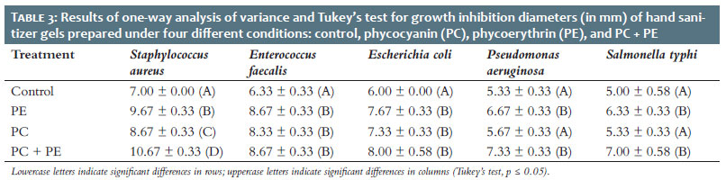
According to one-way ANOVA and Tukey's test of ZOI diameters, the highest antimicrobial activity against S. aureus was observed in gels enriched with PC + PE. For E. faecalis and E. coli, no statistically significant differences in ZOIs were found among the pigment-treated gels. However, the highest inhibition against P. aeruginosa and S. typhi was noted in gels containing PE and PC + PE (Table 3).
One-way ANOVA and Tukey's test revealed that the lowest MIC against S. aureus was observed in gels enriched with PE and PC + PE. For E. faecalis, E. coli, P. aeruginosa, and S. typhi, the lowest MIC values were consistently recorded in gels containing PC + PE.
Analysis of MBC levels using one-way ANOVA and Tukey's test showed that the lowest MBC values against S. aureus, P. aeruginosa, E. faecalis, and S. typhi were found in gels enriched with either PE, PC, or PC + PE. For E. coli, the lowest MBC was observed in formulations containing PC + PE.
Stability was evaluated under various storage conditions (4 ºC, 25 ºC, 40 ºC) by monitoring odor, color, and phase separation. One-way ANOVA and Tukey's test indicated that all samples scored 5 out of 5 for all three parameters at 4 ºC, 25 ºC (up to day 15), and 40 ºC.
Although a significant reduction in odor and color stability was observed over 30 days, no significant differences were found between the control and pigment-enriched gels. For phase separation, significant differences were observed on days 25 and 30 at 25 ºC, 37 ºC, and 40 ºC. However, no statistically significant differences were found between control and treated samples. All gels received a score of 5 for phase separation and color on days 10 and 15 at 40 ºC, and on days 15 and 20 at 25 ºC.
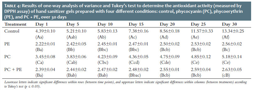
According to one-way ANOVA and Tukey's test, antioxidant activity significantly declined over the 30-day period. On the final day, the highest antioxidant activity was observed in gels enriched with PE and PC + PE (Table 4).
The foaming ability did not differ significantly between control and pigment-treated gels, as indicated by one-way ANOVA and Tukey's test.
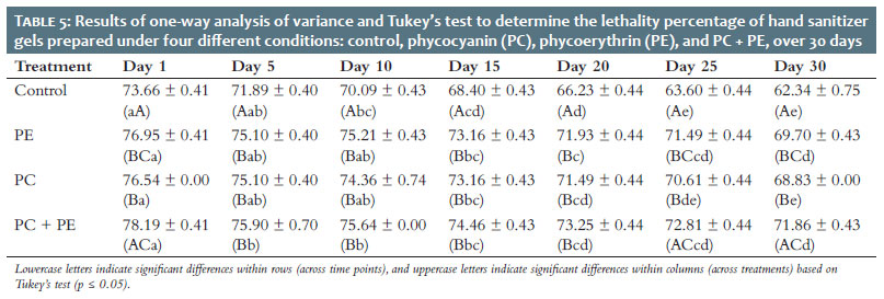
The lethality rate of the sanitizer gel significantly decreased over the 30-day period. The greatest differences were found in gels enriched with PC + PE (Table 5).
The use of natural ingredients for cosmetic purposes dates back to ancient times, long before the formal establishment of the cosmetics industry.44 In recent years, the rise of cosmeceuticals has emphasized the potential of cyanobacteria as a promising source of bioactive components due to their capacity to synthesize a wide variety of functional compounds. Cyanobacteria produce numerous bioactive molecules, including fatty acids, polyphenols, peptides, polysaccharides, and pigments, many of which have applications in health, nutrition, and skincare.45 Among these, cyanobacterial pigments — such as chlorophylls, carotenoids, and PBPs — stand out for their diverse physicochemical properties and broad color range, from blue to red. These pigments have gained increasing attention for their potential applications in food, animal feed, nutraceuticals, and cosmetics.46 In particular, carotenoids and PBPs possess notable antioxidant, moisturizing, and skin-stabilizing properties, making them highly desirable in natural cosmetic formulations.6,47
Carotenoids are commonly incorporated into sunscreens, anti-aging products, and antioxidant-enriched cosmetics due to their excellent free radical scavenging abilities. In cyanobacteria, these pigments help dissipate excess energy generated during photosynthesis, thereby preventing oxidative damage at the cellular level.48 This photoprotective mechanism has clear parallels in human skin, which is similarly susceptible to oxidative stress caused by UV radiation and intense light exposure.49
PBPs are associated with a broad spectrum of bioactivities, including anticancer, antiviral, antimicrobial, and antioxidant effects.50 These properties make them highly valuable as functional additives in cosmetic formulations. In addition to their biological activities, PBPs function as natural colorants, offering a safer alternative to synthetic dyes that are often linked to skin irritation, allergic reactions, and potential toxicity. Their hydrophilic nature and water solubility further enhance their suitability for use in topical skincare applications, particularly in products such as serums and lotions.46
This study aimed to evaluate the effects of chitosan-coated PC and PE on the antioxidant and antimicrobial activity of three cosmetic products: soap, anti-acne face wash, and hand sanitizer gel. The results of the anti-acne face wash gel tests revealed that pigment composition significantly influenced several parameters. Gels enriched with PC and PC + PE exhibited higher viscosity, enhanced antioxidant activity, and stronger antimicrobial activity against acne-causing microorganisms compared to other formulations. In contrast, the control formulation displayed the highest pH.
For the hand sanitizer gel, a decline in both viscosity and pH was observed over time, with the lowest values recorded in the control and PC-enriched formulations, respectively. Antioxidant activity in hand sanitizers was lower overall, particularly in gels containing only PE or the combined pigments, suggesting reduced stability or effectiveness of the pigments in this formulation type. However, all gel formulations maintained full homogeneity throughout the study period. The anti-acne face wash gels were also noted for their appealing physical characteristics — they remained shiny and transparent, and upon application, provided a light and cooling sensation on the skin. Among the tested formulations, the highest antimicrobial lethality in the face wash gel was recorded in those containing PC + PE and PC alone.
However, among the hand sanitizer gel formulations, PC + PE demonstrated the best overall performance. Optimal cultivation conditions are essential for maximizing the production of secondary metabolites in extremophilic cyanobacteria. Zucchi and Necchi51 studied the growth and pigment content of freshwater algal cultures and found that temperature was the primary factor influencing pigment synthesis. In contrast, variations in irradiance, photoperiod, and their interactions contributed less significantly to pigment variation. Their findings also indicated that PC was generally more abundant than PE, and PBPs were more concentrated than chlorophyll a.
In contrast, Rizzo et al.52 reported no variation in total protein content or penicillin-binding protein production under different light conditions. Ma et al.53 showed that PBP content peaked at light intensities below 90 μmol m-2 s-1. However, PC and allophycocyanin levels increased with greater light intensity, while PE levels declined. Furthermore, the study found that exposure to blue and red light led to the highest increases in fresh weight, protein content, and PBP levels, which were associated with higher concentrations of dry matter, PC, and chlorophyll a.
PE has demonstrated versatile applications in the pharmaceutical, antioxidant, and food industries.54-55 However, its limited stability remains a significant barrier to its widespread use. PE is highly sensitive to factors such as pH, salt concentration, temperature, water activity, and light exposure as well as to in vitro processes including extraction, purification, storage, and formulation. These conditions often compromise its structural integrity and bioactivity. Size and protein composition of the PE complex also vary in response to environmental conditions. For instance, low light intensity improves PE synthesis and leads to the elongation of rod-like structures.
Numerous purification and characterization techniques have been developed for isolating PE from various cyanobacterial and red algal strains.14
The stability analysis of color, odor, and consistency for both the anti-acne face wash and hand sanitizer gels revealed that all formulations consistently received the maximum score of 5. In the case of the anti-acne face wash gel, color stability measurements at 25 ºC, as well as at 37 ºC and 40 ºC (from day 20 onward), were highest in the control group. However, no significant differences in odor were observed between treatments and the control across all temperatures over the 30-day evaluation period. Similarly, all hand sanitizer gel formulations maintained full stability — receiving scores of 5 out of 5 for odor, color, and absence of phase separation at 4 ºC, 25 ºC (until day 15), and 40 ºC. The soap formulations also maintained physical integrity and form stability, with all receiving the maximum score of 5.
Foamability remained unchanged throughout the evaluation period for both the anti-acne face wash and hand sanitizer gels. In soaps, parameters such as total fat content, pH, foam formation and height, shelf life, alkalinity, and antioxidant activity gradually declined over time; however, no statistically significant differences were observed. Although insoluble alcohol content increased over the 30 days, this change was not statistically significant.
Cyanobacteria naturally possess protective mechanisms against dehydration, which makes them promising candidates as moisturizing agents in cosmetic formulations. Several studies have demonstrated their ability to improve skin hydration and elasticity.56-57 For example, topical application of microalgae has been shown to enhance skin moisture and elasticity.4 Research on Nostoc commune further revealed that cells containing extracellular polymeric substances (EPS) displayed significantly greater desiccation tolerance, highlighting their superior water-retention capacity when compared to urea and chitosan.58,59
EPS are high-molecular-weight biopolymers composed of hydrated sulfate groups, neutral sugars (e.g., glucose, galactose, mannose, fructose, ribose, xylose, arabinose, fucose, rhamnose), non-carbohydrate components (e.g., phosphate, lactate, acetate, glycerol), and various uronic acids (e.g., glucuronic and galacturonic acids). The strong water-binding ability of these polysaccharides is attributed to the interactions between water molecules and hydrophilic –OH groups. Notably, the composition of EPS varies depending on environmental conditions and species.60 For example, Chroococcidiopsis modifies the composition of its cellular envelope under water stress, producing compounds such as sporopollenin-like materials, proteins, beta-linked polysaccharides, acid sulfates, and lipids. These metabolites contribute to reduced water loss and enhanced regulation of hydration.61
In the current study, both moisture content and saponification rate decreased in soaps over time, with the most substantial reduction in moisture observed in soaps enriched with PE and PC + PE on days 25 and 30. Additionally, the presence of psychrophilic bacteria was highest in the control soap, further emphasizing the protective effect of pigment-enriched formulations against microbial contamination under cold conditions.
Based on the colorimetry results for the soap formulations, the highest brightness levels (L*) were observed in soaps containing PE and PC + PE. The highest a* values, which reflect redness, were found in soaps enriched with PE alone. In contrast, the lowest b* values (indicating yellowness) were associated with PE-treated soaps, and the lowest total color difference (ΔE) values were recorded in soaps containing PC and PC + PE. Regarding the antibacterial properties of the soaps, no significant differences were observed over the 30-day period in the inhibition of E. coli, S. aureus (in control and PE-treated soaps), and P. aeruginosa (in control and soaps enriched with PE and PC). For the hand sanitizer gel formulations, the highest ZOI against S. aureus was found in gels enriched with PC + PE. No significant differences were observed in ZOI diameters for E. faecalis and E. coli among the pigment-enriched gels. However, the highest inhibition rates against P. aeruginosa and S. typhi were recorded in gels containing PE and PC + PE. The MIC results for the hand sanitizer gels showed that the lowest inhibitory concentrations were achieved with PE and PC + PE against S. aureus, and with PC + PE against E. faecalis, E. coli, P. aeruginosa, and S. typhi. Regarding MBC, the lowest bactericidal concentrations against S. aureus, P. aeruginosa, E. faecalis, and S. typhi were observed in gels enriched with the combined pigments (PC + PE), as well as with either PE or PC alone. For E. coli, the lowest MBC values were recorded in gels containing PC + PE.
The increasing focus on skin health, particularly in relation to aesthetics and aging, has driven the demand for innovative cosmetic products derived from natural sources. These products offer the advantages of reduced adverse effects and greater environmental sustainability. Cyanobacteria are promising candidates for the cosmetic and cosmeceutical industries due to their natural ability to resist dehydration, radiation, and oxidative stress through the production of specialized bioactive compounds. This study demonstrated the potential of cyanobacterial pigments, particularly PC and PE, in the formulation of three cost-effective and sustainable cosmetic products: soap, anti-acne face wash, and hand sanitizer gel. These formulations not only exhibited desirable physicochemical and antimicrobial properties but also aligned with biotechnological strategies for enhancing the efficiency and environmental compatibility of cosmetic production. The findings support the use of cyanobacteria as a valuable resource in the development of next-generation, naturally derived cosmetic products.
Bahareh Nowruzi
ORCID: 0000-0001-6656-777X
Statistical analysis, Final approval of the manuscript, Study conception and design, Manuscript drafting and writing, Active participation in research supervision, Intellectual contribution to the clinical management of studied cases, Critical literature review, Critical manuscript revision.
Mahshid Alibabaei
ORCID: 0009-0000-3743-226X
Statistical analysis, Final approval of the manuscript, Study conception and design, Manuscript drafting and writing, Active participation in research supervision, Intellectual contribution to the clinical management of studied cases, Critical literature review, Critical manuscript revision.
1. Spolaore P, Joannis-Cassan C, Duran E, Isambert A. Commercial applications of microalgae. J Biosci Bioeng. 2006;101(2):87-96.
2. Sousa I, Gouveia L, Batista AP, Raymundo A, Bandarra NM. Microalgae in novel food products. In: Papadopoulos K. Food Chemistry Research Developments. New York: Nova Science Publishers; 2008. p. 75-112.
3. Del Campo JA, García-González M, Guerrero MG. Outdoor cultivation of microalgae for carotenoid production: current state and perspectives. Applied Microbiol Biotech. 2007;74(6):1163-74.
4. Mourelle ML, Gómez CP, Legido JL. The potential use of marine microalgae and cyanobacteria in cosmetics and thalassotherapy. Cosmetics. 2017;4(4):46.
5. Berthon J-Y, Nachat-Kappes R, Bey M, Cadoret J-P, Renimel I, Filaire E. Marine algae as attractive source to skin care. Free Radic Res. 2017;51(6):555-67.
6. Joshi S, Kumari R, Upasani VN. Applications of algae in cosmetics: an overview. Int J Innov Res Sci Eng Technol. 2018;7(2):1269.
7. Singh R, Parihar P, Singh M, Bajguz A, Kumar J, Singh S, et al. Uncovering potential applications of cyanobacteria and algal metabolites in biology, agriculture and medicine: current status and future prospects. Front Microbiol. 2017;8:515.
8. Singh S, Kate BN, Banerjee U. Bioactive compounds from cyanobacteria and microalgae: an overview. Critic Rev Biotech. 2005;25(3):73-95.
9. Mourelle M, Gómez CP, Legido J, Legido N. [Innovation in the use of microalgae in thermalism]. Bol Soc Esp Hidrol Méd. 2016;31(1):53-64. Spanish.
10. Lisby S, Gniadecki R, Wulf HC. UV - induced DNA damage in human keratinocytes: quantitation and correlation with long - term survival. Exp Dermatol. 2005;14(5):349-55.
11. Sathasivam R, Radhakrishnan R, Hashem A, Allah EFA. Microalgae metabolites: A rich source for food and medicine. Saudi J Biol Sci. 2019;26(4):709-22.
12. Sonani RR, Rastogi RP, Patel R, Madamwar D. Recent advances in production, purification and applications of phycobiliproteins. World J Biol Chem. 2016;7(1):100.
13. Basheva D, Moten D, Stoyanov P, Belkinova D, Mladenov R, Teneva I. Content of phycoerythrin, phycocyanin, alophycocyanin and phycoerythrocyanin in some cyanobacterial strains: applications. Eng Life Sci. 2018;18(11):861-6.
14. Nowruzi B, Sarvari G, Blanco S. The cosmetic application of cyanobacterial secondary metabolites. Algal Res. 2020;49:101959.
15. Gonzalez-Ramirez E, Andujar-Sanchez M, Ortiz-Salmeron E, Bacarizo J, Cuadri C, Mazzuca-Sobczuk T, et al. Thermal and pH stability of the B-phycoerythrin from the red algae Porphyridium cruentum. Food Biophysics. 2014;9:184-92.
16. Ariede MB, Candido TM, Jacome ALM, Velasco MVR, Carvalho JCM, Baby AR. Cosmetic attributes of algae: a review. Algal Research. 2017;25:483-7.
17. Morone J, Lopes G, Preto M, Vasconcelos V, Martins R. Exploitation of filamentous and picoplanktonic cyanobacteria for cosmetic applications: potential to improve skin structure and preserve dermal matrix components. Marine Drugs. 2020;18(9):486.
18. Liu L, Jokela J, Wahlsten M, Nowruzi B, Permi P, Zhang YZ, et al. Nostosins, trypsin inhibitors isolated from the terrestrial cyanobacterium Nostoc sp. strain FSN. J Nat Prod. 2014;77(8):1784-90.
19. Bhattacharya S, Shivaprakash M. Evaluation of three Spirulina species grown under similar conditions for their growth and biochemicals. J Sci Food Agri. 2005;85(2):333-6.
20. Nowruzi B, Anvar SAA, Ahari H. Extraction, purification and evaluation of antimicrobial and antioxidant properties of phycoerythrin from terrestrial cyanobacterium Nostoc sp. FA1. J Microbial World. 2020;13(2):138-53.
21. Afreen S, Fatma T. Laccase production and simultaneous decolorization of synthetic dyes by cyanobacteria. Int J Innovative Res Sci Eng Technol. 2013;2:3563-8.
22. Mishra S, Mishra D. A novel remote sensing algorithm to quantify phycocyanin in cyanobacterial algal blooms. Environ Res Letters. 2014;9(11):114003.
23. Suzery M, Majid D, Setyawan D, Sutanto H, editors. Improvement of stability and antioxidant activities by using phycocyanin-chitosan encapsulation technique. IOP Conference Series: Earth and Environmental Science; 2017.
24. Kusuma SAF, Abdassah M, Valas BE. Formulation and evaluation of anti-acne gel containing citrus aurantifolia fruit juice using carbopol as gelling agent. Int J App Pharm. 2018;10(4):147-52.
25. Afsar Z, Khanam S. Formulation and evaluation of poly herbal soap and hand sanitizer. Int Res J Pharm. 2016;7(8):54-7.
26. Acharya SB, Ghosh S, Yadav G, Sharma K, Ghosh S, Joshi S. Formulation, evaluation and antibacterial efficiency of water-based herbal hand sanitizer gel. Biorxiv. 2018:373928.
27. Ingle A, Meshram M. Formulation and evaluation of Ayurvedic face wash. Int J Phytopharm. 2018;8(4):26-30.
28. Rasheed A, Reddy AKG, Mohanalakshmi S, Kumar CKA. Formulation and comparative evaluation of poly herbal anti-acne face wash gels. Pharm Biol. 2011;49(8):771-4.
29. Shah MA, Natarajan SB, Gousuddin M. Formulation, evaluation, and antibacterial efficiency of herbal hand wash Gel. Int J Pharm Sci. 2014;25(2):120-4.
30. Kumar L, Verma R. In vitro evaluation of topical gel prepared using natural polymer. Int J Drug Deliv. 2010;2(1).
31. Haneefa MK, Shilpa N, Junise V, Chandran A. Formulation and evaluation of medicated soap of Ixora coccinea root extract for dermal infections. J Pharm Sci Res. 2019;11(8):3094-7.
32. Kamble M, Selwate T, Dhabarde D, Ingole A, Baheti J. Formulation and evaluation of anti-acne face wash gel using guava seed extract. J Drug Deliv Therap. 2019;9(3):5-7.
33. Sharma SK, Singh S. Antimicrobial herbal soap formulation. J Pharma Res Int. 2020;32(36):82-8.
34. Sindhu RK, Chitkara M, Kaur G, Kaur A, Arora S, Sandhu I. Formulation development and antimicrobial evaluation of polyherbal soap. Plant Arch. 2019;19(2):1342-6.
35. Amrutkar SV, Patil AR, Ishikar SK. A review on herbal soap. Res J Topic Cosmet Sci. 2022;13(1):49-54.
36. Hassan M, Kubmarawa D, Modibbo U, NAT. Production of medicated soap from Butyrospermum Paradoxum plant. J Physical Sci Innov. 2010;2:90-6.
37. Sinha SK, Poudyal S, Khatiwada S, Ahmed S, Chatterjee A, Mohanty J, et al. Formulation and evaluation of herbal handwash using neem and reetha extract. J Pharm Phytochem. 2022;11(5):207-10.
38. Hayati S, Rosyida V, Darsih C, Nisa K, Indrianingsih A, Apriyana W, et al. Physicochemical properties, antimicrobial and antioxidant activity of ganoderma transparent soap. IOP Conference Series: Earth and Environmental Science; 2020.
39. Ali MN, Guesmi N, Ali S, Abofard M, Gaber M, Al-Dosari FBA, et al. Evaluation of laboratory formulated hand sanitizing gel in riyadh municipality central area labs. Saudi J Med Pharm Sci. 2020;6(8):548-58.
40. Sari-Chmayssem N, Taha S, Mawlawi H, Guégan J-P, Jeftić J, Benvegnu T. Extracted and depolymerized alginates from brown algae Sargassum vulgare of lebanese origin: chemical, rheological and antioxidant properties. J App Phycol. 2016;28:1915-29.
41. MA W. Methods for dilution antimicrobial susceptibility tests for bacteria that grow aerobically: approved standard. Clsi (Nccls). 2006;26:M7-A.
42. Vieira MV, Pastrana LM, Fuciños P. Microalgae encapsulation systems for food, pharmaceutical and cosmetics applications. Marine Drugs. 2020;18(12):644.
43. Nowruzi B, Khavari-Nejad R, Sivonen K, Kazemi B, Najafi F, Nejadsattari T. Optimization of cultivation conditions to maximize extracellular investments of two Nostoc strains. Arch Hydrobiol Suppl Algol Stud. 2013;142(1):63-76.
44. Scott DA. A review of ancient Egyptian pigments and cosmetics. Studies Conserv. 2016;61(4):185-202.
45. Guedes AC, Amaro HM, Malcata FX. Microalgae as sources of high added - value compounds — a brief review of recent work. Biotech Progress. 2011;27(3):597-613.
46. Pagels F, Guedes AC, Vicente AA, Vasconcelos V. Cyanobacteria-based bioprocess for cosmetic products — cyanobium sp. as a novel source of bioactive pigments. Phycology. 2023;3(1):47-64.
47. Morone J, Alfeus A, Vasconcelos V, Martins R. Revealing the potential of cyanobacteria in cosmetics and cosmeceuticals — a new bioactive approach. Algal Res. 2019;41:101541.
48. Anunciato TP, Rocha Filho PA. Carotenoids and polyphenols in nutricosmetics, nutraceuticals, and cosmeceuticals. J Cosmet Dermatol. 2012;11(1):51-4.
49. Pagels F, Vasconcelos V, Guedes AC. Carotenoids from cyanobacteria: biotechnological potential and optimization strategies. Biomolecules. 2021;11(5):735.
50. Pagels F, Guedes AC, Amaro HM, Kijjoa A, Vasconcelos V. Phycobiliproteins from cyanobacteria: chemistry and biotechnological applications. Biotech Adv. 2019;37(3):422-43.
51. Zucchi MR, Necchi Jr O. Effects of temperature, irradiance and photoperiod on growth and pigment content in some freshwater red algae in culture. Phycol Res. 2001;49(2):103-14.
52. Rizzo RF, Santos BNCd Castro GFPS, Passos TS, Nascimento MA, Guerra HD, et al. Production of phycobiliproteins by Arthrospira platensis under different lightconditions for application in food products. Food Sci Technol. 2015;35:247-52.
53. Ma R, Lu F, Bi Y, Hu Z. Effects of light intensity and quality on phycobiliprotein accumulation in the cyanobacterium Nostoc sphaeroides Kützing. Biotechnol Lett. 2015;37:1663-9.
54. Mishra SK, Shrivastav A, Pancha I, Jain D, Mishra S. Effect of preservatives for food grade C-Phycoerythrin, isolated from marine cyanobacteria Pseudanabaena sp. I J Biol Macromol. 2010;47(5):597-602.
55. Soni B, Visavadiya NP, Dalwadi N, Madamwar D, Winder C, Khalil C. Purified c - phycoerythrin: safety studies in rats and protective role against permanganate - mediated fibroblast - DNA damage. J App Toxicol. 2010;30(6):542-50.
56. Warren - Rhodes KA, McKay CP, Boyle LN, Wing MR, Kiekebusch EM, Cowan DA, et al. Physical ecology of hypolithic communities in the central namib desert: the role of fog, rain, rock habitat, and light. J Geophys Res Biogeosci. 2013;118(4):1451-60.
57. Smith HD, Baqué M, Duncan AG, Lloyd CR, McKay CP, Billi D. Comparative analysis of cyanobacteria inhabiting rocks with different light transmittance in the mojave desert: a mars terrestrial analogue. I J Astrobiol. 2014;13(3):271-7.
58. Li H, Xu J, Liu Y, Ai S, Qin F, Li Z, et al. Antioxidant and moisture-retention activities of the polysaccharide from Nostoc commune. Carbohydrate Polymers. 2011;83(4):1821-7.
59. Tamaru Y, Takani Y, Yoshida T, Sakamoto T. Crucial role of extracellular polysaccharides in desiccation and freezing tolerance in the terrestrial cyanobacterium nostoc commune. App Environ Microbiol. 2005;71(11):7327-33.
60. Nowruzi B, Khavari-Nejad R, Sivonen K, Kazemi B, Najafi F, Nejadsattari T. Optimization of cultivation conditions to maximize extracellular investments of two Nostoc strains. Algologic Stud. 2013;142(1):63-76.
61. Caiola MG, Billi D, Friedmann EI. Effect of desiccation on envelopes of the cyanobacterium Chroococcidiopsis sp. (Chroococcales). Eur J Phycol. 1996;31(1):97-105.
 All content the journal, except where identified, under the Creative Commons Attribution 4.0 International licence - ISSN-e 1984-8773


 Read in Portuguese
 Portuguese PDF
 Print
Print
 Send this article by email
 How to cite this article
 Submit a comment
 Mendeley
 Pocket