


Anna Karoline Gouveia; Guillermo Loda; Marcela Benez; Anna Beatriz Loda
Funding: None.
Conflict of interest: None.
Submitted on: 05/15/2024
Final decision: 07/15/2024
How to cite this article: Gouveia AK, Loda G, Benez M, Loda AB. Transfollicular subcutaneous forehead and brow lift. Surg Cosmet Dermatol. 2025;17:e20250372.
The appearance of the upper third of the face is a key indicator of aging and plays a significant role in communication by conveying non-verbal information. Different surgical techniques have been described for the rejuvenation of this region, with transfollicular subcutaneous forehead and brow lift enabling excellent results by lowering the hairline, reducing forehead height, softening forehead and glabellar rhytids, raising the eyebrows, and improving pseudodermatochalasis. This technique offers the advantages of reduced risk of nerve injury and excellent scalp scar camouflage.
Keywords: Dermatologic Surgical Procedures; Rejuvenation; Rhytidoplasty; Skin Aging; Surgery, Plastic.
The appearance of the upper third of the face is a key indicator of aging, characterized by rhytids, forehead and brow ptosis, and changes in the periocular region, which result from bone resorption, transmission of muscle action to the skin surface,and skin-ligamentous laxity. The upper third of the face also plays a significant role in communication by conveying non-verbal information. Regarding the aesthetics of this region, young men typically have straighter horizontal brows positioned at the level of the orbital rim. In young women, the brows are usually positioned above the level of the orbital rim, featuring a fuller, more quadrangular medial portion and becoming progressively arched and thinner toward the lateral portion, with the brow apex often coinciding with a vertical line drawn from the lateral corneal limbus. Aging leads to a progressive descent of the eyebrows, which can be positioned below the orbital rim, depending on the degree of aging.
Different surgical techniques have been described for the rejuvenation of this region, each with its own advantages and limitations,which,combined with the surgeon's experience,will determine the choice of technique for each patient.While non-surgical treatments can be beneficial and used in combination, the most substantial and long-lasting results are only achieved with surgical lifting.We describe the transfollicular subcutaneous forehead and brow lift (TSFBL), a technique that allows rejuvenation of the eyebrow, glabellar, and forehead regions.
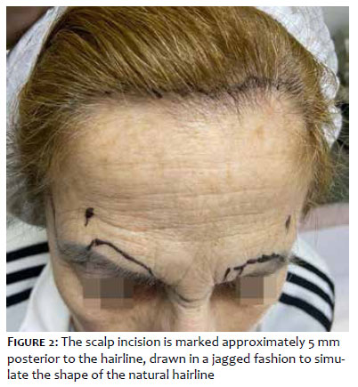
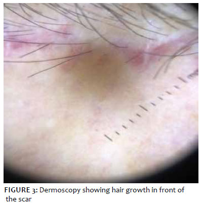
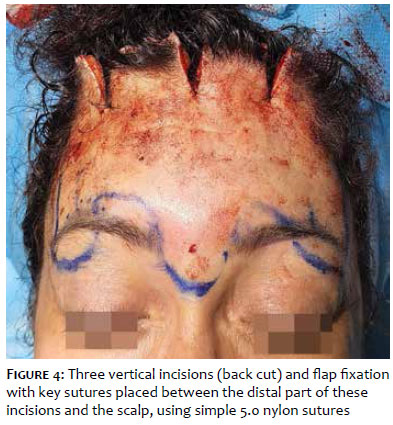
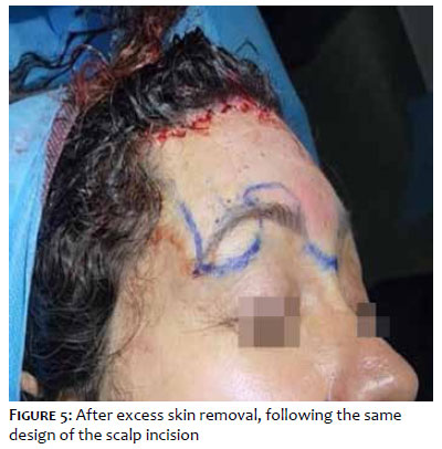
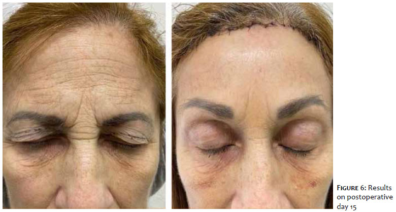
With the patient seated, the incision line is marked. In the lateral third, a line with superior concavity below the eyebrow delineates the area of maximal elevation, where the detachment will be performed in the subcutaneous plane. In the medial region, where the supraorbital and supratrochlear nerves are located, a line with inferior concavity is marked above the eyebrow, serving as the lower limit of the detachment, with the aim of reducing the risk of nerve injury and avoiding over-elevation of the medial brow portion. In the glabellar region, a line with superior concavity is marked as a reference point for the lower limit of the detachment (Figure 1). The lateral limits are marked with a vertical line drawn at the level of the temporal crest. The scalp incision is marked approximately 5 mm posterior to the hairline, drawn in a jagged fashion to simulate the shape of the natural hairline (Figure 2). The procedure is performed under tumescent local anesthesia using an anesthetic solution consisting of 80 mL of 0.9% sodium chloride, 20 mL of 2% lidocaine, and 0.4 mL of 1 mg/mL epinephrine. The solution is injected subcutaneously along the previously marked area, and the total volume to be used depends on the extent of the frontal region. In our experience, this volume ranges from 60 to 80 mL. The incision and dissection plane give the procedure its name, TSFBL. The incision is made using a No. 11 blade, angled between 10 and 20 degrees relative to the horizontal plane. This angle allows for the hair follicles to be sectioned at different heights, preserving part of the bulbs, which will allow for the most anterior follicles to grow in front of the scar (Figure 3), promoting effective scalp scar camouflage. These incision characteristics also produce very thin wound edges, contributing to a better aesthetic outcome. The dissection is performed in the subcutaneous plane, maintaining fat both on the roof and floor, thus creating an adipocutaneous flap and avoiding damage to the underlying frontalis muscle. By respecting the uppermost marking line in the proximal region of the eyebrows, over-elevation of their medial portion is avoided, while the lower dissection of the glabella and lateral portions allows elevation of the tail of the brow and correction of glabellar ptosis, restoring a more you thful appearance. Because this subcutaneous technique allows direct visualization, there is a lower risk of injury to the primary sensory and motor nerves in the glabellar region compared with endoscopic and supraperiosteal techniques. The lateral limit of the detachment should not exceed the temporal crest to avoid injury to the temporal nerve. The superficial musculoaponeurotic system (SMAS) fibrous septa connect the frontalis muscle to the forehead skin. During dissection in the subcutaneous plane, most of these septa are disrupted, resulting in a significant improvement in forehead and glabellar rhytids. While the depressor supercilii muscles can be sectioned under direct visualization, we have chosen not to approach them due to the lack of superiority in the observed results, as well as not to increase the risk of complications. Once the dissection is complete, the flap is pulled over the scalp and 3 vertical incisions are made (back cut): one in the central region of the forehead, which corresponds inferiorly to the glabella; and two paramedian incisions, which correspond to the point of maximal brow elevation, often coinciding with a vertical line drawn from the lateral corneal limbus. The length of these incisions ranges from 15 to 30 mm, depending on the eyebrow position, the distance between the scalp and eyebrows, and the surgeon’s experience. In our practice, a length between 20 and 25 mm is adequate in most cases. The flap is secured with key sutures placed key sutures placed between the distal part of these incisions and the scalp, using simple 5.0 nylon sutures (Figure 4). Excess skin is then removed using a No. 11 blade, following the same design and angle of the scalp incision, and the wound edges are gently closed using simple 5.0 nylon sutures (Figure 5). The procedure is completed with the application of a pressure dressing, and the patient is reassessed within 48 hours. After this period, dressings are no longer required. The sutures are removed after 10 to 14 days (Figure 6).
There are several surgical options for the treatment of brow ptosis, which can be didactically divided into direct visualization, transblepharoplasty, and endoscopic techniques. The direct visualization techniques vary based on the incision location, including excisions performed just above the eyebrows (Castañares technique), in the mid-forehead, pretrichial, coronal, and transfollicular. Endoscopic brow lift is performed in the subgaleal or subperiosteal plane and uses bone fixation materials, resulting in smaller incisions and reduced postoperative edema, dysesthesia, and alopecia. However, this technique requires specialized equipment, is more expensive, has a steeper learning curve, and may offer less long-lasting results compared with direct methods, since the amount of elevation is determined by flap release and fixation, without direct skin excision.1 It is particularly indicated when there is brow asymmetry.2The transpalpebral approach involves securing the brow tissue, muscles, and subcutaneous fat to the periosteum through an upper blepharoplasty incision. This can be performed intraoperatively during an upper blepharoplasty when a traditional brow lift is not planned. Mokhtarzadeh et al., evaluating 98 patients who underwent internal fixation during blepharoplasty, identified an average elevation in central/lateral brow position of 1.47 to 2.29 mm over a follow-up of 4-5 months.3 The direct brow lift includes variations of bilateral elliptical incisions just above the eyebrows (including the Castañares technique), hiding the scar along the hairline, or midfrontal incisions to camouflage scars within horizontal forehead rhytids.
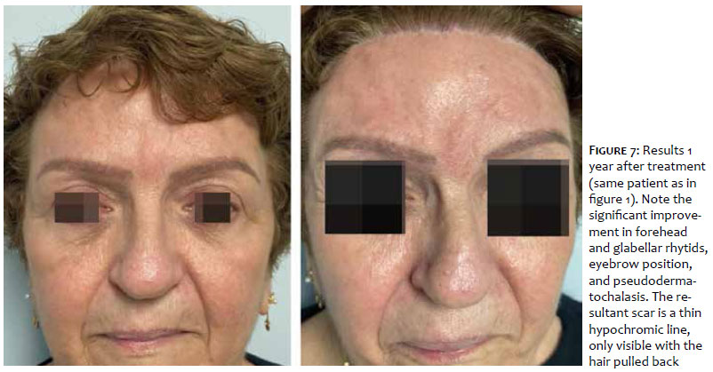
These two types are particularly suitable for patients with unstable hairlines. The technique can be modified to address lateral ptosis alone or to compensate for asymmetries. These techniques may also lower the hairline by shortening the forehead. While dissection in the subcutaneous plane can be performed medially to improve glabellar rhytids, it does not improve forehead rhytids, and the scar may remain visible for a long period.3 The coronal brow lift, an open technique using an ear-to-ear scalp incision, elevates the hairline but can cause alopecia and nerve damage. The pretrichial approach involves an incision on the forehead, just outside and below the scalp. Unlike the coronal brow lift, this technique lowers the hairline by shortening the forehead, but it can lead to the same complications as the other methods, with the added disadvantage of leaving a more visible scar. The transfollicular approach can lower the hairline, reduce forehead height, and soften forehead and glabellar rhytids, elevating the entire upper third of the face, with improvement in pseudodermatochalasis. If excess upper eyelid skin persists after TSFBL, an upper blepharoplasty can be performed in the same surgical procedure to complement the results. In our experience, we prefer performing blepharoplasty at a later stage to obtain a more accurate assessment of the amount of excess skin. The appearance of the resultant scar is satisfactory, resulting in a thin hypochromic line, only visible with the hair pulled back (Figure 7). Overall, all surgical brow elevation techniques have a low incidence of complications, with the most common being hematoma, damage to sensory and motor nerves leading to dysesthesia, paresis, asymmetry, alopecia, and unsightly scarring. Currently available data do not demonstrate the superiority of one technique over another, nor of endoscopic techniques over direct visualization techniques.4,5
The various brow lifting techniques have their own peculiarities regarding specific indications, execution methods, and contraindications, which will determine the choice of the optimal procedure for each patient. In patients with high hair density in the frontotemporal region, our technique of choice is the transfollicular subcutaneous approach due to the satisfactory and long-lasting results in lifting the entire upper third of the face, associated with improvements in rhytids, excellent scalp scar camouflage, safe dissection in the subcutaneous plane under direct visualization, and the possibility to be performed with low-cost materials,compared with endoscopic techniques. The main contraindications to this approach are unstable hairlines and alopecia in the frontotemporal region.
Anna Karoline Gouveia
ORCID: 0000-0001-6258-9824
Conception and design of the study; preparation and writing of the manuscript; intellectual participation in propaedeutic and/or therapeutic approach to studied cases; critical review of the literature.
Guillermo Loda
ORCID: 0000-0003-0511-0025
Approval of the final version of the manuscript;conception and design of the study;effective participation in the conduct of the study;intellectual participation in propaedeutic and/or therapeutic approach to studied cases; critical revision of the manuscript.
Marcela Benez
ORCID: 0000-0003-0289-5656
Approval of the final version of the manuscript; intellectual participation in propaedeutic and/or therapeutic approach to studied cases.
Anna Beatriz Loda
ORCID: 0009-0000-7319-2531
Intellectual participation in propaedeutic and/or therapeutic approach to studied cases.
1. Holck DE, Ng JD, Wiseman JB, Foster JA. The endoscopic browlift for forehead rejuvenation. Semin Ophthalmol. 1998;13(3):149-57.
2. Karimi N, Kashkouli MB, Sianati H, Khademi B. Techniques of eyebrow lifting: a narrative review. J Ophthalmic Vis Res. 2020;15(2):218-35.
3. Mokhtarzadeh A, Massry GG, Bitrian E, Harrison AR. Quantitative efficacy of external and internal browpexy performed in conjunction with blepharoplasty. Orbit. 2017;36(2):102–9.
4. Graham DW, Heller J, Kirkjian TJ, Schaub TS, Rohrich RJ. Brow lift in facial rejuvenation: a systematic literature review of open versus endoscopic techniques. Plast Reconstr Surg. 2011 Oct;128(4):335e-341e. Erratum in: Plast Reconstr Surg. 2011;128(5):1151. Kirkjian, T Jonathan [corrected to Kurkjian, T Jonathan].
5. Byun S, Mukovozov I, Farrokhyar F, Thoma A. Complications of browlift techniques: a systematic review. Aesthet Surg J. 2013;33(2):189-200.
 All content the journal, except where identified, under the Creative Commons Attribution 4.0 International licence - ISSN-e 1984-8773


 Read in Portuguese
 Portuguese PDF
 Print
Print
 Send this article by email
 How to cite this article
 Submit a comment
 Mendeley
 Pocket