


Ana Maria Costa Pinheiro1; Flávia Vieira Brandão2
Financial support: None.
Conflicts of interest: None.
Submitted on: 18/12/2023
Approved on: 11/03/2024
How to cite this article: Pinheiro AMC, Brandão FV. Confocal reflectance microscopy in basal cell carcinoma with globular dermatoscopic pattern. Surg Cosmet Dermatol. 2024;16:e20240335.
The globular dermoscopic pattern in pigmented lesions of basal cell carcinoma can cause diagnostic confusion, especially in cases in which other more commonly observed structures are absent. Therefore, the use of refletance confocal microscopy becomes important to determine the diagnosis and eliminate the need for biopsies.
Keywords: Carcinoma, Basal Cell; Dermoscopy; Microscopy, Confocal.
On histopathology, the globular pattern found in pigmented lesions of basal cell carcinoma (BCC) corresponds to pigmented basaloid aggregates located at the dermoepidermal junction (DEJ) or in the dermis.1,2 When this pattern is predominantly present or in the simultaneous absence of other dermoscopic structures characteristic of basal cell carcinoma, diagnosis using dermoscopy alone can be challenging. In this context, the use of confocal reflectance microscopy (CRM), which shows structures that correlate directly with histopathology, can be of great value. This communication aims to highlight the importance of CRM in the diagnosis of BCC lesions, especially in cases in which dermoscopy does not show the most prevalent characteristics.
The authors reported two cases of pigmentary lesions in young women. The lesions were examined using dermoscopy and CRM. Dermoscopy was performed using the Fotofinder ATBM skin imaging system and CRM using the Vivascope 1500 device, which displays images of the epidermis, DEJ, and dermis. Diagnosis was confirmed through anatomopathological examination.
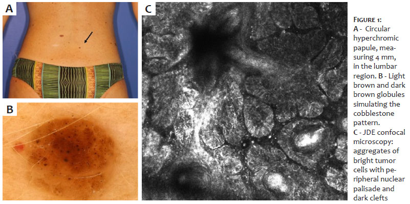
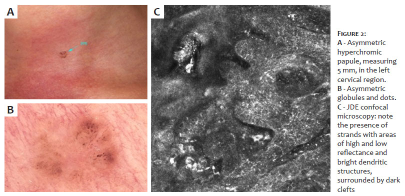
Case 1: A 43-year-old woman, phototype II, with a personal history of melanoma and BCC. During total body mapping, a pigmented lesion, clinically similar to a melanocytic nevus, was identified in the lumbar region. Dermoscopy showed a symmetrical lesion with light and dark brown globules and dots, some of which were aggregated. No other structures were observed. RCM showed the presence of well-demarcated, shiny tumor islands/strands at the DEJ, with a surrounding dark cleft and palisade (Figures 1 and 3).
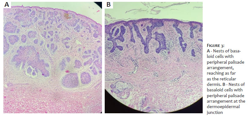
Case 2: A 34-year-old woman, phototype II, complaining of a six-year pigmentary lesion on her neck, with slow growth, and a previous diagnosis of melanocytic nevus. Dermoscopy showed an asymmetrical lesion with brown globules and dots. RCM showed bright strands at the DEJ, with dendritic structures inside and dark cracks around the strands (Figures 2 and 3).
Dermoscopy has a 95% specificity in the diagnosis of BCC.1 In pigmented BCC lesions, characteristics which have been described as blue-grey ovoid nests or globules, concentric structures, leaf-like areas, and brown dots or globules were observed. The latter correspond, in histopathology, to small pigmented basaloid aggregates at the DEJ or superficial dermis, associated with pigmented BCC.2 Although it is common to identify more than one dermoscopic feature in the same lesion, there are cases in which this does not occur, making the differential diagnosis challenging and including other lesions.3,4 RCM shows structures that correlate directly with histopathology from the epidermis to the dermis. In the epidermis, the polarization of the nuclei is oriented along the same axis. The most characteristic structures of basal cell carcinoma stand out at the DEJ, as aggregates of tumor cells, described as shiny strands or nodules, with a peripheral nuclear palisade, often showing dendritic structures inside and dark crevices around them.5-7 These characteristics are more common in pigmented BCC. Therefore, RCM can be a valuable complement to the clinical and dermoscopic evaluation of cutaneous neoplasms, increasing diagnostic accuracy. BCC with a globular pattern, in the absence of dermoscopic features, can be misdiagnosed as a melanocytic nevus. In such circumstances, RCM plays an important role in accurately confirming the diagnosis.
Ana Maria Costa Pinheiro
ORCID: 0000-0002-7804-3567
Approval of the final version of the manuscript; study design and planning; elaboration and writing of the manuscript; data collection, analysis, and interpretation; active participation in research orientation; intellectual participation in the propaedeutic and/or therapeutic conduct of the cases studied; critical literature review; critical review of the manuscript.
Flávia Vieira Brandão
ORCID: 0000-0003-3809-9774
Approval of the final version of the manuscript; preparation and writing of the manuscript; data collection, analysis, and interpretation; critical literature review; critical review of the manuscript.
1. Reiter O, Mimouni I, Dusza S, Halpern AC, Leshem YA, Marghoob AA. Dermoscopic features of basal cell carcinoma and its subtypes: a systematic review. J Am Acad Dermatol. 2021;85(3):653-64.
2. Urun YG, Fiçicioglu S, Urun M, Can Nuray. Clinical, dermoscopic and histopathological evaluation of basal cell carcinoma. Dermatol Pract Concept. 2023;13(1):e202304.
3. Alvarez-Salamanca M, Zaballos MA. Dermoscopy in basal cell carcinoma: an updated review. Actas Dermosifiliogr (Engl Ed). 2021;112(4):330-8.
4. Trigoni A, Lazaridou E, Apalla Z, Vakirlis E, Chrysomallis F, Varytimiadis D, et al. Dermoscopic features in the diagnosis of different types of basal cell carcinoma: a prospective analysis. Hippokratia. 2012;16(1): 29-34.
5. Shahriari N, Grant-Kels JM, Rabinovitz, Oliviero M, Scope A. Reflectance confocal microscopy: diagnostic criteria of common benign and malignant neoplasms, dermoscopic and histopathologic correlates of key confocal criteria, and diagnostic algorithms. J Am Acad Dermatol. 2021;84(1):17-31.
6. Marta AC, Pellacani G, Silva AF, Lellis RF, Maia M. Microscopia confocal reflectante a laser: carcinoma basocelular. Surg Cosmet Dermatol. 2012;4(2): 175-177.
7. Braga JCT, Barcaui CB, Pinheiro AM, Sortino AMF, Abdalla CMZ, Campos GC, et al. Reflectance confocal microscopy consensus terminology glossary terminology in brazilian portuguese for normal skin, melanocytic and non- melanocytic lesions. An Bras Dermatol. 2024;99(1):100-110.
 All content the journal, except where identified, under the Creative Commons Attribution 4.0 International licence - ISSN-e 1984-8773


 Read in Portuguese
 Portuguese PDF
 Print
Print
 Send this article by email
 How to cite this article
 Submit a comment
 Mendeley
 Pocket