


Yana Yemchenko1; Kateryna Bardova2; Oleh Akimov3; Anatolii Levkov4; Heorhii Kostenko3; Viktoriia Kostenko5; Artur Mishchenko3; Natalia Solovyova3; Vitalii Kostenko3
Submitted on: 15/12/2023
Approved on: 07/01/2024
Financial support: None.
Conflict of interest: None.
How to cite this article: Yemchenko Y, Bardova K, Akimov O, Levkov A, Kostenko H, Kostenko V, et al. Probiotic innovations: Bacillus species in dermatology and cosmetology. Surg Cosmet Dermatol. 2024;16:e20240333.
In recent years, the use of probiotics in dermatology and cosmetology has been demonstrating a notable rise, offering both preventive and therapeutic benefits for the skin. The intestinal microbiota plays crucial roles, including enzymatic degradation of dietary fiber, starch, proteins, and fats, as well as the synthesis of vitamins B, K, nicotinic acid, amino acids, and various metabolites. The use of multicomponent probiotics composed of strains of the genus Bacillus is a promising way to optimize the positive effects of these microorganisms in dermatological and cosmetic practice and avoid their undesirable effects.
Keywords: Bacillus; Probiotics; Dermatology; Cosmetic Microbiology.
According to the Consensus Statement proposed by the International Scientific Association for Probiotics and Prebiotics (ISAPP), the term "probiotic" should be applied to living microorganisms that, when consumed in adequate quantities, have a positive effect on the health of the host.1 This definition is also employed by the World Gastroenterological Organization.
In recent years, the use of probiotics in dermatology and cosmetology has been demonstrating a notable rise, offering both preventive and therapeutic benefits for the skin. This trend has been extensively explored in current publications.2 It is noteworthy that these beneficial effects are achieved not only by oral intake of probiotic microorganisms, but also by their direct contact with the skin. It is also worth emphasizing that not only compromised skin, but also healthy skin, exhibits favorable responses to the ingestion of probiotic bacteria.3
The extensive adoption of probiotics in dermatology and cosmetology is supported by basic research findings which have highlighted the presence of functional and metabolic connections between the gut microbiota and skin health (known as the "gut-skin axis"). These studies have also elucidated the antioxidant, anti-inflammatory, antiproliferative, and histoprotective properties of the normal intestinal and skin microbiota, as well as its ability to safeguard connective tissue biopolymers and impede aging of the skin.
The human gastrointestinal tract harbors more than 100 trillion microorganisms primarily consisting of bacteria, although viruses, fungi, and protozoa are also present.4 In the colon, the density of bacterial cells is estimated to range from 1011 to 1012 per ml, making this segment of the intestine one of the most densely populated microbial ecosystems on Earth. Almost 10 million genes have been identified in the gut microbiome, while the human genome consists of approximately 23,000 genes.4
The intestinal microbiota plays several crucial roles, including enzymatic degradation of dietary fiber, starch, proteins, and fats, as well as the synthesis of vitamins B and K, nicotinic acid, amino acids, and various metabolites. It also serves to safeguard the host organism against pathogenic microorganisms through mechanisms such as microbial antagonism, pH regulation, production of antimicrobial compounds, and modulation of cell signaling. Thus, the intestinal microbiota has a profound impact on both innate and adaptive immunity.5
Alteration in the species composition and spatial distribution of the intestinal microbiota, known as dysbiosis, can result in disruption of the barrier function of the colon6 and is implicated in the pathogenesis of various conditions, including pseudomembranous (or "antibiotic-associated") colitis, ulcerative colitis, colorectal cancer, obesity, type 2 diabetes mellitus, atherosclerosis, steatohepatitis, autoimmune diseases, osteoarthritis, and disorders of the nervous system such as multiple sclerosis, neurodegenerative diseases, epilepsy, depression, autism, and schizophrenia.6-9
Microorganisms are known to colonize the human intestine at birth. During early development, the composition of the gut microbiome undergoes changes until it reaches a relatively stable state. The human intestine contains approximately 1000 different species of bacteria classified into phyla such as Bacteroidetes, Firmicutes, Actinobacteria, Proteobacteria, Verrucomicrobia, Fusobacteria, Tenericutes, Spirochaetes, Cyanobacteria, and Saccharibacteria.10 One of the most notable alterations in the gut microbiome is the Firmicutes/Bacteroidetes ratio, as an increase in Firmicutes has been reported in cases of obesity.
It has been established that the gut microbiome synthesizes a minimum of 30 bioregulatory compounds, including short-chain fatty acids, secondary bile acids, trimethylamine, cortisone, glucagon-like peptide-1, peptide YY, ghrelin, and leptin, as well as several neurotransmitters such as gamma-aminobutyric acid, serotonin, dopamine, and norepinephrine.11 Certain members of the intestinal microbiota can respond to host-secreted hormones. These bioregulators, generated by the gut microbiota, enter the bloodstream and can influence distant organs and systems, including the skin.12
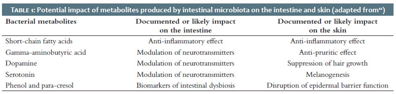
Table 1 provides an inventory of metabolic products originating from the intestinal microbiota that possess the capability to traverse the intestinal barrier, access the systemic circulation, and impact the skin.12
Numerous studies have shown the mutual relations between the colon microbiota and the functional and metabolic state and structure of the skin through the impact of the former on the immune system.
Short-chain fatty acids (monocarboxylic acids with a chain length of up to 6 carbon atoms) are the byproducts of the fermentation of undigested polysaccharides by intestinal bacteria. Among these compounds, acetate, propionate, and butyrate dominate in the gastrointestinal tract, constituting more than 95% of the total, with formate, valerate, caproate, and others comprising the remaining fraction.13 Acetate and propionate are primarily produced by representatives of the Bacteroidetes phylum, while bacteria of the Firmicutes phylum, including representatives of the Bacillales and Lactobacillales, are the main sources of butyrate14, a key enhancer of epithelial barrier function.15 Excessive fat and sugar consumption with insufficient fiber intake, as is typical in the Western diet, disrupts the balanced Firmicutes/Bacteroidetes ratio. This is accompanied by an increase in the permeability of the intestinal barrier that contributes to the development of inflammatory and immune diseases.16 The amount of short-chain fatty acids also decreases with the development of intestinal dysbiosis associated with the use of broad-spectrum antibiotics.17
Recent research has demonstrated that dietary fiber and short-chain fatty acids are able to modulate the immune response in various inflammatory conditions, extending their impact beyond the intestine to distant organs like the lungs18 and skin.19 The anti-inflammatory effect of these acids is attributed to the inhibition of the histone deacetylase enzyme by butyrate and propionate20, as well as the activation of metabotropic G-protein-coupled receptors such as GPR109A (known as the niacin receptor) by butyrate, and GPR41 (commonly referred to as the free fatty acid receptor 3, FFAR3) and GPR43 (FFAR2) by acetate, propionate, and butyrate. The inhibition of histone deacetylase with concurrent activation of histone acetyltransferase results in epigenetic post-translational modifications, accompanied by a reduction in the expression of proinflammatory cytokines, consequently restraining the systemic inflammatory response.
It has been revealed that GPR109A can activate colon macrophages and dendritic cells, promoting the differentiation of T-regulatory lymphocytes, which are responsible for producing the anti-inflammatory cytokine interleukin (IL)-10.21 Additionally, this receptor is able to block lipopolysaccharide (LPS)-induced activation of the transcription factor kappa B (NF-κB).22 The signaling pathway associated with this receptor plays a key role not only in colon inflammation but also in the development of various skin conditions, including psoriasis, inflammatory disorders such as incontinentia pigmenti, sunburn, allergic contact dermatitis, autoimmune diseases, and skin cancer.23 This suggests that GPR109A could be a promising therapeutic target for the treatment of skin diseases.
Recent evidence highlights that children and infants with dermatitis or a predisposition to allergic sensitization have a gut microbiota with diminished capacity for producing short-chain fatty acids, notably butyrate.24 These findings support the hypothesis that a low fiber intake, typical of Western lifestyles, may contribute to impairment of the skin barrier and subsequent susceptibility to early allergen sensitization. In line with this, Trompette et al.25, using an experimental model of atopic dermatitis, demonstrated that a diet enriched in fermented dietary fiber reduced systemic allergen sensitization and disease severity. The authors attribute this effect to the production of short-chain fatty acids, particularly butyrate, which enhances the functionality of not only the intestinal barrier but also the skin barrier through the induction of epidermal keratinocyte differentiation and the production of essential structural components of the epidermis.
Other metabolites produced by the intestinal microbiota have been found to impact skin function. For instance, bacterially produced gamma-aminobutyric acid (GABA), similarly to its endogenous counterpart, acts as an inhibitory neurotransmitter and thus possesses the ability to suppress neurons responsible for signaling skin itching.26 In a mouse model of atopic dermatitis, GABA demonstrated the ability to ameliorate skin lesions by rebalancing the levels of T helper cells of the Th1 and Th2 types, with a shift towards a predominance of Th1 cells.27 GABA is also able to inhibit matrix metalloproteinase-1 (MMP-1), an enzyme involved in the breakdown of type I collagen, and increase the expression of human type I collagen (COL1A1 and COL1A2). These processes play an important for maintaining skin elasticity.28
In turn, dopamine directly influences human hair follicles, inhibiting hair growth by inducing the catagen (resting stage) that is important for prevention of hirsutism and hypertrichosis.29 Serotonin is able to enhance melanogenesis through the activation of 5-HT2A receptors.30
The development of intestinal dysbiosis alters the systemic impact of gut microbiota metabolites. In such conditions, the blood plasma concentration of bioactive toxins, specifically phenol and para-cresol, which are byproducts of aromatic amino acids, rises. At present, these compounds are considered biomarkers for intestinal dysbiosis. Recent research has demonstrated their ability to reduce skin hydration and disrupt the epidermal barrier function due to derangement of keratinocyte differentiation.31
Regulating interactions between the host and microbiota stands as a fundamental role of the immune system, and regions inhabited by commensals, such as the skin and intestine, house a substantial portion of the body's immune cells. Given the predominant activity of the immune system, commensal microbial communities significantly influence mucosal immunity. Limiting the contact between microorganisms and the intestinal epithelial membrane to minimize inflammatory reactions and microbial translocation is crucial for maintaining the host's homeostatic balance. To achieve this segregation, the intestinal epithelial cell barrier, mucus layer, T cells, secretory immunoglobulin A, and dendritic cells collaborate to establish a protective structure termed the mucosal firewall. This structure restricts the movement of commensal bacteria to lymphoid tissues, thus preventing the development of inflammation.10 Furthermore, intestinal cells in their normal state demonstrate relatively low expression of Toll-like receptors, particularly types 2 and 4; this is associated with insensitivity to bacterial LPS enhanced by the production of IL-10 and TLR-inhibitory peptide by the colon mucosa.
The ability of the symbiotic microbiota to inhibit the translocation of NF-κB to the nucleus and thus eliminate the expression of several proinflammatory and prooxidant proteins, as well as histolytic enzymes, contributes to the protection of the host against inflammatory reactions to commensal microorganisms.32
However, any alteration in the gut microbial diversity can increase the vulnerability of the host and impair the immune tolerance of the intestinal mucosa,33 potentially impacting skin health.34
This condition significantly diminishes the anti-inflammatory properties of the normal microbiota and creates an environment for the passage of molecular structures associated with microorganisms – known as microbe-associated molecular patterns (MAMPs), such as LPS, peptidoglycan, flagellin, bacterial DNA, etc. – across the compromised intestinal epithelial cell barrier and into the systemic bloodstream. This can subsequently lead to the onset of a systemic inflammatory response.35
For instance, the DNA of intestinal microbiota representatives has been detected in plasma samples from patients with psoriasis. In a study involving 54 patients and 27 healthy controls, bacterial DNA was identified in 16 out of 54 patients with psoriasis, while none was observed in the control group. Furthermore, patients with psoriasis were found to have an increase in markers of systemic inflammatory response (γ-interferon, IL-1β, IL-6, IL-12, and tumor necrosis factor-α) compared to healthy controls. Bacterial DNA sequencing revealed the presence of the same type of microorganisms commonly found in the intestinal microbiota.10 Thus, the intake of MAMPs into skin tissues results in the development of inflammation, structural damage, and compromised epidermal barrier function.12 Based on contemporary theories, the development of intestinal dysbiosis involves a series of pathophysiological events that contribute to skin damage10,36: increased permeability of the intestinal barrier permitting the passage of microorganisms and their byproducts, leading to B-cell hypersensitivity, T-cell deterioration, and reduced secretion of secretory immunoglobulins A; dysbiotic intestinal microbiota, toxic products, neurotransmitters, and altered immune cells reach the skin tissue through the circulatory system. This transition shifts the skin condition from a state of health, characterized by a balanced microorganism composition and an appropriate level of antimicrobial peptides (from both human and bacterial sources) to a dysbiotic state; finally, MAMPs from the dysbiotic intestinal and skin microbiota trigger signaling cascades, such as NF-κB-dependent pathways, resulting in the degradation of skin connective tissue, disruption of the epidermal barrier function, inflammatory and immune-mediated skin damage, and development of skin diseases and/or aging.
It is important to note the bidirectional nature of the gut-skin axis. Exposure of the skin to ultraviolet B (UVB) radiation has been shown to enhance the diversity of the gut microbiome, likely mediated by vitamin D production.37 Indeed, the concentration of vitamin D in human serum was found to correlate with the relative abundance of the genera Lachnospira and Fusicatenibacter. Furthermore, disruptions in the skin barrier can potentially contribute to pathological processes in the intestine that are not directly linked to dysbiosis. For instance, sensitization of the body to epicutaneous exposure to peanut protein can result in immunoglobulin E-mediated intestinal infiltration by mastocytes.38
The probiotics most commonly used in dermatology and cosmetology include some species of Lactobacillus, Bifidobacterium, Enterococcus, and certain representatives of the Bacillus genus. Notably, a systemic course of probiotics is emerging as a novel approach to preserving skin health and function.
In recent years, several randomized, placebo-controlled clinical trials have yielded compelling evidence for the efficacy of oral probiotics in managing conditions such as atopic dermatitis39, 40 and psoriasis.41 These studies report that the use of probiotic microorganisms significantly improves the quality of life of patients, reduces disease severity and the risk of relapse, and normalizes inflammatory biomarker levels. Furthermore, a study has provided supporting evidence for a therapeutic benefit of a topical preparation containing Enterococcus faecalis fermentation products on the skin microbiome and the management of acne vulgaris.42 The Italian dermatologist Christian Diehl conducted a comprehensive analysis of the mechanisms responsible for anti-inflammatory and antioxidant effects of probiotics on the human and mammalian body.2 These mechanisms encompass: the production of various metabolites with anti-inflammatory and antioxidant properties, including butyrate, folate, and glutathione, by probiotic microorganisms such as Bifidobacteria and Lactobacillus fermentum; inhibition of NF-κB-dependent production of pro-inflammatory cytokines, matrix metalloproteinases, and reactive oxygen and nitrogen species by Bacillus spp. activation of the Nrf2-Keap1 signaling pathway – an antioxidant response element (Bacillus spp. strain LBP32); a histoprotective effect, which entails the inhibition of matrix metalloproteinase expression (seen in L. acidophilus and L. plantarum), along with anti-elastase and anti-collagenase activity (notably in L. casei, L. diolivorans, L. rhamnosus, and lactobacillus exopolysaccharides); inhibition of enzymes responsible for the production of reactive oxygen species, including NADPH oxidase and cyclooxygenase-2 (seen in L. fermentum CECT5716, L. coryniformis CECT5711, and L. gasseri CECT5714); chelation of metal ions, specifically Fe2+ and Cu2+ cations (Streptococcusthermophilus 821, L. casei KCTC3260, and L. helveticus CD6); and expression of enzymatic antioxidants such as superoxide dismutase, catalase, and glutathione peroxidase (seen in L. fermentum and L. lactis). In animal experiments, probiotic bacteria have demonstrated the ability to provide photoprotective effects when applied to the skin (e.g., Bifidobacterium breve, L. johnsonii, L. plantarum HY7714, and L. acidophilus), accelerate the healing of skin wounds (as observed with L. plantarum), improve skin barrier function (Streptococcus thermophilus, L. plantarum, and Bifidobacterium breve), and enhance skin hydration (Bifidobacterium).2 The ability of probiotics to improve skin hydration and prevent photoaging was confirmed in a randomized, double- blind, placebo-controlled clinical trial in which L. plantarum HY7714 was taken orally by participants with dry skin and wrinkles.43 The laboratory of Theofilos Poutahidis discovered a rejuvenating effect of probiotic bacteria on the skin and fur of elderly mice.44 According to the investigators, consumption of probiotic yogurt causes a shift in the anagen phase (the active phase of hair growth) with sebocytogenesis that leads to the formation of thick and shiny fur due to induction of the anti-inflammatory cytokine IL-10 and the neurohormone oxytocin by bacteria. In older male mice treated with probiotics, there was an observed increase in subcuticular folliculogenesis when compared to the control group.44 Additionally, the shinier hair in the female experimental group was considered by the researchers as a sign that correlates with fertility. However, it is important to note that probiotic bacteria can have side effects when taken orally, including septic conditions (in cases of intestinal barrier dysfunction and immunodeficiency), immune and metabolic disorders, and outcomes resulting from horizontal gene transfer.45 There have been reported cases of bacteremia and sepsis associated with the consumption of probiotics containing L. acidophilus,L. casei, S. boulardii, L. rhamnosus, Bifidobacterium breve, and Bacillus subtilis.
In the context of using probiotics for inflammatory skin diseases, lactobacilli may pose a certain risk due to their ability to activate the production of proinflammatory cytokines by type 1 T-helper cells.46 Moreover, L. reuteri has been found to stimulate autoimmune responses in a mouse model of lupus erythematosus.47
Modern literature sources criticize the idea that probiotic microorganisms can be effectively introduced to a stable biofilm formed by the resident microbiota. Furthermore, species within the Lactobacillus and Bifidobacterium genera are known for their very slow growth and marked sensitivity to gastric juice when taken orally, which impedes their passage through the gastrointestinal tract.48 It is widely accepted that the impacts of probiotic bacteria are primarily related to their direct effect on epithelial and immune cells, as well as the maintenance of problematic microbiota through the exchange of gases and metabolites.49
Thus, the current scientific literature substantiates a need to search for effective probiotic agents suitable for oral and topical administration in dermatology and cosmetology, while maintaining a high safety profile. In this context, the use of representatives of the transient microbiota – in particular, spore-forming bacteria within the Bacillus genus – as probiotics appears quite promising. These microorganisms, despite their brief residence in the intestine and their inability to integrate into the biofilm, have the potential to positively influence its function, fortify epithelial barriers, and counteract immune, inflammatory, and metabolic disorders associated with intestinal and skin dysbiosis, as well as underlying diseases and age-related changes.
The genus Bacillus comprises 77 species, constituting a substantial group of gram-positive, rod-shaped microorganisms that are primarily aerobic, but can tolerate anaerobic conditions, and form heat-resistant endospores.50 These bacilli, alongside lactobacilli, constitute the major components of the colon microbiota. By producing catalase and subtilisin, bacilli can promote the growth and viability of Lactobacillus culture. It is noteworthy that certain bacilli, such as B. subtilis var. natto, have been used in the fermentation of Asian foods since time immemorial. Most commercial Bacillus-based probiotics contain B. subtilis, B. polyfermenticus, or B. clausii, and some include B. cereus, B. coagulans, B. pumilus, and B. licheniformis, whose spores exhibit stability during storage and resistance to temperature variations, gastric acidity, and bile.51
Upon reaching the mucous membranes of the oral cavity and pharynx and encountering the gastric milieu, these spores become activated and begin vegetative growth. Analysis of fecal samples has revealed that probiotic strains of B. cereus, B. clausii, and B. pumilus can persist in the gastrointestinal tract of mice for up to 16 days.50 A number of reviews and original publications highlight the benefits of probiotic strains of Bacillus, the most important being their safety even at high concentrations, their antagonism to a wide range of pathogenic and opportunistic microorganisms, their ability to synthesize useful biologically active compounds, their positive impact on the immune status of the host organism, and their antimutagenic, antioxidant, anti-inflammatory, histoprotective, and antiproliferative properties. Bacillus strains also exhibit resistance to lytic enzymes, ensuring high viability in the gastrointestinal tract, and are environmentally safe.51
According to recent studies, about 800 substances that can be produced by bacilli possess antibacterial properties. Among these compounds are bacteriocins, which are ribosomal peptides or proteins undergoing post-translational modifications (subtilin, erycin S, coagulin, and megacin), as well as antibiotics (bacillisin and surfactin).52
The prospects for applying Bacillus bacteria as probiotics in dermatology and cosmetology have significantly expanded, primarily due to their remarkable antioxidant, anti-inflammatory, immunomodulatory, histoprotective, and antiproliferative properties. Many of these properties can be attributed to the ability of bacilli to produce exopolysaccharides (EPS), high-molecular-weight byproducts of bacterial metabolism.53 For instance, EPS derived from B. subtilis have been shown to effectively regulate cytokine production by T-helper types 1 and 254, promote the polarization of macrophages toward the M2 phenotype55, and reduce the expression of key components in proinflammatory signaling pathways, including transcription factors NF-κB and STAT6, as well as Janus kinase 1 (JAK1).56 These transcription factors play pivotal roles in the pathogenesis of psoriasis and other inflammatory skin disorders, as well as age-related disorders. When B. subtilis was introduced into enterocyte cultures, a notable reduction in the production of proinflammatory IL-8 and inducible NO synthase isoforms in response to various stimuli (IL-1β, deoxynivalenol, and flagellin) was observed. This reduction was attributed to the inhibition of NF-κB activation, achieved by disrupting the degradation of the inhibitory protein IκB.57 This study also demonstrated that certain strains of B. subtilis are able to enhance the integrity of the intestinal barrier by upregulating the expression of tight junction proteins. Moreover, probiotic strains of this species demonstrate the ability to attenuate the degradation of connective tissue components within the extracellular matrix.58
A useful trait that favorably distinguishes B. subtilis is its ability to slow aging and extend life expectancy, as evidenced in a study using the nematode Caenorhabditis elegans as a model organism.59 It is important to emphasize that this effect of B. subtilis primarily resulted from downregulation of the insulin-like growth factor signaling pathway, which is characteristic of the healthy longevity observed in centenarians. EPS derived from B. amyloliquefaciens can also reduce the expression of pro-inflammatory cytokines, phagocytic activity, and oxidative stress, effects associated with the inhibition of NF-κB signaling and extracellular signal-regulated protein kinase 1/2.60 Moreover, EPS activates the nuclear factor erythroid 2-related factor 2 (Nrf2)-regulated antioxidant-response element, an NF-κB-antagonistic signaling pathway. Collectively, these mechanisms considerably reduce the manifestations of oxidative stress and the severity of inflammation.
Recently, the ability of Bacillus spp. probiotics to block the signaling system of pathogenic microorganism colonies has been discovered, which is implemented through the mechanism known as quorum sensing, a means of maintaining the "social behavior" of bacteria. This capability of bacilli is highly important for correcting skin dysbiosis and creating the prerequisites for incorporating specific strains of the Bacillus genus into topical probiotics and personal care products (body sprays, soaps, skin creams, toothpastes, toothbrush cleaners, etc.). At present, such species as B. subtilis, B. licheniformis, and B. pumilus are already being used for these purposes.52
One of the most promising approaches to maximize the positive impact of bacilli while mitigating potential side effects associated with their consumption (the risk of enterotoxin formation, antibiotic resistance, and biogenic amine production) is the development and application of multi-component probiotic formulations. Based on research conducted by Ukrainian scientists, the most balanced probiotic composition providing antioxidant, anti-inflammatory, immunomodulatory, histoprotective, and antiproliferative effects with a high safety profile is the product Remedium™, a formulation which includes five multidrug-resistant strains of the genus Bacillus (B. subtilis, B. amyloliquefaciens,B. licheniformis, B. pumilus, and B. megaterium)50. The safety of this probiotic composition has been validated by the U.S. Food and Drug Administration (FDA). One dose of the product contains 1.7×10⁹ CFU/vial of live probiotic bacteria.
It is noteworthy that this medication contains two bacillus species, B. subtilis and B. amyloliquefaciens, which have proven pharmacological action associated with their impact on the NF-κB, STAT, and Nrf2 signaling systems. Another important effect of this preparation is its antibacterial properties, attributed to the antagonistic behavior of probiotic strains against a broad spectrum of opportunistic pathogens, particularly bacteria of the genera Staphylococcus and Proteus, as well as Candida fungi.50
The commensal microbiota in the colon maintains constant functional communication with skin cells and elements of its microbiocenosis, a phenomenon known as the gut-skin axis. This interaction occurs through the production of bioregulatory compounds (short-chain fatty acids, GABA, serotonin, dopamine, and others) and metabolites and involves the participation of both the innate and adaptive immune effectors. The development of intestinal dysbiosis alters the systemic impact of metabolites produced by the intestinal microbiota and increases the permeability of the intestinal barrier, allowing microorganisms, toxic substances, neurotransmitters, and modified immune cells to enter the bloodstream. These components then reach the skin through the circulatory system, disrupting its microbial community, connective tissue structure, and epidermal barrier function. These disruptions contribute significantly to the pathogenesis of skin diseases and hasten aging.
Contemporary literature consistently supports the practicality of using both oral and topical probiotics in dermatology and cosmetology. This approach yields notable benefits, such as improving patients' quality of life and ameliorating the course of inflammatory, immune, and hyperproliferative skin conditions (acne, atopic dermatitis, psoriasis, hidradenitis suppurativa, rosacea, seborrheic dermatitis, focal alopecia, and skin cancer), reducing the severity of these conditions and the likelihood of recurrence. Probiotics also provide protective effects against environmental factors and accelerate the process of skin wound healing.
Modern literature extensively substantiates the expediency of using probiotic strains of spore-forming bacteria of the genus Bacillus in dermatology and cosmetology based on their properties to restore the normal functioning of the gut-skin axis, suppress the growth of pathogenic microorganisms, decelerate aging, and provide significant antioxidant, anti-inflammatory, immunomodulatory, histoprotective, and antiproliferative effects, including those associated with impact on intracellular signaling systems.
The use of multicomponent probiotics composed of the most suitable strains of the genus Bacillus is a promising way to optimize the positive effects of these microorganisms in dermatological and cosmetic practice and avoid their undesirable effects.
The authors declare that there is no known conflict of interest regarding this article.
Yana Yemchenko
ORCID: 0000-0003-1207-6777
Collecting, analyzing, and interpreting data; effective participation in research guidance; critical review of the literature; critical review of the manuscript
Kateryna Bardova
ORCID: 0000-0002-1765-7549
Author's contribution: Approval of the final version of the manuscript; study design and planning; preparation and writing of the manuscript; intellectual participation in propaedeutic and/or therapeutic conduct of studied cases; critical review of the manuscript
Oleh Akimov
ORCID: 0000-0002-4958-3695
Approval of the final version of the manuscript; preparation and writing of the manuscript; critical review of the literature; critical review of the manuscript
Anatolii Levkov
ORCID: 0000-0003-0596-440X
Preparation and writing of the manuscript; effective participation in research guidance; intellectual participation in propaedeutic and/or therapeutic conduct of studied cases
Heorhii Kostenko 0009-0002-6361-9305
Collecting, analyzing, and interpreting data; intellectual participation in propaedeutic and/or therapeutic conduct of studied cases; critical review of the literature
Viktoriia Kostenko
ORCID: 0000-0001-9077-2191
Collecting, analyzing, and interpreting data; effective participation in research guidance; critical review of the literature; critical review of the manuscript
Artur Mishchenko
ORCID: 0000-0001-8521-956X
Approval of the final version of the manuscript; effective participation in research guidance; intellectual participation in propaedeutic and/or therapeutic conduct of studied cases; critical review of the literature
Natalia Solovyova
ORCID: 0000-0001-5167-2729
Preparation and writing of the manuscript; collecting, analyzing, and interpreting data; intellectual participation in propaedeutic and/or therapeutic conduct of studied cases; critical review of the literature
Vitalii Kostenko
ORCID: 0000-0002-3965-1826
Approval of the final version of the manuscript; study design and planning; preparation and writing of the manuscript; intellectual participation in propaedeutic and/or therapeutic conduct of studied cases; critical review of the literature; critical review of the manuscript
1. Hill C, Guarner F, Reid G, Gibson GR, Merenstein DJ, Pot B, et al. Expert consensus document. The International Scientific Association for Probiotics and Prebiotics consensus statement on the scope and appropriate use of the term probiotic. Nat Rev Gastroenterol Hepatol. 2014;11(8):506-14.
2. Diehl C. Probiotics in dermatology. Ukr J Dermatol Venereol Cosmetol. 2019;(2):99-108.
3. Holma RM, Kekkonen RA, Hatakka K, Poussa T, Vaarala O, Adlercreutz H, et al. Consumption of Galactooligosaccharides together with probiotics stimulates the In Vitro peripheral blood mononuclear cell proliferation and IFNγ production in healthy men. ISRN Immunology. 2011;2011:584682.
4. Valdes AM, Walter J, Segal E, Spector TD. Role of the gut microbiota in nutrition and health. BMJ. 2018;361:k2179.
5. Cristofori F, Dargenio VN, Dargenio C, Miniello VL, Barone M, Francavilla R. Anti- inflammatory and immunomodulatory effects of probiotics in gut inflammation: a door to the body. Front Immunol. 2021;12:578386.
6. Belizário JE, Faintuch J. Microbiome and gut dysbiosis. Exp Suppl. 2018;109:459-476.
7. Martel J, Chang SH, Ko YF, Hwang TL, Young JD, Ojcius DM. Gut barrier disruption and chronic disease. Trends Endocrinol Metab. 2022;33(4):247-265.
8. Yoo JY, Sniffen S, McGill Percy KC, Pallaval VB, Chidipi B. Gut dysbiosis and immune system in Atherosclerotic Cardiovascular Disease (ACVD). Microorganisms. 2022;10(1):108.
9. Strati F, Pujolassos M, Burrello C, Giuffrè MR, Lattanzi G, Caprioli F, et al. Antibiotic-associated dysbiosis affects the ability of the gut microbiota to control intestinal inflammation upon fecal microbiota transplantation in experimental colitis models. Microbiome. 2021;9(1):39.
10. Mahmud MR, Akter S, Tamanna SK, Mazumder L, Esti IZ, Banerjee, et al. Impact of gut microbiome on skin health: gut- skin axis observed through the lenses of therapeutics and skin diseases. Gut Microbes. 2022;14(1):2096995.
11. Clarke G, Stilling RM, Kennedy PJ, Stanton C, Cryan JF, Dinan TG. Minireview: Gut microbiota: the neglected endocrine organ. Mol Endocrinol. 2014;28(8):1221-1238.
12. De Pessemier B, Grine L, Debaere M, Maes A, Paetzold B, Callewaert C. Gut-skin axis: current knowledge of the interrelationship between microbial dysbiosis and skin conditions. Microorganisms. 2021;9(2):353.
13. Den Besten G, Van Eunen K, Groen AK, Venema K, Reijngoud DJ, Bakker BM. The role of short-chain fatty acids in the interplay between diet, gut microbiota, and host energy metabolism. J Lipid Res. 2013;54(9):2325-2340.
14. Levy M, Thaiss CA, Elinav E. Metabolites: messengers between the microbiota and the immune system. Genes Dev. 2016;30(14):1589-1597.
15. Zheng L, Kelly CJ, Battista KD, Schaefer R, Lanis JM, Alexeev EE, et al. Microbial-Derived Butyrate Promotes Epithelial Barrier Function through IL-10 receptor-dependent repression of claudin-2. J Immunol. 2017;199(8):2976-2984.
16. Richards JL, Yap YA, McLeod KH, Mackay CR, Mariño E. Dietary metabolites and the gut microbiota: an alternative approach to control inflammatory and autoimmune diseases. Clin Transl Immunology. 2016;5(5):e82.
17. Scott NA, Andrusaite A, Andersen P, Lawson M, Alcon-Giner C, Leclaire C, et al. Antibiotics induce sustained dysregulation of intestinal T cell immunity by perturbing macrophage homeostasis. Sci Transl Med. 2018;10(464):eaao4755.
18. Trompette A, Gollwitzer ES, Pattaroni C, Lopez-Mejia IC, Riva E, Pernot J, et al. Dietary fiber confers protection against flu by shaping Ly6c- patrolling monocyte hematopoiesis and CD8+ T cell metabolism. Immunity. 2018;48(5):992-1005.e8.
19. Schwarz A, Bruhs A, Schwarz T. The short-chain fatty acid sodium butyrate functions as a regulator of the skin immune system. J Invest Dermatol. 2017;137(4):855-864.
20. Hinnebusch BF, Meng S, Wu JT, Archer SY, Hodin RA. The effects of short-chain fatty acids on human colon cancer cell phenotype are associated with histone hyperacetylation. J Nutr. 2002;132(5):1012-1017.
21. Singh N, Gurav A, Sivaprakasam S, Brady E, Padia R, Shi H, et al. Activation of Gpr109a, receptor for niacin and the commensal metabolite butyrate, suppresses colonic inflammation and carcinogenesis. Immunity. 2014;40(1):128-139.
22. Thangaraju M, Cresci GA, Liu K, Ananth S, Gnanaprakasam JP, Browning DD, et al. GPR109A is a G-protein-coupled receptor for the bacterial fermentation product butyrate and functions as a tumor suppressor in colon. Cancer Res. 2009;69(7):2826-2832.
23. Sur I, Ulvmar M, Toftgård R. The two-faced NF-kappaB in the skin. Int Rev Immunol. 2008;27(4):205-223.
24. Ta LDH, Chan JCY, Yap GC, Purbojati RW, Drautz-Moses DI, Koh YM, et al. A compromised developmental trajectory of the infant gut microbiome and metabolome in atopic eczema. Gut Microbes. 2020;12(1):1-22.
25. Trompette A, Pernot J, Perdijk O, Alqahtani RAA, Domingo JS, Camacho-Muñoz D, et al. Gut-derived short-chain fatty acids modulate skin barrier integrity by promoting keratinocyte metabolism and differentiation. Mucosal Immunol. 2022;15(5):908-926.
26. Akiyama T, Carstens MI, Carstens E. Transmitters and pathways mediating inhibition of spinal itch-signaling neurons by scratching and other counterstimuli. PLoS One. 2011;6(7):e22665.
27. Hokazono H, Omori T, Ono K. Effects of single and combined administration of fermented barley extract and gamma-aminobutyric acid on the development of atopic dermatitis in NC/Nga mice. Biosci Biotechnol Biochem. 2010;74(1):135-139.
28. Uehara E, Hokazono H, Sasaki T, Yoshioka H, Matsuo N. Effects of GABA on the expression of type I collagen gene in normal human dermal fibroblasts. Biosci Biotechnol Biochem. 2017;81(2):376-379.
29. Langan EA, Lisztes E, Bíró T, Funk W, Kloepper JE, Griffiths CE, et al. Dopamine is a novel, direct inducer of catagen in human scalp hair follicles in vitro. Br J Dermatol. 2013;168(3):520-525.
30. Lee HJ, Park MK, Kim SY, Choo HYP, Lee AY, Lee CH. Serotonin induces melanogenesis via serotonin receptor 2A. Br J Dermatol. 2011;165(6):1344-1348.
31. Miyazaki K, Masuoka N, Kano M, Iizuka R. Bifidobacterium fermented milk and galacto- oligosaccharides lead to improved skin health by decreasing phenols production by gut microbiota. Benef Microbes. 2014;5(2):121-128.
32. Neish AS. The gut microflora and intestinal epithelial cells: a continuing dialogue. Microbes Infection. 2002;4(3):309-317.
33. Renz H, Brandtzaeg P, Hornef M. The impact of perinatal immune development on mucosal homeostasis and chronic inflammation. Nat Rev Immunol. 2011;12(1):9-23.
34. Pascal M, Perez-Gordo M, Caballero T, Escribese MM, Longo MNL, Luengo O, et al. Microbiome and Allergic Diseases. Front Immunol. 2018;9:1584.
35. Li X, Watanabe K, Kimura I. Gut microbiota dysbiosis drives and implies novel therapeutic strategies for diabetes mellitus and related metabolic diseases. Front Immunol. 2017;8:1882.
36. Ratanapokasatit Y, Laisuan W, Rattananukrom T, Petchlorlian A, Thaipisuttikul I, Sompornrattanaphan M. How microbiomes affect skin aging: the updated evidence and current perspectives. Life (Basel). 2022;12(7):936.
37. Bosman ES, Albert AY, Lui H, Dutz JP, Vallance BA. Skin exposure to narrow band ultraviolet (UVB) light modulates the human intestinal microbiome. Front Microbiol. 2019;10:2410.
38. Bartnikas LM, Gurish MF, Burton OT, Leisten S, Janssen E, Oettgen HC, et al. Epicutaneous sensitization results in IgE-dependent intestinal mast cell expansion and food-induced anaphylaxis. J Allergy Clin Immunol. 2013;131(2):451-460.e1-6.
39. Aldaghi M, Tehrani H, Karrabi M, Abadi FS, Sahebkar M. The effect of multistrain synbiotic and vitamin D3 supplements on the severity of atopic dermatitis among infants under 1 year of age: a double-blind, randomized clinical trial study. J Dermatolog Treat. 2022;33(2):812-817.
40. Prakoeswa CRS, Bonita L, Karim A, Herwanto N, Umborowati MA, Setyaningrum T, et al. Beneficial effect of lactobacillus plantarum IS-10506 supplementation in adults with atopic dermatitis: a randomized controlled trial. J Dermatolog Treat. 2022;33(3):1491-1498.
41. Moludi J, Khedmatgozar H, Saiedi S, Razmi H, Alizadeh M, Ebrahimi B. Probiotic supplementation improves clinical outcomes and quality of life indicators in patients with plaque psoriasis: a randomized double-blind clinical trial. Clin Nutr ESPEN. 2021;46:33-39.
42. Han HS, Shin SH, Choi BY, Koo N, Lim S, Son D, et al. A split face study on the effect of an anti-acne product containing fermentation products of Enterococcus faecalis CBT SL-5 on skin microbiome modification and acne improvement. J Microbiol. 2022;60(5):488-495.
43. Lee DE, Huh CS, Ra J, Choi ID, Jeong JW, Kim SH, et al. Clinical evidence of effects of Lactobacillus plantarum HY7714 on skin aging: a randomized, double blind, placebo-controlled Study. J Microbiol Biotechnol. 2015;25(12):2160-2168.
44. Levkovich T, Poutahidis T, Smillie C, Varian BJ, Ibrahim YM, Lakritz JR, et al. Probiotic bacteria induce a 'glow of health'. PLoS One. 2013;8(1):e53867.
45. Zawistowska-Rojek A, Tyski S. Are probiotic really safe for humans?. Pol J Microbiol. 2018;67(3):251-258.
46. Dong H, Rowland I, Yaqoob P. Comparative effects of six probiotic strains on immune function in vitro. Br J Nutr. 2012;108(3):459-470
47. Zegarra-Ruiz DF, El Beidaq A, Iñiguez AJ, Lubrano Di Ricco M, Manfredo Vieira S, Ruff WE, et al. A diet-sensitive commensal lactobacillus strain mediates TLR7-dependent systemic autoimmunity. Cell Host Microbe. 2019;25(1):113-127.e6.
48. Penaloza-Vazquez A. Bacillus species are superior probiotic feed-additives for poultry. J Bacteriol Mycol. 2016;3:00023.
49. Wieërs G, Belkhir L, Enaud R, Leclercq S, Philippart de Foy JM, Dequenne I, et al. How Probiotics Affect the Microbiota. Front Cell Infect Microbiol. 2020;9:454.
50. Golovach IY, Rekalov DG, Kostenko VO. Prospects for applying probiotics based on spores of bacillus species in the integral therapy for diseases of the musculoskeletal system. Ukr Rheumatol J. 2022;(3):1-9.
51. Sorokulova I. Modern status and perspectives of bacillus bacteria as probiotics. J Prob Health. 2013;1(4):1000e106.
52. Jeżewska-Frąckowiak J, Seroczyńska K, Banaszczyk J, Jedrzejczak G, Żylicz-Stachula A, Skowron PM. The promises and risks of probiotic Bacillus species. Acta Biochim Pol. 2018;65(4):509-519.
53. Sathishkumar R, Kannan R, Jinendiran S, Sivakumar N, Selvakumar G, Shyamkumar R. Production and characterization of exopolysaccharide from the sponge-associated bacillus subtilis MKU SERB2 and its in-vitro biological properties. Int J Biol Macromol. 2021;166:1471-1479.
54. Bang MA, Seo JH, Seo JW, Jo GH, Jung SK, Yu R, et al. Bacillus subtilis KCTC 11782BP-produced alginate oligosaccharide effectively suppresses asthma via T-helper cell type 2-related cytokines. PLoS One. 2015;10(2):e0117524.
55. Li R, Kou X, Tian J, Meng Z, Cai Z, Cheng F, et al. Effect of sulfur dioxide on inflammatory and immune regulation in asthmatic rats. Chemosphere. 2014;112:296-304.
56. Zhang L, Yi H. An exopolysaccharide from bacillus subtilis alleviates airway inflammatory responses via the NF-κB and STAT6 pathways in asthmatic mice. Biosci Rep. 2022;42(1):BSR20212461.
57. Rhayat L, Maresca M, Nicoletti C, Perrier J, Brinch KS, Christian S, et al. Effect of bacillus subtilis strains on intestinal barrier function and inflammatory response. Front Immunol. 2019;10:564.
58. Sojan JM, Raman R, Muller M, Carnevali O, Renn J. Probiotics enhance bone growth and rescue BMP inhibition: new transgenic zebrafish lines to study bone health. Int J Mol Sci. 2022;23(9):4748.
59. Ayala FR, Bauman C, Cogliati S, Leñini C, Bartolini M, Grau R. Microbial flora, probiotics, bacillus subtilis and the search for a long and healthy human longevity. Microb Cell. 2017;4(4):133-136.
60. Sung WW, Lin YY, Huang SD, Cheng HL. Exopolysaccharides of bacillus amyloliquefaciens amy-1 mitigate inflammation by inhibiting ERK1/2 and NF-κB pathways and activating p38/Nrf2 pathway. Int J Mol Sci. 2022;23(18):10237.
 All content the journal, except where identified, under the Creative Commons Attribution 4.0 International licence - ISSN-e 1984-8773


 Read in Portuguese
 Portuguese PDF
 Print
Print
 Send this article by email
 How to cite this article
 Submit a comment
 Mendeley
 Pocket